RESULTS AND DISCUSSION
 The CT and NT images differ from each other strongly in quality and displayed contrasts. In the CT images boundaries between cavities filled with sediment and surrounding fossil bone are sharply marked (Figure 2,
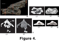
Figure 4,
Figure 5). A significant contrast appears between darkly depicted sediment matrix and brightly depicted fossil bone. Comparatively in the NT images the contrast between sediment matrix and fossil bone is low and boundaries between different materials are blurred (Figure 3,
Figure 4,
Figure 5). Sediment-filled cavities (e.g., canals and foramina within the vertebrae) are easy to identify in the CT images and difficult to trace in the NT images (Figure 4c-d,
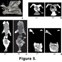
Figure 5c).
The CT and NT images differ from each other strongly in quality and displayed contrasts. In the CT images boundaries between cavities filled with sediment and surrounding fossil bone are sharply marked (Figure 2,
Figure 4,
Figure 5). A significant contrast appears between darkly depicted sediment matrix and brightly depicted fossil bone. Comparatively in the NT images the contrast between sediment matrix and fossil bone is low and boundaries between different materials are blurred (Figure 3,
Figure 4,
Figure 5). Sediment-filled cavities (e.g., canals and foramina within the vertebrae) are easy to identify in the CT images and difficult to trace in the NT images (Figure 4c-d,
Figure 5c).
 The high contrast between sediment matrix and fossil bone in the CT images mirrors the differences in density and material Z between the two materials (Figure 1,
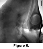
Table 1). In the NT images, the low contrast between fossil bone and sediment matrix is a response to the small difference in the linear attenuation coefficient for thermal neutrons of calcium (i.e., the main constituent of apatite) and silica (i.e., the main constituent of sediment around and within the vertebrae) (Table 1). The additional blur in the NT slices is due to a higher scattering background induced by the hydrogen content of resin material and the use of a neutron area detector. Because both neutron and X-ray attenuation are dependent on the type of sediment, the contrast between fossil bone and sediment would probably be higher with a different sediment composition. For example, in the neutron radiography of an ichthyosaur head embedded in a shale matrix, the bone structures are clearly visible and distinguishable from the sediment matrix (Figure 6).
The high contrast between sediment matrix and fossil bone in the CT images mirrors the differences in density and material Z between the two materials (Figure 1,
Table 1). In the NT images, the low contrast between fossil bone and sediment matrix is a response to the small difference in the linear attenuation coefficient for thermal neutrons of calcium (i.e., the main constituent of apatite) and silica (i.e., the main constituent of sediment around and within the vertebrae) (Table 1). The additional blur in the NT slices is due to a higher scattering background induced by the hydrogen content of resin material and the use of a neutron area detector. Because both neutron and X-ray attenuation are dependent on the type of sediment, the contrast between fossil bone and sediment would probably be higher with a different sediment composition. For example, in the neutron radiography of an ichthyosaur head embedded in a shale matrix, the bone structures are clearly visible and distinguishable from the sediment matrix (Figure 6).
 The
gray level contrast between fossil bone and larger amounts of polyester cast
resin is high in both CT and NT images (Figure 2,
Figure 3,
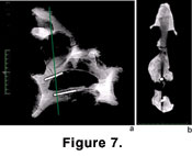
Figure 5). In the CT images, glued fractures and smaller cracks in the vertebrae appear dark like the background and are not distinguished from the latter by the X-rays, and very thin fractures are not displayed. However, this effect can be overcome by a different calibration of the CT scanner to bone density and/or using a high-resolution industrial X-ray scanner (Ketcham and Carlson 2001). In contrast to adhesives or resin, the presence of metal inclusions like iron (e.g., for stabilisation of the bones), would lead to a strong attenuation of the X-rays. Therefore, metal within objects causes a significant noise and decrease of CT image quality (Figure 7). High atomic number elements like metals can be better penetrated by neutrons (Figure 1), resulting in a much smaller decrease of image quality than in a CT image.
The
gray level contrast between fossil bone and larger amounts of polyester cast
resin is high in both CT and NT images (Figure 2,
Figure 3,
Figure 5). In the CT images, glued fractures and smaller cracks in the vertebrae appear dark like the background and are not distinguished from the latter by the X-rays, and very thin fractures are not displayed. However, this effect can be overcome by a different calibration of the CT scanner to bone density and/or using a high-resolution industrial X-ray scanner (Ketcham and Carlson 2001). In contrast to adhesives or resin, the presence of metal inclusions like iron (e.g., for stabilisation of the bones), would lead to a strong attenuation of the X-rays. Therefore, metal within objects causes a significant noise and decrease of CT image quality (Figure 7). High atomic number elements like metals can be better penetrated by neutrons (Figure 1), resulting in a much smaller decrease of image quality than in a CT image.
In the NT images, filling of cracks and fractures with glue is detectable by its high neutron attenuation shown as very bright regions (Figure 5b). The most blurred boundaries between areas filled with glue and fossil bone are due to diffusion of the glue in the bone matrix surrounding the fractures (Figure 4c,
Figure 5b-d) or due to the strongly scattered neutrons in hydrogen.
 Generally, the more light material like glue or polyester cast resin is present in the remains, the stronger the neutrons are deflected, causing significant noise and a decrease of image quality. This effect could be minimized by the use of a collimating linear neutron detector instead of the neutron area detector. The strong display of glue was helpful in the case of the sauropod vertebrae to distinguish between glue-filled cracks and pneumatic structures, and to show additional small, glue-filled pneumatic canals that were not displayed with CT.
The density of the fossil bone and a maximal thickness of the scanned vertebrae (17.9 cm) influenced image quality in the CT scans. Larger objects (up to 20 cm) generally were depicted with less resolution and a stronger blur than the smaller objects (about 10 cm). The imaging of dense material can be optimized by using an industrial X-ray CT with high voltage instead of a medical X-ray CT (Ketcham and Carlson 2001;
Rowe et al. 1999,
2001;
Tykoski 2002). However, as it was mentioned above, X-ray attenuation is additionally influenced by the type of sediment around or within the fossil bone. In NT, the maximal penetration thickness of non-organic material (i.e., rocks, sediment and fossilized bone) is about 10 cm, but we found no problem with penetration of a vertebra of 12 cm thickness. Image quality in NT depends strongly on the material composition of an object, and in the case of fossil bone material (apatite) and siliciclastic sediment (mainly silica and calcareous cement), the minimal difference in the linear neutron attenuation coefficients leads to low contrast.
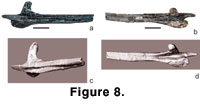 There is no difference between the 3-D polygon-surface models extracted from the volume data of the CT and NT scans. The resulting surface models are of high quality and allow 3-D modelling of flexibility between single vertebrae as well as building models of the objects with stereolithography (Figure 8).
