![]()
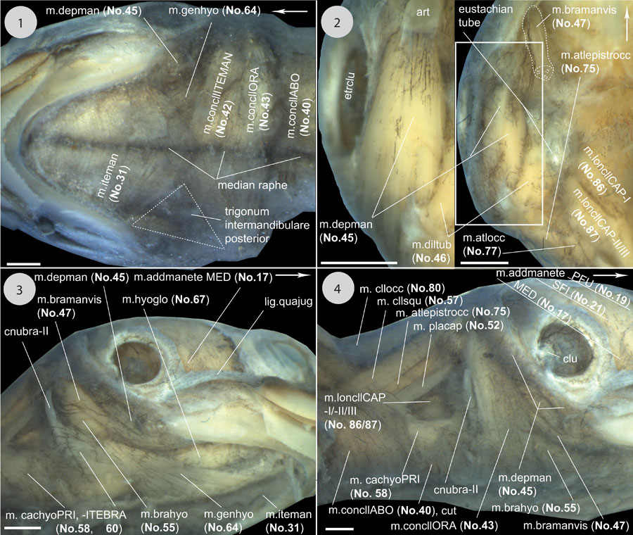
FIGURE 30. Continuation of Figure 28, Figure 29; continued in Figure 31. 1) ventral view to the head. 2) Ventral view to the otic region. Removed: hyoid apparatus, mm. intermandibularis (No. 31) et retrahens capiti collique Pars carapacobasioccipitalis (No. 88). In the left picture a detail is shown and the superficial, ventralmost fibres of m. depressor mandibulae (No. 45) are removed. 3) Ventrolateral view to the head/neck region. Removed (right side): mm. constrictor colli Pars intermandibularis (No. 42), m. intermandibularis (No. 31), and parts of m. constrictor colli Pars aboralis (No. 40). 4) Ventrolateral view to the head/neck region on the left body side. The superficial neck constrictors (No. 40, 42, 43 / m. sphincter colli posterior), m. intermandibularis (No. 31), and m. geniohyoideus (No. 64) are removed. Scale equals 2 mm.