![]()
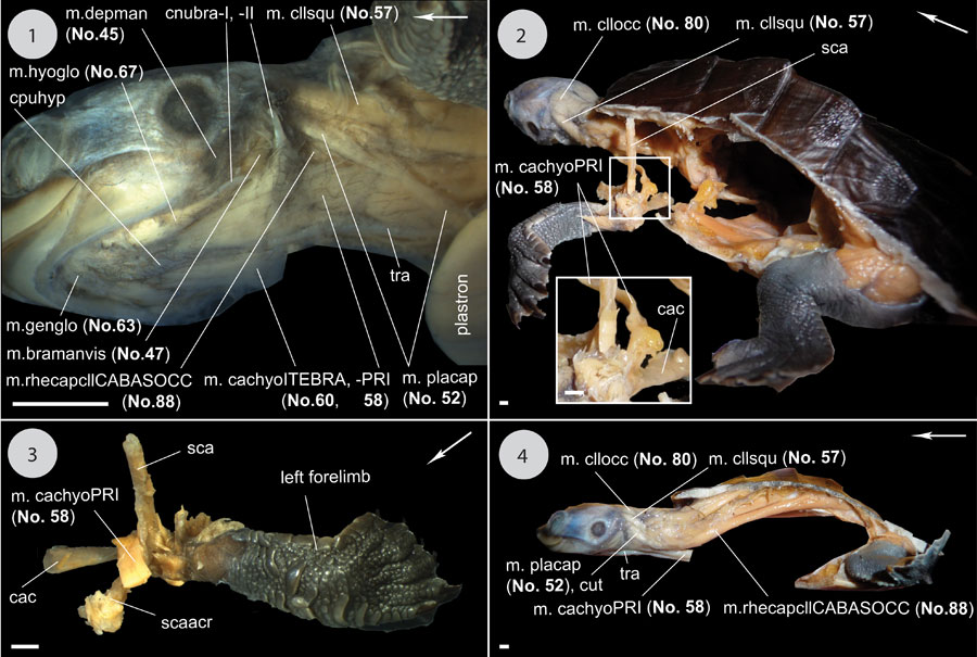
FIGURE 31. Continuation of Figure 28, Figure 29, Figure 30. 1) Ventrolateral view to the head. Removed: mm. constrictor colli (No. 40, 42, 43) and the left half of m. intermandibularis (No. 31). 2) Posterolateral and dorsal view to the body. Removed: parts of the carapace and the plastron, and the intestines. Focus on the insertion site of m. coracohyoideus (No. 58) on the shoulder girdle. 3) Anterodorsal view to the left forelimb and shoulder girdle. All muscles except m. coracohyoideus (No. 58) removed. 4) Lateral view to the whole animal: Removed: Forelimbs and shoulder girdles with the related muscles, most parts of the shell, intestines. Scale equals 2 mm.