INTRODUCTION
Microwear analysis requires images or 3D data to be acquired from tooth surfaces at relatively high magnification, sampling data from very small areas, typically only a few hundred micrometres across. In order for the microwear features and textures to be accurately detected or replicated it is imperative that the tooth surface be thoroughly cleaned prior to imaging or moulding. Any surface contaminant or coating that could potentially mask microwear must be removed. The cleaning process must not abrade or etch the tooth surface or leave residue that might obscure the original microwear.
Quantitative analysis of tooth microwear has been applied extensively to mammals (Walker et al. 1978;
Gordon 1984;
Teaford 1988;
Organ et al. 2005;
Ungar et al. 2007) and is starting to be applied to dinosaurs (Williams et al. 2009); prior studies of dinosaur tooth microwear have been qualitative (e.g.,
Fiorillo 1991,
1998;
Upchurch and Barrett 2000;
Schubert and Ungar 2005). The cleaning methods employed by most of these researchers involve soft brushing or gentle swabbing (using cotton swabs) with either distilled water or a solvent such as acetone or ethanol. Whilst these methods work well on material that has been treated with modern consolidants such as the methacrylate co-polymer Paraloid, they have proven time consuming and laborious where more traditional consolidants such as shellac or glyptal have been used and totally ineffective where the shellac has aged.
Material from the older museum collections (19th and early 20th century), particularly dinosaur material, has often been treated with one or both of two consolidants: shellac and animal resin. As shellac ages it darkens and becomes cross-linked (bonds develop that link one polymer chain to another) making it extremely resistant to solvents. As microwear analysis is increasingly applied to dinosaurs, more researchers are likely to discover this problem.
Problems with Brush-Based Cleaning:
When attempting to remove consolidant from the occlusal surface of a tooth by the brushing on of a solvent and continual cleaning of the brush, or by the use of disposable swabs, the whole tooth and surrounding area tends to become soaked in a combination of the solvent and dissolved consolidant. Given that consolidants like shellac typically form a coating rather than penetrate a surface when they are originally applied, this is a backward step. The brushing process also tends to move consolidant around, smearing it over microwear and making it difficult to determine when all vestiges have been removed. This technique is especially problematic if SEM analysis is to be performed on moulds and casts rather than the original specimens (brush marks in remaining consolidant are a particular hazard). Use of this technique also results in a high rate of solvent evaporation requiring the use of a fume cupboard. It has also been suggested that repeated applications of solvents such as alcohol and acetone can dehydrate enamel and dentine leading to surface damage (Fernandez-Jalvo and Monfort 2008) although this damage is questioned by dental researchers who claim dentine in particular becomes more resistant (e.g.,
Nalla et al. 2005).
The possibility of acetone-caused cracking is of particular concern as the process can be time consuming and requires repeated application of brushed-on solvent where the consolidant is shellac, as the older shellac is, the more resistant it tends to become.
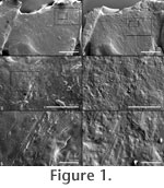 Figure 1 shows a tooth surface after each of two consecutive attempts to remove the consolidant coating via the brushing on of ethanol.
Figure 1.1, 1.3 and 1.5 show the first attempt. Brush strokes are clearly visible in the higher magnification images
Figure 1.3 and 1.5. Figure 1.2, 1.4 and 1.6 show the second attempt. Whilst an underlying pervasive and dominant near vertical microwear
pattern is emerging, sufficient varnish remains to in-fill and partially obscure
this pattern.
Figure 2 shows a tooth surface that was cleaned whilst being viewed under a stereo microscope (at x40 magnification giving a 5 mm field-of-view) by brushing on ethanol until it appeared to be clear of consolidant. Figure 1 shows a tooth surface after each of two consecutive attempts to remove the consolidant coating via the brushing on of ethanol.
Figure 1.1, 1.3 and 1.5 show the first attempt. Brush strokes are clearly visible in the higher magnification images
Figure 1.3 and 1.5. Figure 1.2, 1.4 and 1.6 show the second attempt. Whilst an underlying pervasive and dominant near vertical microwear
pattern is emerging, sufficient varnish remains to in-fill and partially obscure
this pattern.
Figure 2 shows a tooth surface that was cleaned whilst being viewed under a stereo microscope (at x40 magnification giving a 5 mm field-of-view) by brushing on ethanol until it appeared to be clear of consolidant.
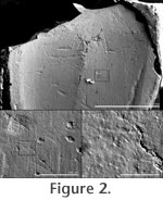 The higher magnification SEM images (Figure 2.2 and 2.3) clearly show that the tooth surface is still coated in consolidant. The higher magnification SEM images (Figure 2.2 and 2.3) clearly show that the tooth surface is still coated in consolidant.
It is both time consuming and frustrating to complete a sequence of brush cleaning, moulding, casting and SEM imaging only to discover that a tooth surface is not clean, especially if the original tooth is in a remote museum collection and a return visit must be arranged. A more reliable cleaning method is needed.
Solvent Gels
Art conservators wanting to clean varnish from paintings without damaging the oil paint beneath discovered that by suspending the solvent in a gel, they could limit evaporation and control both contact time and the pH. The addition of soaps and detergents to the gel allowed the dissolved varnish to be sequestered by the gel and thus easily removed from the painting (Southall 1988). They found that overpainted areas could be dealt with by the addition of xylene to the gel, causing partial dissolution and swelling of the paint layer (Wolbers et al. 1990). It is this solvent gel formulation, used for varnish on paintings, which we have adapted for the removal of various consolidants from dinosaur teeth, including aged shellac.
This paper describes the cleaning method used by the authors, developed from a technique pioneered by museum conservators. This technique, for the removal of varnish from oil paintings via the application of solvent gels, is not widely known, and papers describing its use (Hedley 1980;
Burnstock and White 1990;
Eastaugh 1990;
Wolbers et al. 1990;
Wolbers 1992) are not widely available.
|