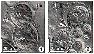
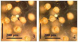
Figures 1 and 2. Nomarski optical images of early stages of binary division in central capsules (CCa and CCb) of a colonial radiolarian before the shells have been deposited, showing the silica-secreting cytokalymma (Ck), surrounding gelatinous coat of the colony (Gn), and algal symbionts (Sy). This is evidence of simultaneous shell deposition since all of the central capsules divide before the shells are deposited nearly simultaneously. Scale bar = 200 microns. Adapted from Anderson and Swanberg, 1981.

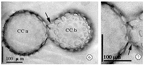
Figures 3 to 5. Light microsocopic images of a living colony of Acrosphaera showing a spherical bud-like mass (Figure 3, arrow) at the side of a shell-enclosed central capsule. The protrusion eventually separates at a greater distance (Figure 4, arrow) but is connected in the rhizopodial network. At other places in the colony, mature central capsules surrounded by a shell (Figure 5) are united as a pair resembling the fossil shells in Figure 6. This may be evidence of successive shell deposition; i.e., additional central capsules are produced after most have matured.


Figures 6 and 7. Light microscopic views of a united pair (arrows) of shells from fossil material showing the bud-like form of shell CCb attached to shell CCa.


