

| Figure 1. Shrew upper molar from TVOR Site, Miocene of Fort Polk, Louisiana. Occlusal view. Scanning electron micrograph by Xiaogang Xie and Suyin Ting. |
 |
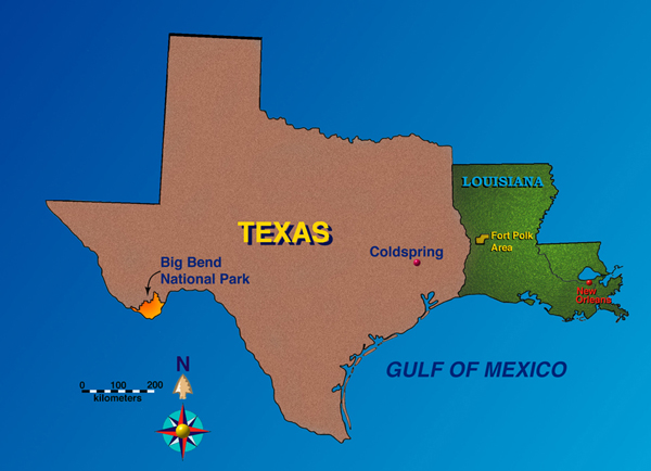
| Figure 2. Location map showing Fort Polk in Louisiana, and Big Bend National Park and Coldspring in Texas. Permits are required for research on Fort Polk and in Big Bend National Park. |
 |
| Figure 3. Lateral view of merychippine horse mandible encased in pedogenic nodular material from DISC Site, Miocene of Fort Polk, Louisiana. Photograph by Kerry Lyle. |
 |

| Figure 5. Judy's conglomerate, Late Cretaceous, Aguja Formation, Big Bend National Park, Texas, with Julia Sankey. Photograph by Jean Sankey. |
 |
| Figure 6. LSU field crew wet screening untreated mudstone in the Rio Grande, Big Bend National Park, Texas. |
 |
| Figure 7. Robert Rainey (L) and Judith Schiebout (R) bag conglomerate at Joe's Bonebed, Big Bend National Park, in 1970. Photograph by John A. Wilson. |
 |
| Figure 8. Helicopter carries bags for screening from remote sites in Big Bend National Park, Texas in 1984. |
 |
| Figure 9. Bags of rock from the Louisiana Miocene as brought in from the field, closed with duct tape. Airless jackhammer and conglomerate piece from Stonehenge Site rest on the bags. |
 |

| Figure 11. Dumping a screen on a drying tray. |
 |
| Figure 12. Dried screening residue from Stonehenge Site showing nodules darkened with iron and manganese oxides. Dr. Schiebout indicates a bone fragment. |
 |
| Figure 13. Dried screening residue from DISC Site showing nodules larger and lighter colored than those from Stonehenge Site. |
 |
| Figure 14. Dr. Ting in goggles and respirator, facial protective gear worn during handling of the glacial acetic acid. |
 |


| Figure 17. Judy's conglomerate, cross sectional view, Late Cretaceous, Aguja Formation, Big Bend National Park, Texas. Nodules and bone pieces show as white flecks. Photo by Julia Sankey. |
 |
| Figure 18. Dry (L) and wet (R) pieces of Julia's conglomerate from the Late Cretaceous, Aguja Formation, Big Bend National Park. Photo by Julia Sankey. |
 |
| Figure 19. Weathered, man-made surface of the main conglomerate at DISC Site, Miocene of Fort Polk. It will be broken up with sledge or airless jackhammer and shoveled into bags. |
 |
| Figure 20. Incisor on weathered surface of the main DISC conglomerate. |
 |
| Figure 21. Cleaned cross sectional surface showing contact of main conglomerate from DISC site and underlying overbank mudstone. |
 |
| Figure 22. Erosion at DISC Site revealing several minor layers of pedogenic nodule conglomerate. |
 |

| Figure 24. Close up, cross sectional view, of the conglomerate at TVOR Site showing both fresh dark surfaces which have been broken and lighter colored weathered surface. Cross bedding is evident. |
 |

| Figure 26. Thin section under polarized light, cut a few cm higher than Figure 25, from the upper part of the main conglomerate at DISC Site, showing more quartz sand. Scale = 1 mm. Photograph by Julitta Kirkova. |
 |

| Figure 28. Sawed, cross sectional surface of a conglomerate boulder from a site near Coldspring, Texas at which mammals of the Miocene Cold Spring Local Fauna had been recovered. |
 |
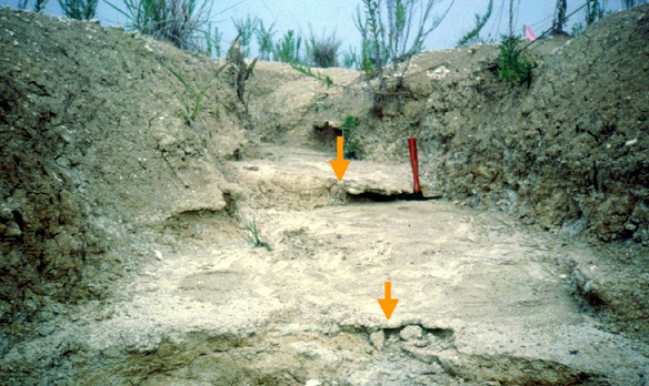
| Figure 29. Erosional gully, TVOR S Site in the Miocene of Fort Polk, shows a pedogenic nodule conglomerate, indicated by an arrow. This site is a kilometer from TVOR. Photo by Megan Jones. |
 |
| Figure 30. Close up of conglomerate seen in Figure 29. Photo by Megan Jones. |
 |
| Figure 31. Nodule encrusted and cracked large mammal bone in place at Joe's Bonebed in mudstone of the Paleocene Black Peaks Formation in Big Bend National Park, Texas. |
 |


| Figure 34. All mammal teeth from a session of picking at TVOR Site. A geomyoid rodent tooth is indicated by the arrow. |
 |
| Figure 35. 300-counts of kinds of fossils from TVOR Site (A, B, C) and DISC (D). Figure 35C is modified from figure 8 and 35D is modified from figure 7 of Schiebout (1997b). | ||
 |
||
 |
||
 |
||
 |
||

