FIGURE 1. Pseudogarypus synchrotron sp.n., holotype, right chelal fingers, PPC-SRµCT reconstruction of the pedipalp, at 0.667 µ of voxel size and 50 mm of propagation distance. 1: paraxial; 2: dorsal; 3:ventral; 4: antiaxal; 5: Pseudogarypus extensuswith brood pouch.
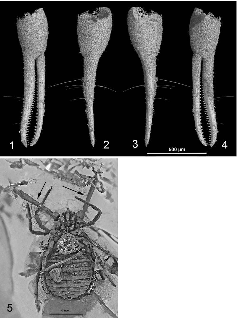
FIGURE 2. Pseudogarypus synchrotron sp.n., holotype. 1: inclusion in matrix (Baltic amber); 2: magnification in visible light; 3: PPC-SRµCT 3D reconstruction at PPC-SRµCT 3D reconstruction at 5.06 µm of voxel size, dorsal view; 4: ventral view.
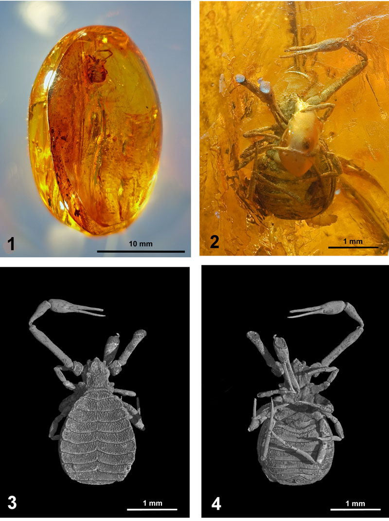
FIGURE 3. Pseudogarypus synchrotron sp.n., holotype. 1: left chela (mirror view); 2: Pseudogarypus hemprichii, right chela, (according BEIER, 1937); 3: Pseudogarypus synchrotron sp.n., paratype, right chela; 4: Pseudogarypus minor, right chela, (according BEIER, 1947); 5: Pseudogarypus extensus, left chela, (according to BEIER, 1937); 6: Pseudogarypus pangaea, left chela. The dimensions are given in mm.
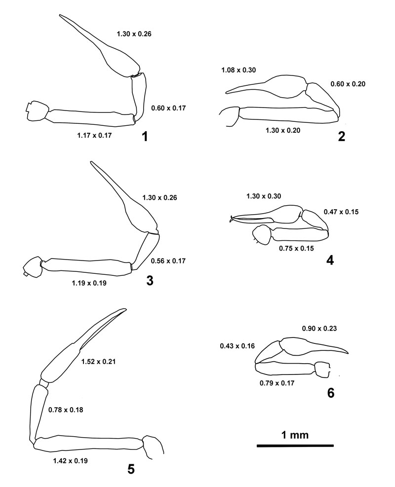
FIGURE 4. Pseudogarypus synchrotron sp.n., holotype, µPPC-SRµCT reconstruction at 2.14 µm of voxel size and 120 mm propagation distance. 1: right, 2: left, 3: front, 4: rear.
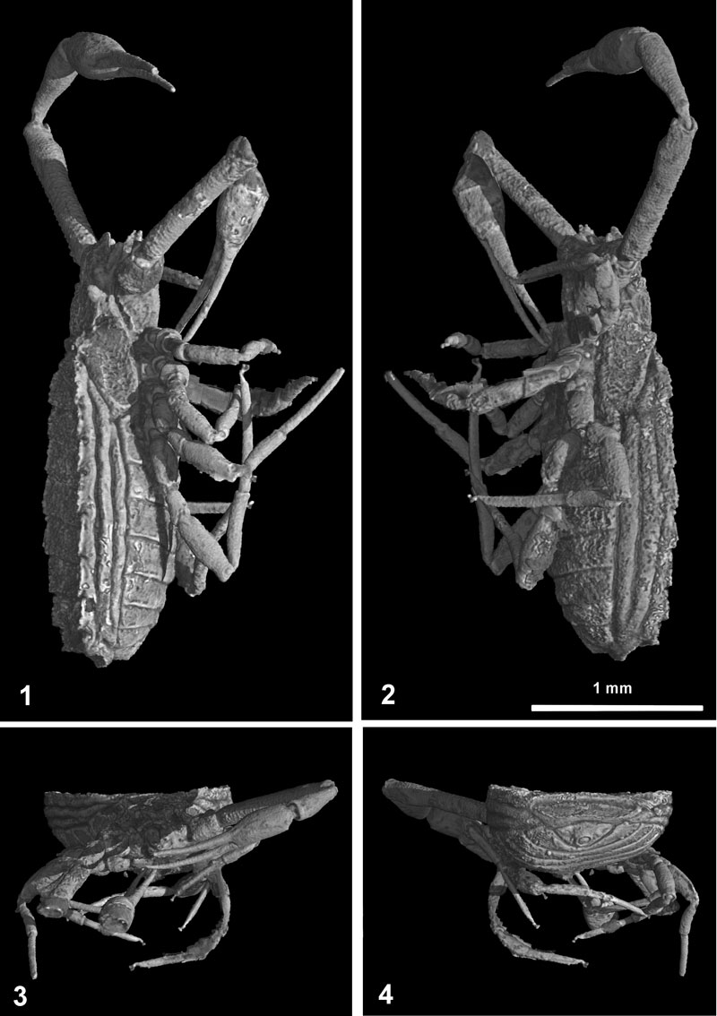
FIGURE 5. Pseudogarypus synchrotron sp.n., holotype, habitus dorsal.
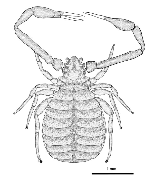
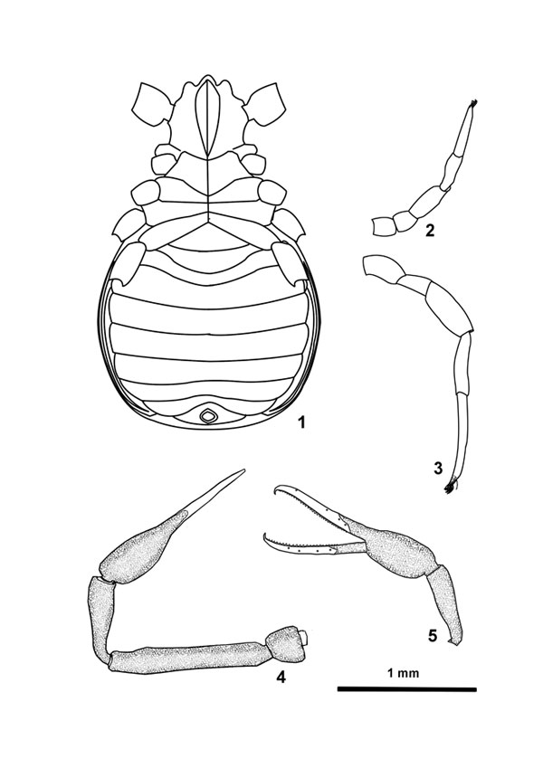
FIGURE 6. Pseudogarypus synchrotron sp.n., holotype. 1: ventral reconstruction with coxae, 2: leg I; 3: leg IV; 4: pedipalp left; 5: P. synchrotron sp.n., paratype, left chela with position of trichobothria.