FIGURE 1. Topographic map of the Haasgat fossil site relative to other nearby South African Pliocene and Pleistocene fossil localities. Contour lines equal 20 m.
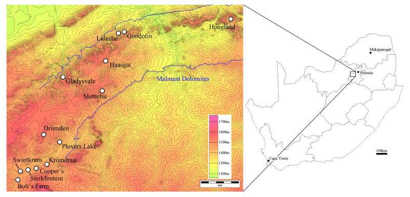
FIGURE 2. HGD Colobini: (1) HGD 1165A and B, Cercopithecoides haasgati partial cranium and mandible, lateral view; (2) HGD 2452, cf. Cercopithecoides right mandible, lateral (above), oblique (middle), and occlusal (below) views. Scale bar equals 1 cm.
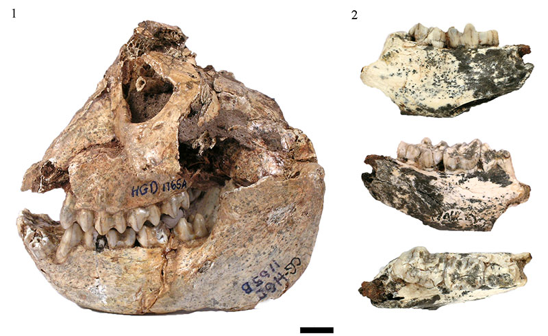
FIGURE 3. HGD Papio angusticeps: (1) HGD 606 male maxilla, occlusal view (2) HGD 1246 female mandible, occlusal view. Scale bar equals 1 cm.
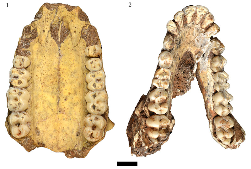
FIGURE 4. Haasgat Alcelaphini: (1) HGD 43, Connochaetes gnou right mandible, occlusal view; (2) HGD 44, Damaliscus dorcas maxilla, occlusal view; (3) HGD 311, Megalotragus sp. left m3, occlusal view. Scale bar equals 1 cm.
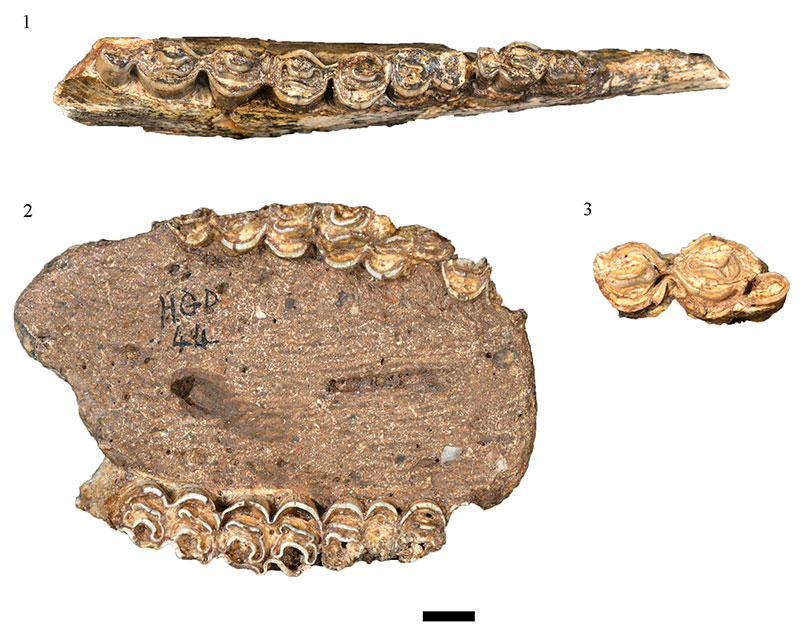
FIGURE 5. Swartkrans Hanging Remnant cf. Makapania sp. (SK) and Haasgat indeterminate alcelaphins: (1) SK 3005 right maxilla with M2 and M3, occlusal view; (2) HGD 73 right M1 or M2, occlusal view; (3) HGD 2193 left M3, occlusal view; (4) SK 2693 left m1, occlusal view; (5) SK 2965 left mandible with m3, occlusal view; (6) HGD 90 right mandible with p3-m3, occlusal [above] and buccal [below] views; (7) HGD 51 left mandible with m2 and m3, occlusal view; (8) HGD 100 right mandible with m2 and m3, occlusal view. Scale bar equals 1 cm.
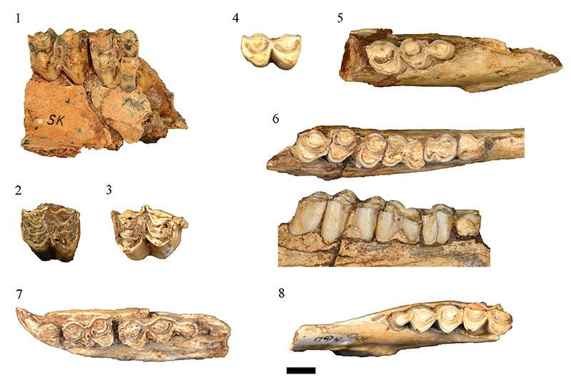
FIGURE 6. HGD 326, Antidorcas bondi left maxilla, occlusal and lingual views. Scale bar equals 1 cm.
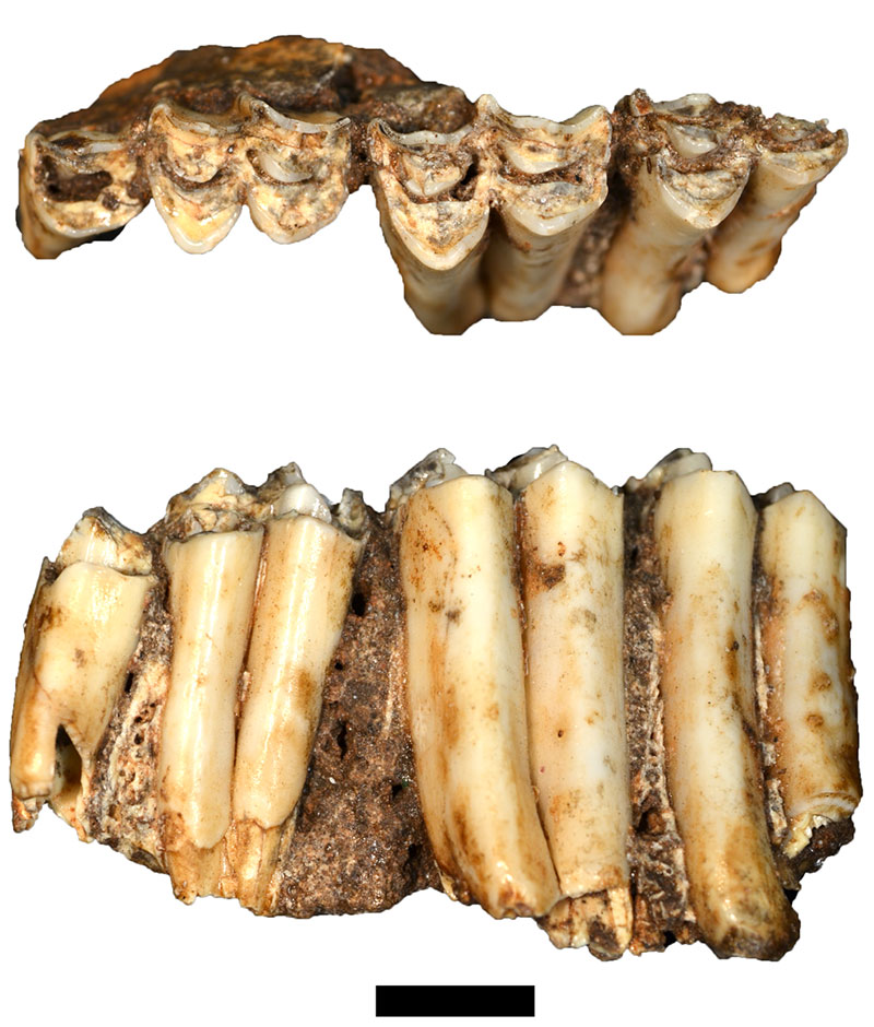
FIGURE 7. Haasgat Hippotragini: (1) HGD 89, Hippotragus sp. right M3, occlusal view; (2) HGD 106, Hippotragus sp. left deciduous p3, occlusal view. Scale bar equals 1 cm.
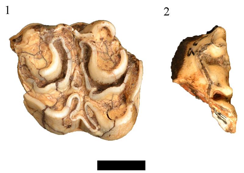
FIGURE 8. Occlusal views of Haasgat and comparative Oreotragus left maxillae: (1) TM 14845 modern Oreotragus oreotragus; (2) G 2700, Gondolin GD 2 right maxilla (reversed); (3) M 949, Makapansgat Member 3; (4) HGD 1301. Scale bar equals 1 cm.
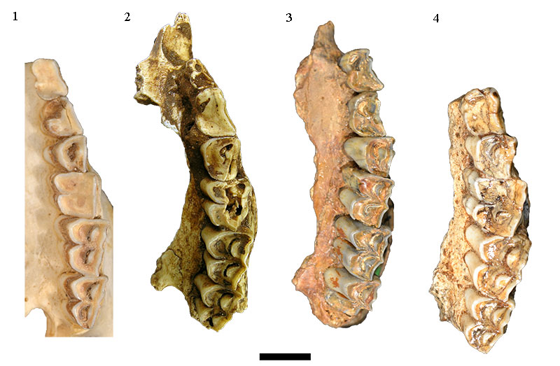
FIGURE 9. Occlusal views of Haasgat and comparative Oreotragus left mandibles: (1) HGD 298 right mandible (reversed); (2) M 997, Makapansgat Member 3; (3) G 4940, Gondolin GD 2; (4) TM 11720 modern Oreotragus oreotragus. Scale bar equals 1 cm.
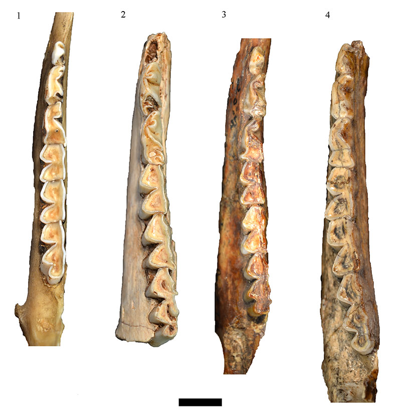
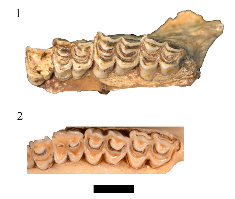
FIGURE 10. Haasgat indeterminate Reduncini: (1) HGD 38 left mandible with p2-m3, occlusal view; (2) HGD 37 left mandible with m2 and m3, occlusal view; (3) HGD 41 maxilla, detail of left P3-M3, occlusal view. Scale bar equals 1 cm.
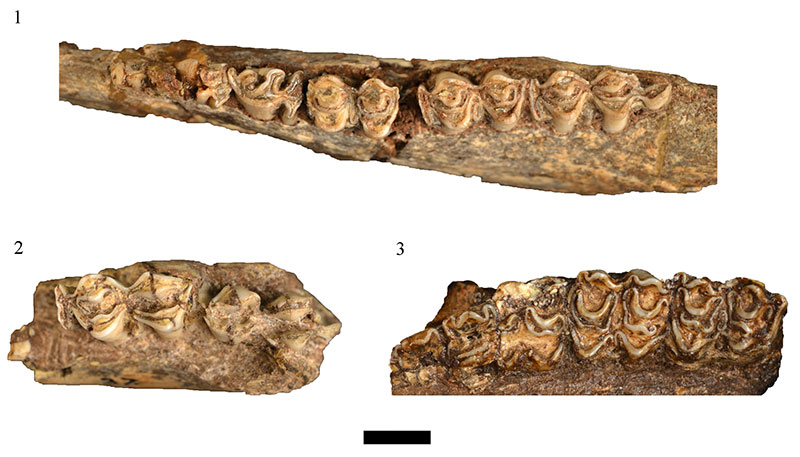
FIGURE 11. Haasgat Tragelaphini: (1) HGD 126, Taurotragus sp. right maxilla with partial M1 and M2, occlusal view; (2) HGD 96, Tragelaphus sp. left probable M1, occlusal view; (3) HGD 123, Tragelaphus sp. right mandible, occlusal (above) and lateral (below) views. Scale bar equals 1 cm.
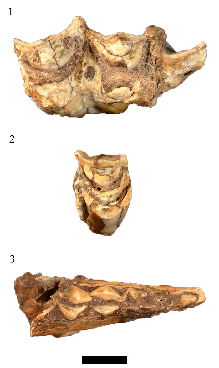
FIGURE 12. Side views of the HGD 1323 reconstructed indeterminate suid third molar. Scale bar equals 1 cm.
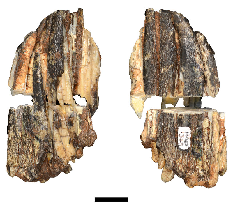
FIGURE 13. Haasgat cf. Dinofelis sp. (HGD 950) (above) right fourth metatarsal, Kromdraai B Megantereon cultridens (KB 5339c) (below) left fourth metatarsal (mirrored), and AZ 420 Panthera pardus right fourth metatarsal, proximal articular surface and medial views. Scale bar equals 1 cm.
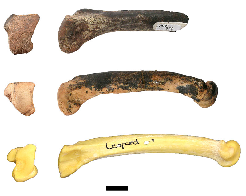
FIGURE 14. HGD Equus specimens: (1) HGD 1105, Equus capensis partial left mandible, occlusal view; (2) HGD 1090 Equus cf. quagga right maxillary premolar or molar, occlusal view; (3) HGD 1109 Equus cf. quagga left maxillary second premolar, occlusal view. Scale bar equals 1 cm.
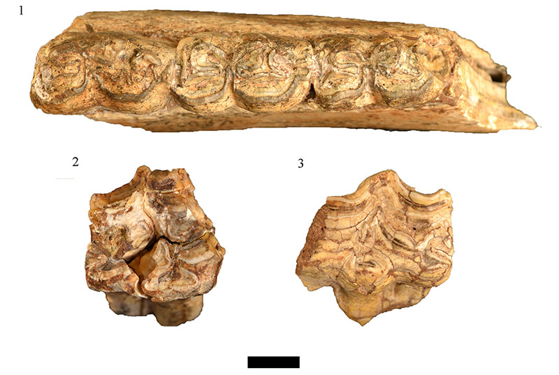
FIGURE 15. HGD Procavia specimens: (1) HGD 1116 Procavia antiqua partial cranium, inferior view; (2) HGD 1134 Procavia antiqua right mandible, lateral view; (3) HGD 1118, Procavia transvaalensis partial cranium, inferior views. Scale bar equals 1 cm.
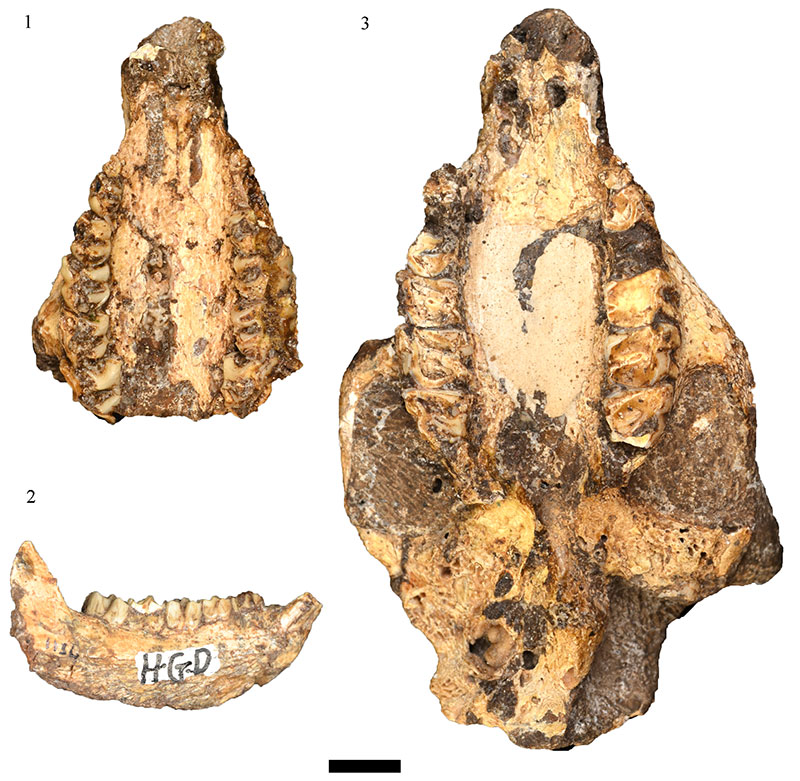
FIGURE 16. HGD Hystrix africaeaustralis specimens: (1) HGD 1342 left mandible, medial and superior views; (2) HGD 1352 partial cranium, inferior view. Scale bar equals 1 cm.
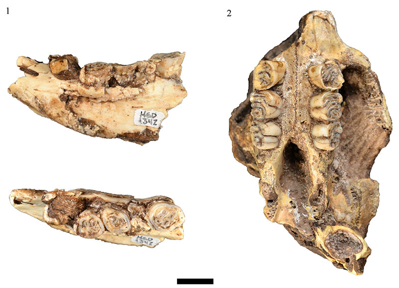
FIGURE 17. The 'articular surface' of the HGD 585 specimen (left) and proximal articular surface of a Giraffa camelopardalis intermediate phalanx (right). Scale bar equals 1 cm.
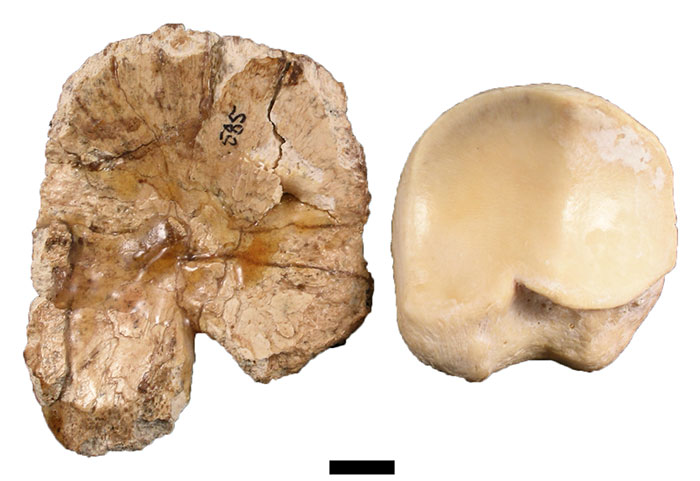
FIGURE 18. Occlusal views of the (1) HGD 31 right maxilla and (2) AZ 10120 Sylvicapra grimmia right M2 and M3. Scale bar equals 1 cm.
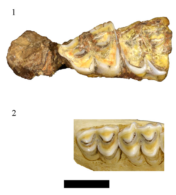
FIGURE 19. Occlusal views of the (1) HGD 27 left maxilla and (2) FMNH 127972 Ourebia ourebi left P4-M3. Scale bar equals one cm.