FIGURE 1. Illustrations of a dragon that allegedly lived in the marshes around Rome in 1691, from a 1696 book by Cornelius Meyer. 1.1. A double illustration from the last page in Meyer's book, showing the skeleton (at bottom) and a fleshed-out reconstruction (at top). 1.2. An illustration from the cover of Meyer's book, showing another fleshed-out reconstruction of the dragon.
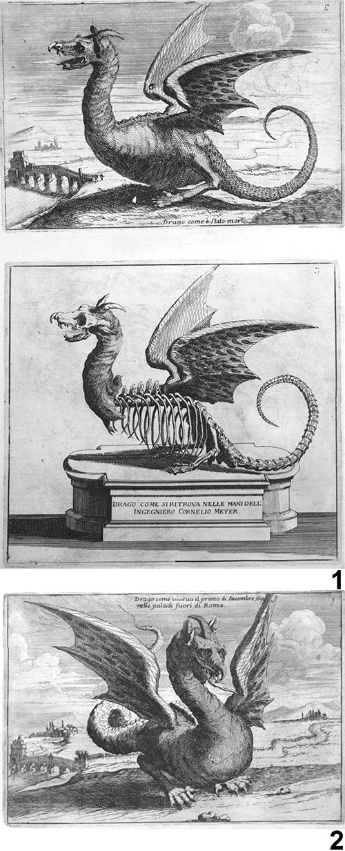
FIGURE 2. Skull and lower jaw of Meyer's dragon, a domestic dog, and the pterosaur Scaphognathus crassirostris. 2.1. Head of Meyer's dragon. 2.2. Skull of a domestic dog (Canis familiaris). 2.3. Mandible of the same domestic dog. 2.4. Articulated skull and mandible of domestic dog, showing that the lower canine tooth is anterior to the upper canine tooth. 2.5. Skull of an adult specimen of Scaphognathus crassirostris, from Goldfuss (1831). 2.6. Skull of a juvenile specimen of S. crassirostris, modified from Wellnhofer (1991) and with gray shading on bones from the right side of the skull that have been displaced dorsally; Goertzen (1998) mistook the displaced right frontal bone in an illustration in Wellnhofer (1991) for a crest. ap = angular process, cp = coronoid process, lf = left frontal bone, rf = right frontal bone.
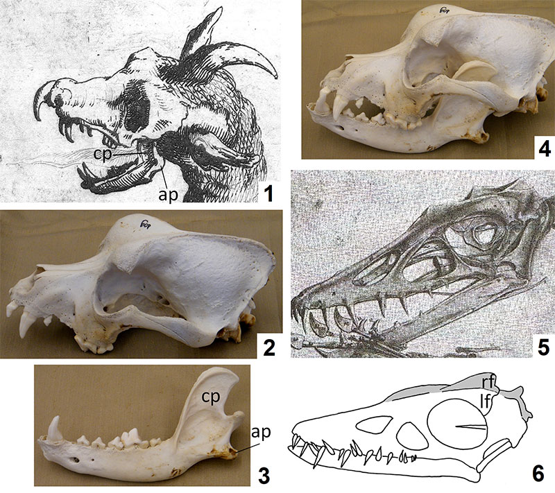
FIGURE 3. Seventeeth-century engravings of dragons, showing the hooked proboscis typical of seventeenth-century dragon depictions. These figures are from Allen and Griffiths (1979) (left), Hogarth and Clery (1979) (middle), and Huxley (1979) (right).
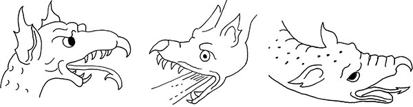
FIGURE 4. Ribcages of Meyer's dragon and extant vertebrates, showing that the tips of the ribs are tapered in Meyer's dragon and bony fishes but squared-off in amniotes. 4.1. Meyer's dragon. 4.2. A bony fish, the common carp (Cyprinus carpio). 4.3. A reptile, the saltwater crocodile (Crocodylus porosus). 4.4. A mammal, the sun bear (Helarctos malayanus).
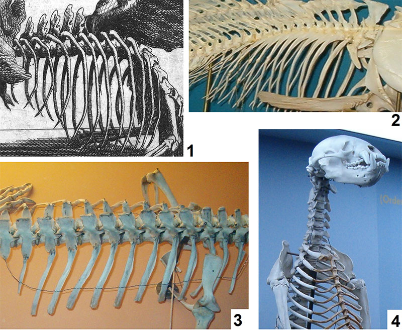
FIGURE 5. Wings of Meyer's dragon, a bat, and the pterosaur Scaphognathus crassirostris. 5.1. Meyer's dragon. 5.2. Jamaican fruit bat (Artibeus jamaicensis). 5.3. S. crassirostris, from Goldfuss (1831). 5.1. White arrows mark the expected location for wing claws. 5.2 and 5.3. Broken white ovals indicate wing claws.
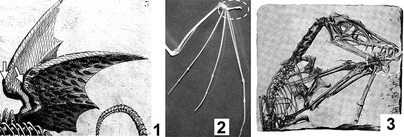
FIGURE 6. "Hindlimb" of Meyer's dragon and two pterosaurs, and forelimb skeletons of a bear and a wolverine. 6.1. Meyer's dragon. 6.2. The pterosaurs Rhamphorhynchus gemmingi (left) and Pterodactylus kochi (right), from Arthaber (1919). 6.3. Brown bear (Ursus arctos), drawn from a photo of a mounted skeleton. 6.4. Wolverine (Gulo gulo). 6.5. Meyer's dragon, brown bear, and wolverine, with a red arc connecting the tips of the metacarpals of each, showing that the tips of the metacarpals make a shallow arc in Meyer's dragon and the bear and an acute arc in the wolverine.
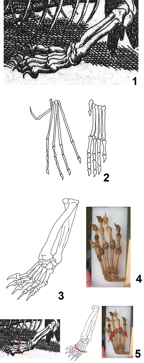
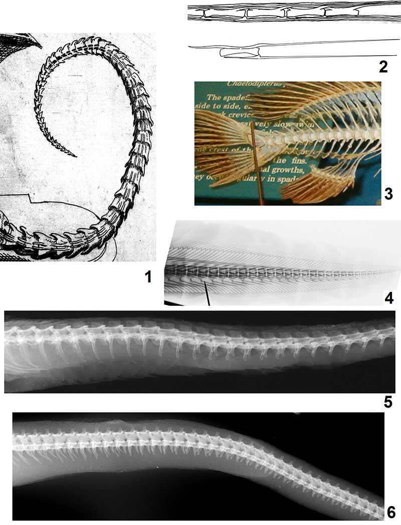
FIGURE 7. Tail of Meyer's dragon, a long-tailed pterosaur, and extant vertebrates. 7.1. Meyer's dragon. 7.2. Vertebrae of the long-tailed pterosaur Rhamphorhychus gemmingi in right lateral view, modified from Wellnhofer (1975); above: series of caudal vertebrae, showing the collection of elongate dorsal and ventral processes that are absent in Meyer's dragon; below: a single caudal vertebra and hemal arch, showing the elongate dorsal and ventral processes. 7.3. Tail of a sea bass (Centropristis striatus) showing that the caudal vertebrae of a typical bony fish differ from those of Meyer's dragon in that they do not gradually taper to a point. 7.4. Tail of a Mediterranean moray eel (Muraena helena), showing that vertebral morphology differs from that of Meyer's dragon. 7.5. Tail of a snake of the viper family (Viperidae), a puff adder (Bitis arietans), showing that the bony processes of the tail differ from those of Meyer's dragon. (F)—Tail of a snake of the python family (Pythonidae), an African rock python (Python sebae), showing that the bony processes of the tail differ from those of Meyer's dragon.