FIGURE 1. Geological settings of the Montagne Noire (after Clausen and Álvaro, 2007). 1- Location of the Massif Central and the Montagne Noire in France. 2- Structural sketch of the Montagne Noire. 3- Geological sketch of the Northern flank Avène-Mendic and Mélagues Units with location of the studied sections.
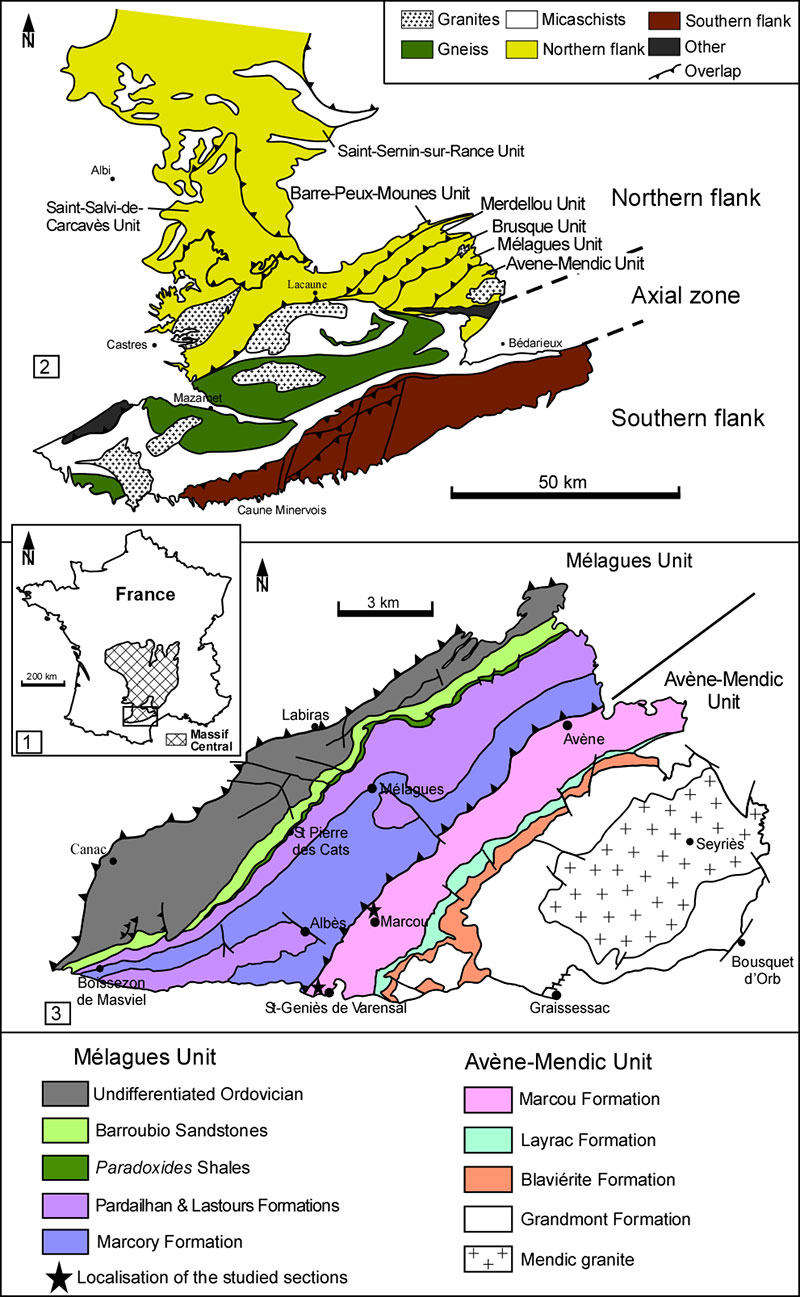
FIGURE 2. Attempt of lithological correlation between northern (Clausen and Álvaro, 2007) and southern flank of the Montagne Noire (Álvaro et al. 2010) and chronostratigraphic divisions of the southern flank (Álvaro et al. 2010) and of the northern flank (Kerber, 1988; Clausen and Álvaro, 2007).
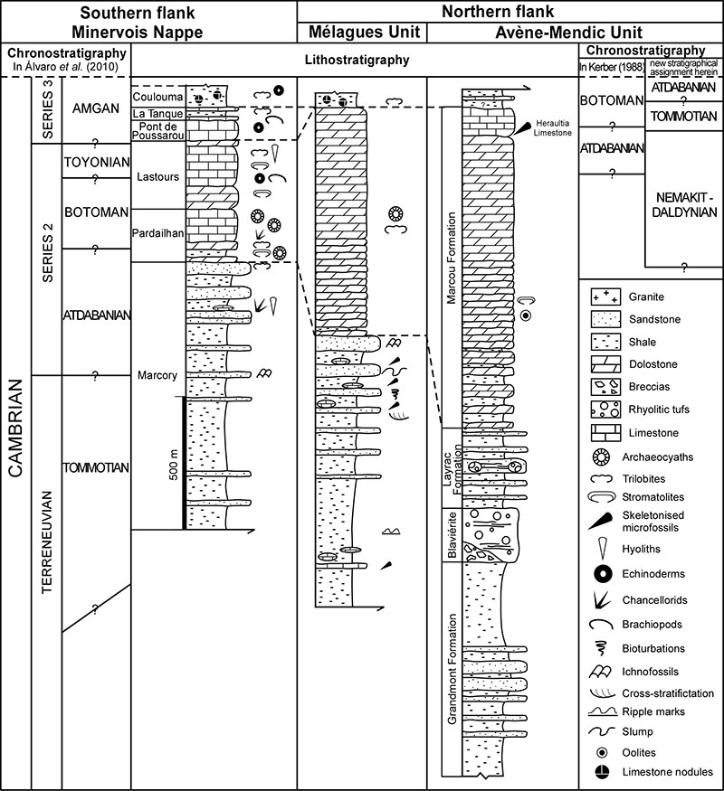
FIGURE 3. Cross-section of the Montagne Noire detailling its general structural pattern. The Catalan domain corresponds to the Gondwana precambrian basement cropping in the Axial Zone. The Para-autochtonous Units (mendic-Sorezois domain) thrusts above the Axial Zone. Finally, the Occitan domaine comprises paleozoic sediments supposed to have deposited at the margin of the Catalan craton (Demange, 2001).
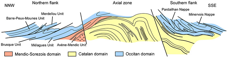
FIGURE 4. Oelandiella korobkovi, Vostokova 1962. 1-4 USTL1235/1: 1. Right lateral view, arrow shows external coating of the shell on a comarginal rib; 2. Left lateral view; 3. Upper lateral view, arrow shows intermediate rib; 4. Anterior view showing ribs crossing the dorsum. 5-10 USTL1259/5: 5. Right lateral view with square showing location of 7; 6. Left lateral view, arrow shows the external coating; 7. Detail of sharp comarginal rib; 8. Upper view; 9. Anterior view showing ribs crossing the dorsum; 10. Posterior view showing planispiral complete whorl with smooth apex. 11-15 USTL1241/9: 11. Detail of co-marginal ribs slightly tapering and fading close to the umbilicum; 12. Left lateral view; 13. Upper view; 14. Anterior view with ribs crossing the dorsum; 15. Posterior view, arrow shows external coating of the shell. 16-21 USTL1257/3: 16, 17. Anterior and posterior views respectively; 18. Upper view; 19. Posterior-lateral view, arrow shows intermediate rib; 20. Right lateral view of the internal mold with external coating; 21. Left lateral view, arrow shows intermediate rib. 22. USTL1258/10, left lateral view showing sinuous apertural margin. 23. USTL1255/13, left lateral view showing incomplete whorl and uplifted apex from the apertural plan. 24. USTL1246/5, apertural view showing aperture elongated along the antero-posterior axis. Scale bars equal: 7, 100 µm; 4, 9, 10, 15, 200 µm; 1-3, 11, 16, 17, 300 µm; 5, 6, 8, 12, 13, 14, 18-24, 500 µm.

FIGURE 5. 1-11. Protoconus orolgainicus (Zhegallo in Esakova and Zhegallo, 1996). 1-6. USTL1215/13; 1. Upper view, upper arrow shows lateral furrows borded by faint constrictions, lower arrows show space between internal and external molds (shell thickness); 2. Left lateral view, square shows location of 3; 3. Detail of polygonal structures in the furrows framing the dorsal median buttress; 4. Right lateral view; 5, 6. Anterior and posterior views respectively. 7-9. USTL1264/11: 7. Upper view; 8. Right lateral view, square shows location of 9; 9. Detail of subapical shelf. 10-11. USTL1241/11: 10. Right lateral view, square shows location of 11; 11. Detail of subapical shelf. 12-18. Bemella cf. simplex Yu, 1979. 12-14. USTL1223/15: 12. Upper view with comarginal striations; 13. Left lateral view; 14. Posterior view. 15-17. USTL1236/6: 15. Upper view with apex shifted to the right; 16. Posterior view; 17. Left lateral view. 18. USTL1256/11: right lateral view of a specimen with curved apex and striated external coating. 19-23. Prosinuites tripartitus Kerber, 1988. 19-21 USTL1257/2: 19. Upper view; 20. Anterior view with dorsal furrows and anterior apertural margin sinus; 21. Right lateral view with apex overhanging the posterior apertural margin. 22-23. USTL1235/4: 22. Left lateral view; 23. Upper view of the dorsal field with sinusoidal growth lines. Scale bars equal: 3, 50 µm; 5, 6, 11, 12-14, 100 µm; 1, 2, 4, 9, 16, 18, 22, 23, 200 µm; 15, 17, 300 µm; 7, 8, 10, 19-21, 500 µm.
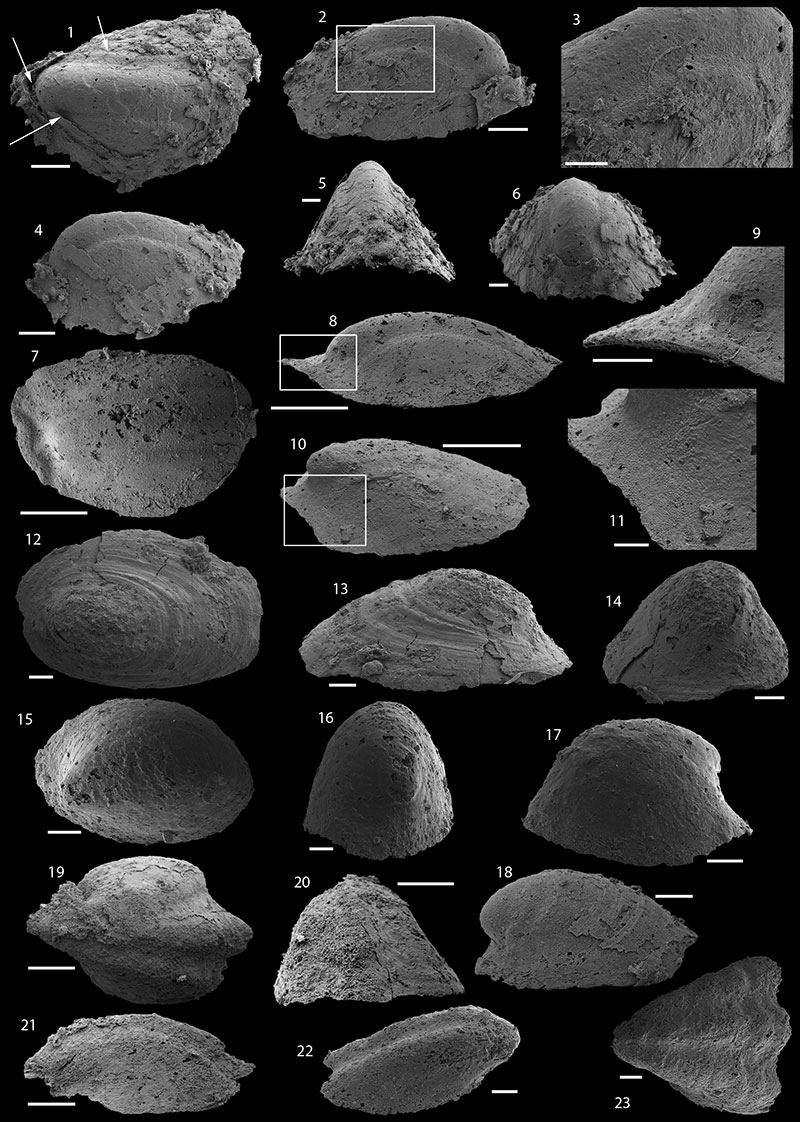
FIGURE 6. 1-7 Dorispira lauta? (Yu, 1979). 1-7: USTL1245/6: 1. Left lateral view; 2. Right lateral view, square shows location of 3. 3. Detail of latero-dorsal furrow with polygonal imprints; 4. Upper view with prominent subapical shelf; 5. Anterior view showing mid-dorsal buttress with wavy structures; 6. Posterior view, square shows location of 7; 7. Detail of subapical field. 8-15. Obscurania tormoi Devaere, sp. nov. 8-12 USTL1223/7: 8. Upper view; 9. Right lateral view; 10. Left lateral view; 11. Posterior view, square shows location of 12; 12. Detail of the apex with mammilated papillae. 13-15. USTL1223/20: 13. Upper view of internal mold with external coating on anterior part; 14. Left lateral view, square shows location of 15; 15. Detail of apically curved mammilated papillae. Scale bars equals: 15, 50 µm; 3, 7, 12, 14, 100 µm; 9, 10, 11, 13, 200 µm; 5, 6, 8, 300 µm; 1, 2, 4, 500 µm.
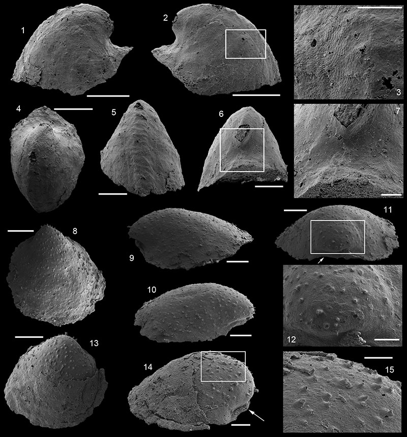
FIGURE 7. 1-10 Igorella sp. 1-5. USTL1227/5: 1. Right lateral view showing loose coiling; 2. Left lateral view; 3, 4. Posterior and anterior views respectively, showing lateral compression; 5. Upper-lateral view. 6-10. USTL1250/1: 6. Left lateral view; 7. Anterior views with ribs crossing the dorsum; 8. Right lateral view; 9. Upper-lateral view; 10. Posterior view. Scale bars equal: 1, 2, 5, 6, 8, 9, 500 µm; 3, 4, 7, 10, 200 µm.
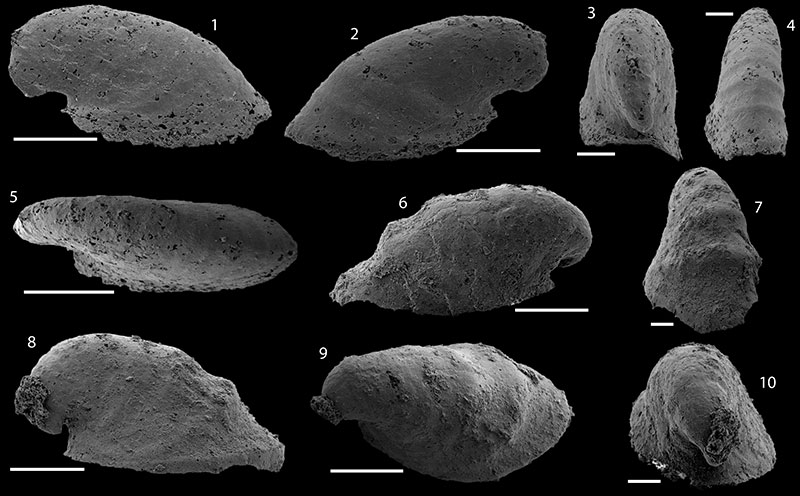
FIGURE 8. 8.1-16. Halkieria sacciformis Meshkova, 1969. 1-4. USTL1257/8: 1. Dorsal view with longitudinal ribs; 2. Lateral view; 3. Ventral view; 4. Proximal view. 5-8. USTL1252/2: 5. Dorsal view with internal cavity cisible; 6. Ventral view, square shows location of 8; 7. Lateral view, arrow shows internal cavity; 8. Detail of the triangular base. 9-13. USTL1249/10: 9. Lateral view; 10. Ventral view; 11. Apical view; 12. Dorsal view, square shows location of 13; 13. Detail of the ornamentation of dorsal side. 14-16 USTL1249/6: 14. Dorsal view; 15. Lateral view showing the S-shape of sclerites; 16. Ventral view with distal tip. 17-24. Halkieria operculus (Qian, 1984). 17-19. USTL1250/8: 17. Dorsal view with visible lateral flanges; 18. Ventral view, square shows location of 19; 19. Detail of the base. 20-22. USTL1250/9: 20. Dorsal view with fine transverse striations; 21. Ventral view; 22. Lateral view. 23-24. USTL1250/7: 23. Dorsal view, square shows location of 24; 24. Detail of the transversal striations. Scale bars equal: 13, 19, 100 µm; 8, 17, 18, 20-22, 24, 200 µm; 1-7, 9-12, 14-16, 23, 500 µm.
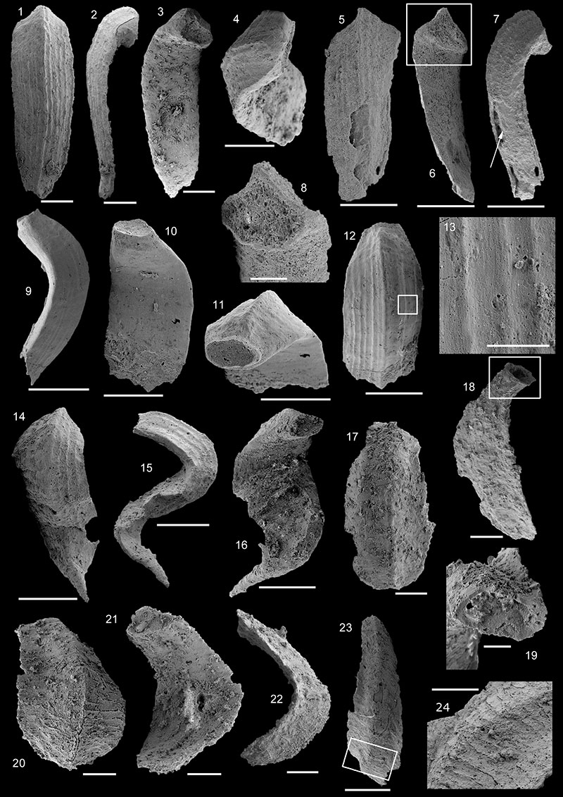
FIGURE 9. 1-11. Siphogonuchites triangularis Qian, 1977. 1-3. USTL1254/23: 1. Dorsal view; 2. Lateral view; 3. Distal view. 4-6. USTL1227/4: 4. Dorsal view; 5. Lateral view; 6. Ventral view. 7-11. USTL1255/9: 7. Dorsal view; 8. Ventral view; 9. Lateral view; 10. Distal view, square shows location of 11; 11. Detail of cross-section. 12-17. Lomasulcachites macrus Qian and Jiang, 1980; USTL1244/4: 12. View of the sulcus; 13. Lateral view; 14. Lateral view, square shows location of 16; 15. Distal view with cross section; 16. Details of ornamentations; 17. Proximal view with cross-section. Scale bars equal: 1, 2, 3, 11, 100 µm; 10, 16, 200 µm; 4-9, 12-15, 17, 500 µm.
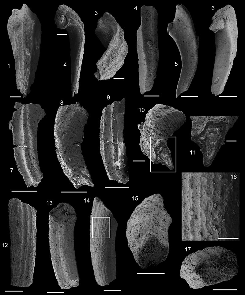
FIGURE 10.Ocruranus cf. subpentaedrus (Jiang, 1980). 1-6 USTL1228/5: 1. Upper view; 2. Posterior view; 3. Anterior view; 4. Left lateral view; 5. Right lateral view, square shows location of 6; 6. Detail of parallel striations on external surface of internal molds. 7-9. USTL1251/1: 7. Upper view; 8. Anterior view; 9. Left lateral view. Scale bars equal: 6, 50 µm and1-5, 6-9, 500 µm
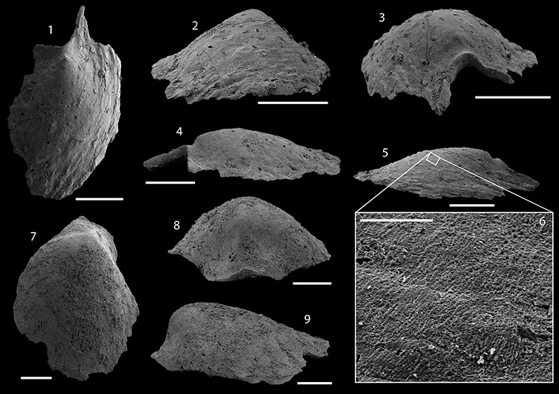
FIGURE 11.Purella gracilis Zhegallo, 1982. 1-5 USTL1211/5: 1. Upper view of internal mold with external coating; 2, 3. Lateral views; 4. Proximal view, square shows location of 5 and arrow spiny scales that are symmetrically arranged on both sides of the subapical field; 5. Detail of external coating, arrow points spiny scale. 6-7. USTL1213/6: 6. Lateral view of internal mold, square shows location of 7; 7. Detail of polygonal imprints on lateral fields. 8-13. USTL1243/5: 8. Upper-lateral view of internal mold with external coating; 9. Lateral view, square shows location of 13; 10. Lateral view; 11. Proximal view, arrow shows subapical spiny scales; 12. Distal view showing median, longitudinal buttress; 13. Detail of internal mold and scaly external coating. 14-19. USTL1244/3: 14. Upper-lateral view; 15. lateral view, external coating continuously scaly (no slits); 16. Lateral view, square shows location of 19; 17. Proximal view, square shows location of 18; 18. Detail of subapical external coating, arrow points spiny scale; 19. Detail of polygonal imprints on internal mold. Scale bars equal: 19, 20 µm; 5, 7, 18, 100 µm; 1-4, 13, 200 µm; 17, 300 µm; 6, 8-12, 14-16, 500 µm.
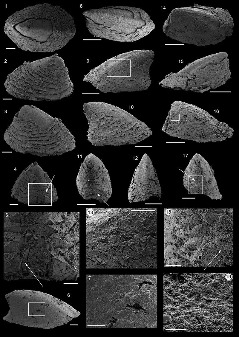
FIGURE 12.Purella layracensis (Kerber, 1988). 1-3. USTL1238/6: 1. Upper view, large subapical shelf; 2. Lateral view, square shows location of 3; 3. Detail of polygonal imprints and granules on the internal mold. 4-6, 8. USTL1253/4: 4. Lateral view; 5. Upper view, square shows location of 6; 6. Detail of granulated internal mold and scaly external coating; 8. Distal view showing mid-dorsal buttress. 7, 10-11. USTL1235/5: 7. Upper-lateral view; 10. Lateral view, square shows location of 11; 11. Detail of internal mold granules situated in the centre of eye-shaped openings of subtriangular external coating papillae. 9- USTL1233/9, upper view of subrectangular specimen. 12-16. USTL1220/7: 12. Proximal view, square shows location of 16; 13. lateral view, square shows location of 14; 14. Detail of high granules passing through openings of subtriangular papillae; 15. Upper view of low form; 16. Detail of highly-developped internal mold granules. 17-19. USTL1245/1: 17. Upper view of morphotype with subapical spines, square shows location of 19; 18. Lateral view; 19. Detail of subapical spines. Scale bars equal: 16, 20 µm; 15, 50 µm; 3, 11, 12, 13, 15, 19, 100 µm; 1, 2, 6, 9, 200 µm; 7, 10, 300 µm; 4, 5, 8, 17, 18, 500 µm.
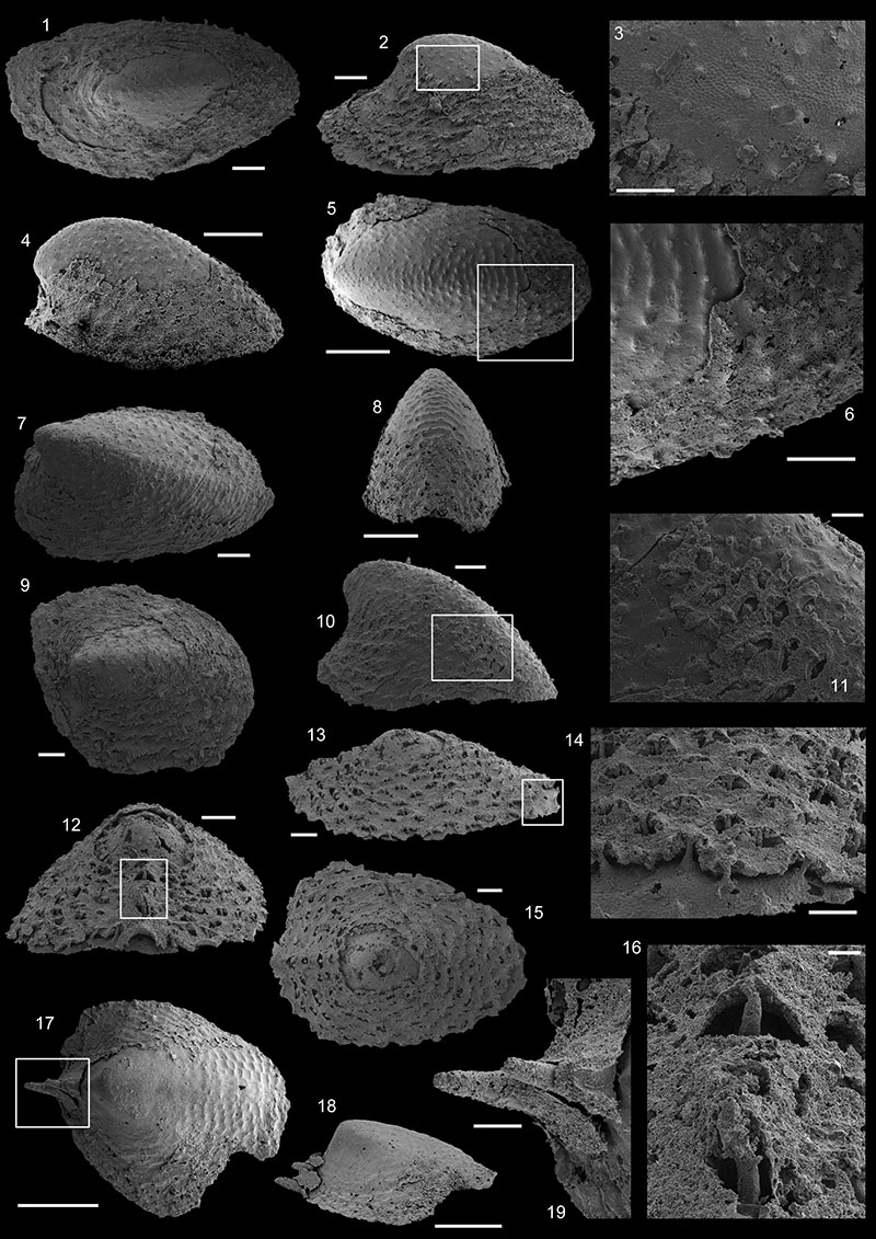
FIGURE 13. Sketch of Watsonella crosbyi Grabau, 1900 highlighting key anatomical features and terms used herein. pp: posterior part; ap: anterior part; d: dorsum; mf: medial dorsal furrow; vs: ventral side; aa: anterior part of the aperture: pa: posterior part of the aperture; a: apex; b: beak; ls: lateral shields; su: subapical shield. Schematic interpretation of internal mold, shell and external coating connections.
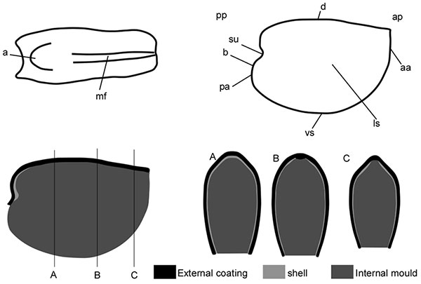
FIGURE 14.Watsonella crosbyi Grabau, 1900. 1-5. USTL1226/10: 1. Right lateral view, arrow shows concave subapical field; 2. Posterior view; 3. Left lateral view; 4. Anterior view; 5. Dorsal view. 6-12. USTL1241/7: 6. Right lateral view, square shows location of 11; 7. Anterior view; 8. Detail of polygonal imprints developed in the median dorsal furrow; 9. Left lateral view; 10. Posterior view; 11. Detail of concave subapical field with external mold; 12. Dorsal view, square shows location of 8. 13-20. USTL1236/4: 13. Left lateral view, lower right square shows location of 18, lower right, location of 19 and upper square, location of 20; 14. Posterior view; 15. Right lateral view; 16. Anterior view, 17. Dorsal view; 18. Detail of apical area with external coating; 19. Detail of lateral fields with imprints of lamello-fibrillar shell structure; 20. Detail of dorsal furrow with thick external coating. 21-27. USTL1243/2: 21. Right lateral view, square shows location of 23; 22. Anterior view; 23. Detail of dorsal furrow filled with thick external coating; 24. Left lateral view; 25. Posterior view, square shows location of 26; 26. Detail of apex with polygonal imprints; 27. Dorsal view. 28-29. USTL1209/8: 28. Right lateral view; 29. Posterior-lateral view; 30. 30-33. USTL1225/12: 30. Right lateral view, square shows location of 31; 31. Detail of large but faint polygonal imprints on lateral fiels; 32. Dorsal view, square shows location of 33; 33. Detail of dorsal furrow with external coating. Scale bars equal: 31, 33, 50 µm; 8, 11, 18-20, 23, 26, 29 100 µm; 2, 4, 14, 16, 17, 28, 30, 32, 200 µm; 1, 3, 5, 7, 10, 300 µm; 13, 15, 6, 9, 12, 21, 22, 24, 25, 27, 500 µm.
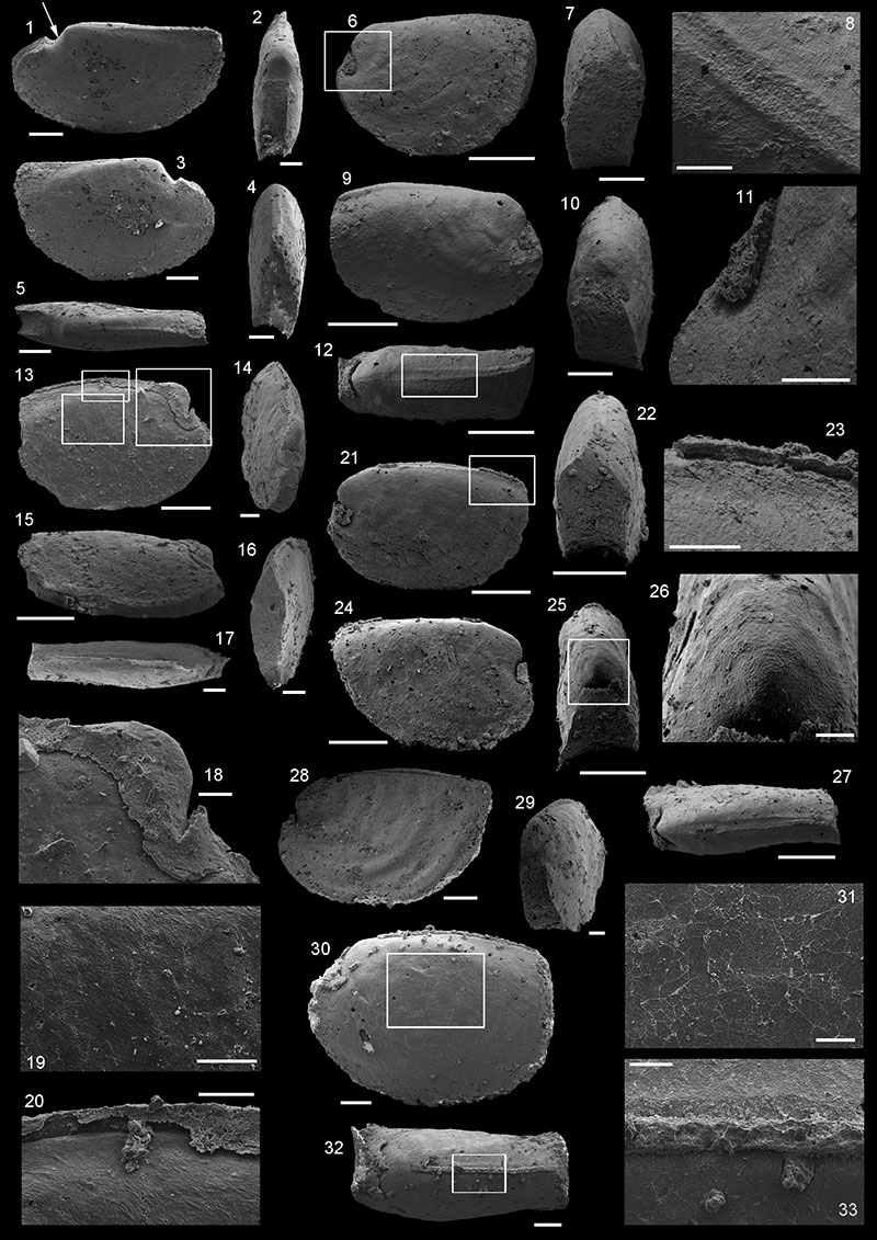
FIGURE 15.Watsonella crosbyi Grabau, 1900. 1-8. USTL1227/8: 1. Right lateral of specimen with phosphatised shell preserved; 2. Posterior view; 3. Left lateral view, upper square shows location of 6, lower left square, location of 7 and lower right square shows location of 8: 4. Anterior view; 5. Dorsal view; 6. Detail of shell with growth lines, internal mold visible; 7. Detail of shell with growth lines; 8. Detail of shell with growth lines, internal mold visible. 9. Left lateral view of specimen PIN 4664/1525 of Watsonella crosbyi Grabau, 1900 in Gravestock et al. 2001. 10. Left lateral view of USTL L12_067. 11. Right lateral view of specimen NIGP Mo 131363 identified as Watsonella yunnanensis (He and Yang, 1982) in Feng and Sun, 2003. 12. Right lateral view of USTL1214/15. Left lateral view of specimen N 3593/743 identified as Heraultipegma sibirica Missarzhevsky, 1974 in Missarzhevsky, 1989. 14. Left lateral view of USTL1243/2. Scale bars equal: 6-10, 12, 100 µm; 2, 4, 300 µm; 1, 3, 5, 11, 13, 14, 500 µm,
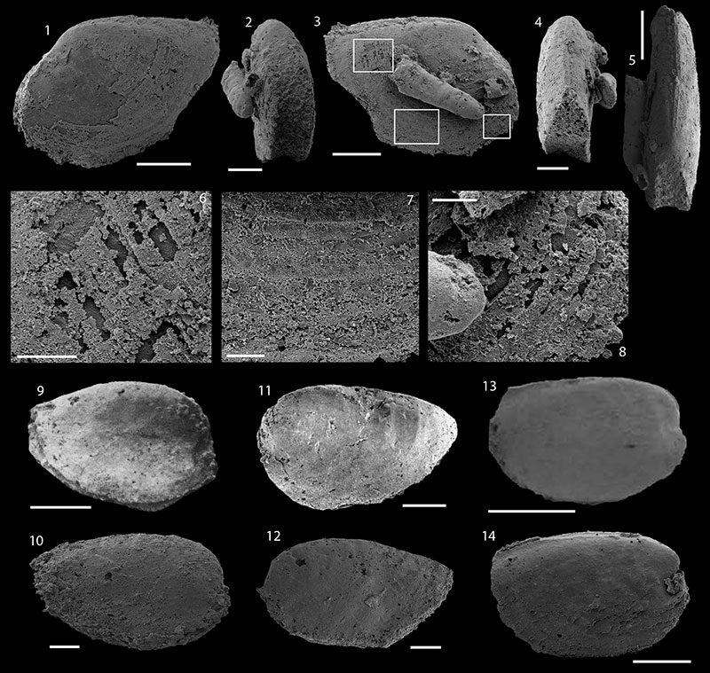
FIGURE 16. 1-6 Xianfengella prima He and Yang, 1982. 1-3. USTL1246/1: 1. Apertural view; 2. Lateral view, arrow shows subapical, comarginal notch in the internal molds; 3. Apical view. 4-6. USTL1211/14: 4. Apical view; 5. Lateral view; 6. Upper view. 7-12 Merismoconcha sp. 7-9. USTL1234/1: 7. Upper view, arrow shows the internal mold transverse-furrow; 8. Lateral view; 9. Apical view. 10-12. USTL1215/16: 10. Upper view; 11. Lateral view, arrow shows the internal mold transverse-furrow; 12. Apical view. Scale bars equal: 1, 4, 9, 11, 12, 200 µm; 5-8, 10, 300 µm.
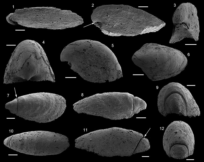
FIGURE 17. 1-9 Morphotype A. 1-7 USTL1258/8: 1. Upper view; 2. Abapical view, square shows location of 5; 3. Lateral-apical view, square shows location of 6; 4. Lateral view, square shows location of 7; 5. Detail of comarginal ribs of internal mold with flat external coating; 6. Detail of subapical sinus; 7. Detail of prismatic imprints between comarginal ribs of internal mold. 8-9. USTL1247/1: 8. Upper view; 9. Lateral view. 10-14 Morphotype B, specimen USTL1214/11: 10. Upper view; 11. Lateral view, square shows location of 12; 12. Detail of striated external coating; 13. Detail of distance between external coating and internal mold (shell thickness); 14. apical view, square shows location of 13. 15-20. Morphotype C, specimen USTL1215/10: 15. Upper view, square shows location of 18; 16. Lateral view; 17. Apical view, square shows location of 20; 18. Detail of the surface of the internal mold with circular granules, square shows location of 19; 19. Detail of pierced circular granules; 20. Detail of polygonal imprints on the subapical field. Scale bars equal: 19, 5 µm; 13, 20 µm; 7, 12, 18, 20, 50 µm; 5, 10, 11, 14-17, 200 µm; 6, 8, 9, 300 µm 1-4, 500 µm.
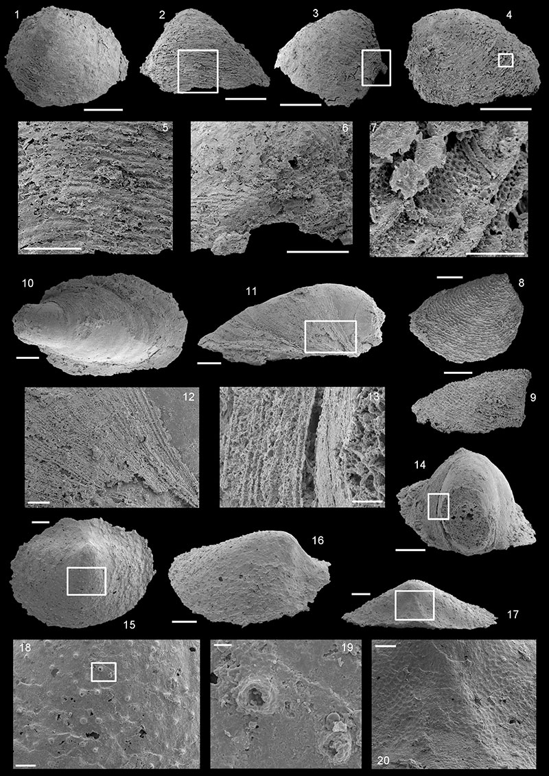
FIGURE 18. 1-5. Brachiopod indet. specimen USTL1223/2: 1. Upper view of dorsal valve, square shows location of 5; 2, 4. Lateral views; 3. Posterior view; 5. Detail of comarginal step-like structures corresponding to shell lamellae. 6-13. Khasagtina? specimen USTL1234/10: 6. Upper view, lower square shows location of 11, upper square shows location of 12; 7. Anterior view; 8, 10. Lateral views; 9. Posterior view, square show location of 13; 11. Detail of radial ribs; 12. Detail of groove parallel to the subapical apertural margin; 13. Detail of subapical area with sinus, arrow shows groove parallel to the subapical apertural margin. Scale bars equal: 5, 20 µm; 1-4, 11-13, 100 µm; 8, 10, 200 µm, 6-9, 300 µm.
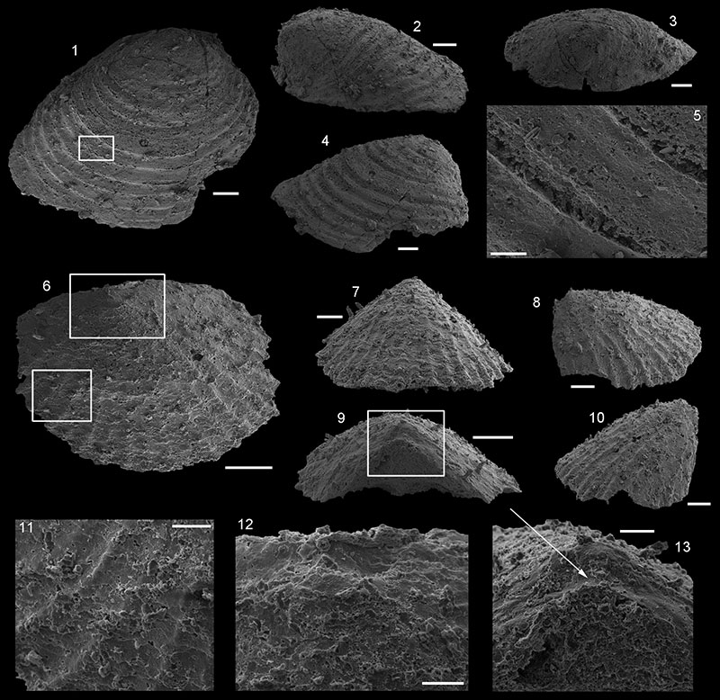
FIGURE 19. 1-30. Conotheca subcurvata (Yu, 1974). 1-3. USTL1261/2: 1, 2. Longitudinal views; 3. Apertural view. 4-5. USTL1210/9: 4. Longitudinal view; 5. Apertural view. 6-7. USTL1221/6: 6. Longitudinal view; 7. Apertural view. 8-11. USTL1253/1: 8, 9. Longitudinal views; 10. Apertural view; 11. Apical view. 12-15. USTL1264/3: 12, 13. Longitudinal views; 14. Apertural view; 15. Apical view. 16-19. USTL1253/2: 16, 17. Longitudinal views; 18. Apertural view; 19. Apical view. 20-30. USTL1251/13: 20. Detail of apical part of preserved digestive track; 21. Detail of apertural part of preserved digestive track with operculum; 22. Longitudinal view of broken internal coating with visible digestive track, square shows location of 21 and 30; 23. Longitudinal view of broken internal coating with visible digestive track, square shows location of 20; 24. Longitudinal view; 25. Apertural view, arrow shows eccentric apex of operculum; 26. View of the operculum, square shows location of 29; 27. Apertural view, arrow shows faint concentric depressions between apex and abapical apertural margin; 28. Apical view with digestive track visible; 29. Detail of apertural margin; 30. Lateral view of operculum. 31-36. Paragloborilus subglobosus He in Qian, 1977. 31-34. USTL1221/7: 31,32. Longitudinal views; 33. Longitudinal view, arrow shows annulations; 34. Apertural view. 35-36. USTL1222/10: 35. Apertural view; 36. Longitudinal view. Scale bars equal: 29, 50 µm; 4, 6, 7, 10, 18, 35, 36, 100 µm; 1-3, 5, 8, 9, 11, 14-17, 19-21, 25-28, 30-34, 200 µm; 12, 13, 22-24, 500 µm.
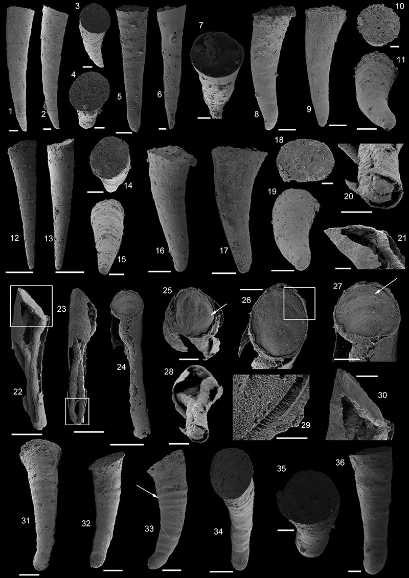
FIGURE 20.Cobboldiella transalpina Kerber, 1988. 1-4. USTL1217/19: 1. Ventral view, arrow points ventral fine growth lines; 2. Dorsal view, arrow points dorsal transverse rib; 3. Apertural view; 4. Lateral view, arrow shows lateral transverse ribs. 5-6. USTL1211/2: 5. Ventro-lateral view of internal mold; 6. Apertural view. 7-9. USTL1220/3: 7. Lateral view of conch with embryonic chamber preserved; 8. Dorsal view, arrow shows dorsal transverse ribs; 9. Apertural view. Scale bars equal: 1-6, 200 µm; 7-9, 100 µm.
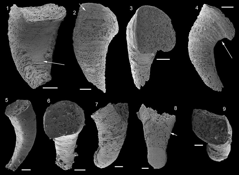
FIGURE 21. 1-13. Pseudorthotheca rotundicincta Cobbold 1935. 1-7. USTL1258/5: 1. Longitudinal view; 2. Apertural view; 3. Longitudinal view; 4. Apical view; 5. Longitudinal view, lower square shows location of 6, upper square shows location of 7; 6. Detail of transverse ribs; 7. Detail of transverse ribs near the aperture. 8-11. USTL1240/9: 8. Longitudinal view, arrow shows sinusoidal transverse rib; 9, 10. Longitudinal views; 11. Apical view with bluntly pointed apex. 12-13. USTL1231/1: 12. Longitudinal view, square shows location of 13; 13. Detail of sharp transverse ribs. 14-30. Pseudorthotheca acuticincta Cobbold 1935. 14-16. USTL1217/20: 14. Longitudinal view; 15. Longitudinal view, square shows location of 16; 16. Detail of swollen and round apex. 17-20. USTL1257/6: 17. Inclined longitudinal view, square shows location of 20; 18. Longitudinal view; 19. Apertural view; 20. Detail of discontinuous and shifted transverse rib. 21-24. USTL1253/10: 21, 22. Longitudinal views; 23. Apical view with swollen round apex; 24. Apertural view. 25-26. USTL1247/7: 25. Longitudinal view, internal mold with external coating; 26. Longitudinal view, internal mold with partly preserved external coating. 27. USTL1221/5, specimen with low ribs. 28-30. USTL1229/2: 28, 29. Longitudinal view; 30. Apertural view. Scale bars equal: 6, 16, 100 µm; 7, 11, 13-15, 20-24, 19, 30, 200 µm; 25-27, 300 µm; 1-5, 8-10, 12, 17, 18, 28, 29, 500 µm.
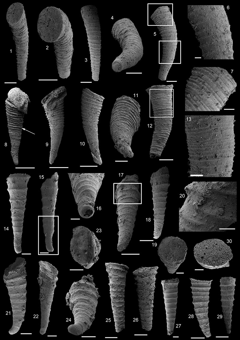
FIGURE 22. 1-17. Paracarinachites sinensis Qian and Jiang, 1982. 1-7. USTL1228/6: 1. Outer side view, square shows location of 7; 2, 3. Lateral views, square shows location of 6, arrow shows location of 7; 4. Abapical view; 5. Apical view; 6. Detail of oblique, slightly undulating transverse striations; 7. Detail of denticles. 8-11 USTL1225/3: 8. Outer side view, arrow shows lateral flange; 9, 10. Lateral views; 11. Abapical view. 12. Specimen USTL1211/15, outer side of internal mold. 13-17. USTL1225/1: 13. Outer side view, arrow shows lateral flange; 14, 15. Lateral views; 16. Abapical view;17. Lateral abapical view. 18-20. Paracarinachites sp. USTL1216/9: 18. Outer side view of one growth increment; 19, 20. Lateral views. Scale bars equal: 4-7, 18-20, 100 µm; 1-3, 8-17, 200 µm.
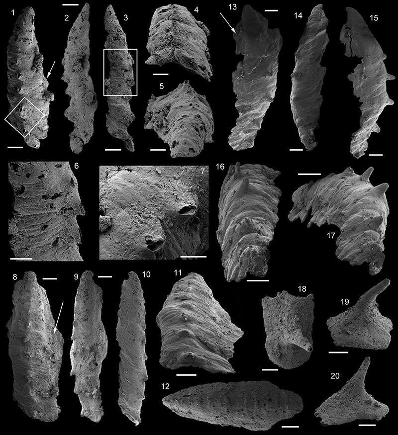
FIGURE 23.Aculopileus aculeatus Kerber, 1988. 1-8. USTL1220/8: 1. Upper view, square shows location of 4; 2. Lateral view, square shows location of 5 and 8, arrow shows furrow parallel to fold associated with subapical apertural margin; 3. Lateral view; 4. Detail of densely spaced papillae of the internal mold; 5. Detail of projecting hollow tube; 6; 6. Abapical view; 7. Apical view; 8. Detail of projecting hollow tube. 9-10. USTL1220/18: 9. Detail of irregular polygonal pattern of external coating; 10. Lateral view with hollow tube visible; square shows location of 9. Scale bars equal: 5, 8, 20 µm; 4, 9, 100 µm; 1-3, 6, 7, 10, 200 µm.
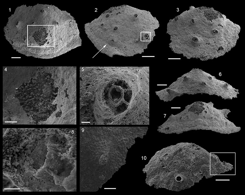
FIGURE 24.Alaconcha rugosa Devaere, sp. nov. 1-3, 5, 6, 9. USTL1247/3: 1. Upper view; 2. Apical view, square shows location of 3; 3. Detail of subapical projections; 5. Lateral view; 6. Abapical view; 9. Apical view. 4, 8, 10, 11. USTL1225/11: 4. Upper view; 8. Lateral view; 10. Abapical view; 11. Apical view. 7. USTL1238/9, upper view. Scale bars equal: 3, 100 µm; 4, 8, 10, 11, 200 µm; 7, 300 µm; 1,2, 5, 6, 9, 500 µm.
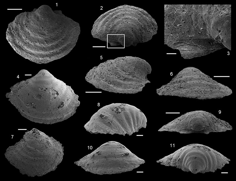
FIGURE 25.Aetholicopalla adnata Conway Morris, 1990. 1-2. USTL1211/6: 1. External view; 2. Arrow shows attachment area. 3-4. USTL1262/13. 5-13. USTL1258/13: 5. Cross section showing internal cavity, lower square shows location of 9; upper square shows location of 13; 6. External view, square shows location of 10; 7, 8. External view, internal and external phosphatic layers, square shows location of 11; 9. Detail of the two phosphatic layers with interconnecting pillars; 10. Detail of external surface of inner phosphatic layer; 11. Detail of external phosphatic layer; 12. Detail of possible bacterial filament; 13. Partly filled internal cavity and empty space next to phosphatic layers, square shows location of 12. 14-16. USTL1236/1: 14, 15. Lateral views of specimen attached to a clast; 16. Upper view. 17-19. USTL1258/11: 17. Arrow shows attachment area; 18. Lateral view; 19. Side opposite to attachment. 20-23. USTL1243/1: 20. Side opposite to attachment; 21. Attachment side, arrow shows deformation due to attachment; 22, 23. Lateral views. Scale bars equal: 12, 20 µm; 9, 50 µm;13, 100 µm, 1-8, 10, 11, 17-19, 200 µm, 14-16, 20-23, 500 µm.
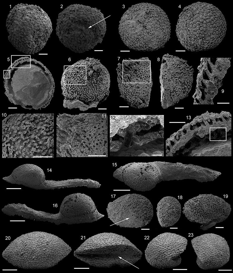
FIGURE 26. 1-3. USTL1209/3: 1. Dorsal view; 2. Lateral view; 3. Apical view, arrow shows slightly developed subapical shelf. 4-5. USTL1109/4: 4. Dorsal view; 5. Lateral view. 6-8. USTL1260/17: 6. Apertural view, square shows location of 7; 7. Detail of subapical field with polygonal imprints; 8. Dorsal view. 9-11. USTL1259/12: 9. Dorsal view; 10. Lateral-subapical view, square shows location of 11; 11. Detail of subapical area (internal mold and external coating preserved). 12-14. USTL1261/5: 12. Dorsal view; 13. Abapical view, square shows location of 14; 14. Detail of densely spaced papillae of the internal mold. 15-16. USTL1220/17: 15. Dorsal view; 16. Lateral view, arrow shows folded upwards subapical margin. 17-18. USTL1220/22: 17. Dorsal view; 18. Abapical view, sinusoidally striated external coating. 19-20. USTL1213/16: 19. Abapical view; 20. Dorsal view. Scale bars equal: 14, 60 µm; 7, 11, 15, 16, 100 µm; 1-5, 9, 10, 17-20, 200 µm; 6, 8, 12, 13, 300 µm.
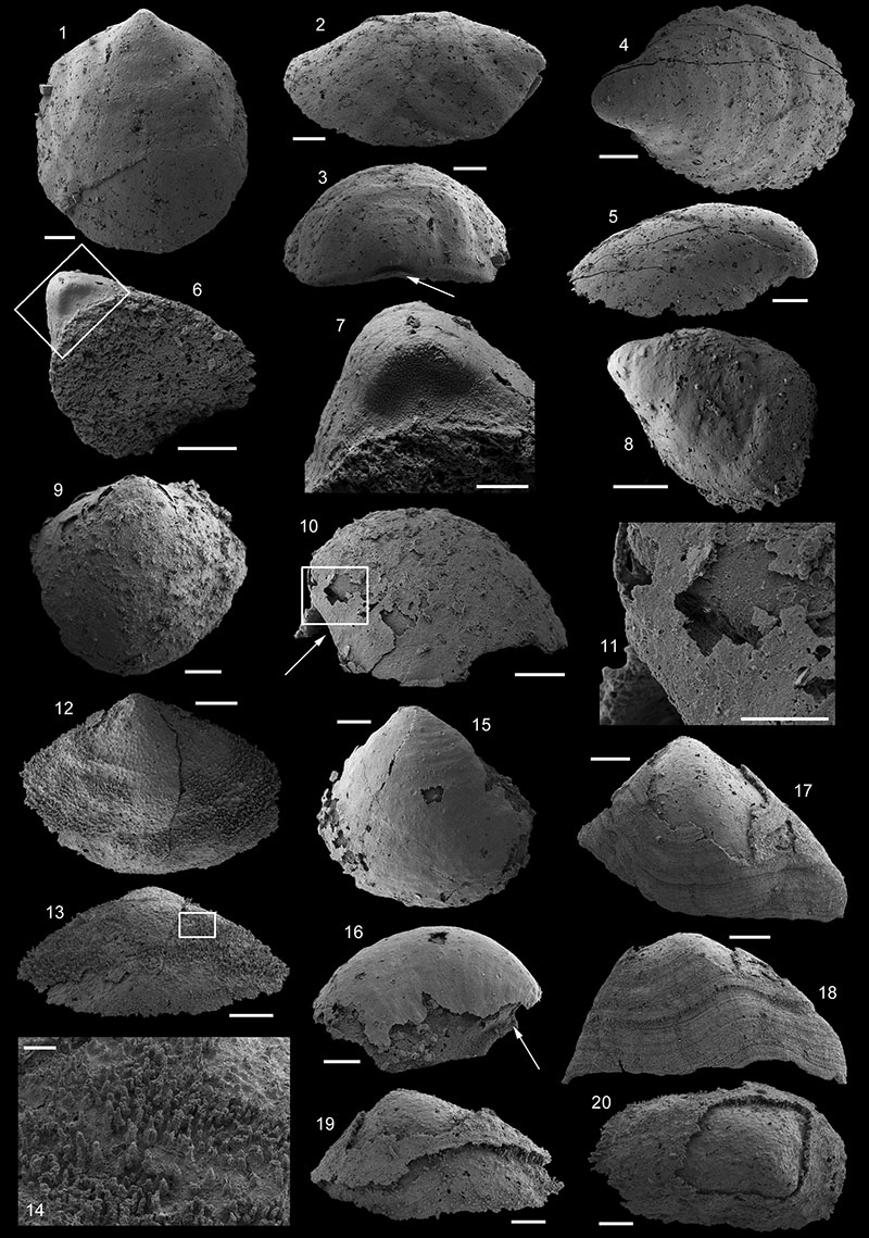
FIGURE 27. 1-2. USTL1235/13 L15_048-049: 1. Dorsal view; 2. Abapical view, arrow shows slit. 3. USTL1238/5, dorsal view. 4-5. USTL1223/13: 4. Lateral view; 5. Apical view, arrow shows sinus in apertural margin. 6-7. USTL1220/14: 6. Dorsal view; 7. Lateral view. 8-10. USTL1240/10: 8. Detail of internal mold an external coating; 9. Upper view; 10. Lateral view, square shows location of 8. 11-12. USTL1217/22: 11. Dorsal view, square shows location of 12; 12. Detail of external coating comarginal ribs. 13-14. USTL1243/4: 13. Lateral view; 14. Dorsal view. 15-16. USTL1241/6: 15. Lateral view; 16. Dorsal view. 17-18. USTL1241/5: 17. Lateral view; 18. Dorsal view. Scale bars equal: 8, 12, 50 µm; 4-7, 11, 100 µm; 1-3, 200 µm; 9, 10, 13-18, 500 µm.
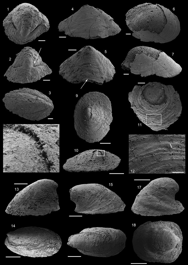
FIGURE 28. 1-2. USTL1243/6, Scale bar equals 500 µm. 1. Lateral view; 2. Abapical view. 3-4. USTL1242/3, scale bar: 300 µm. 3. Abapical view; 4. lateral view.
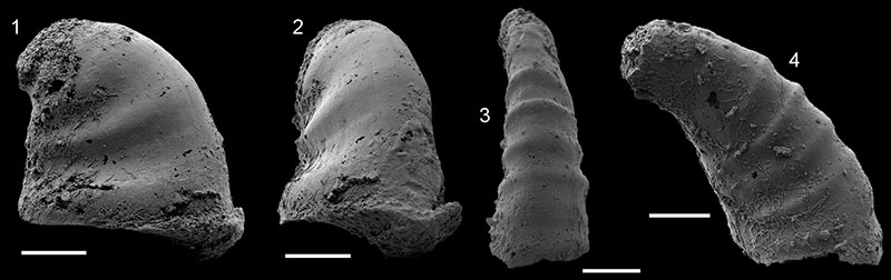
FIGURE 29. Stratigraphic range of taxa identified in section K2 (Marcou) and new biozone reported.
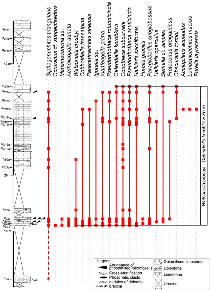
FIGURE 30. Stratigraphic range of taxa identified in section K3B (Marcou) and new biozone reported.
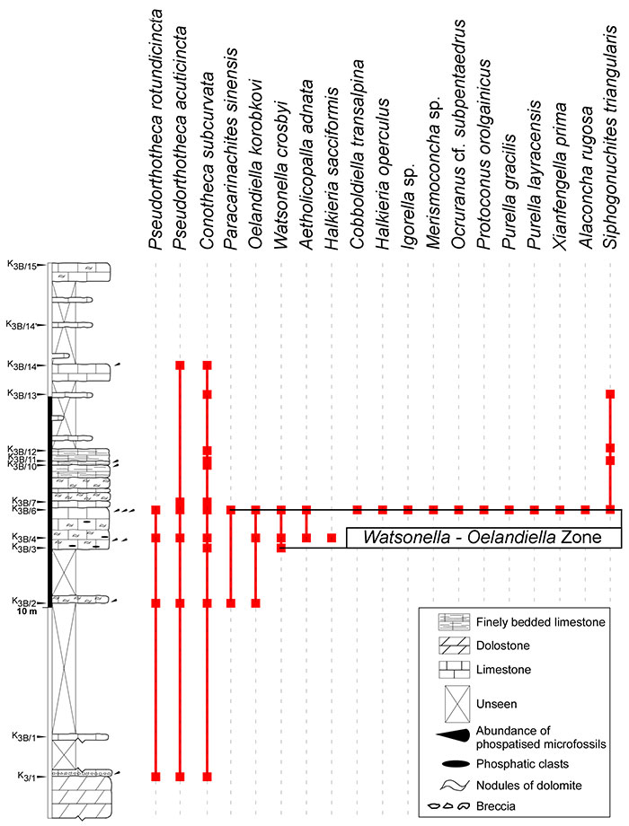
FIGURE 31. Stratigraphic range of taxa identified in section K3T (Marcou) and new biozone reported.
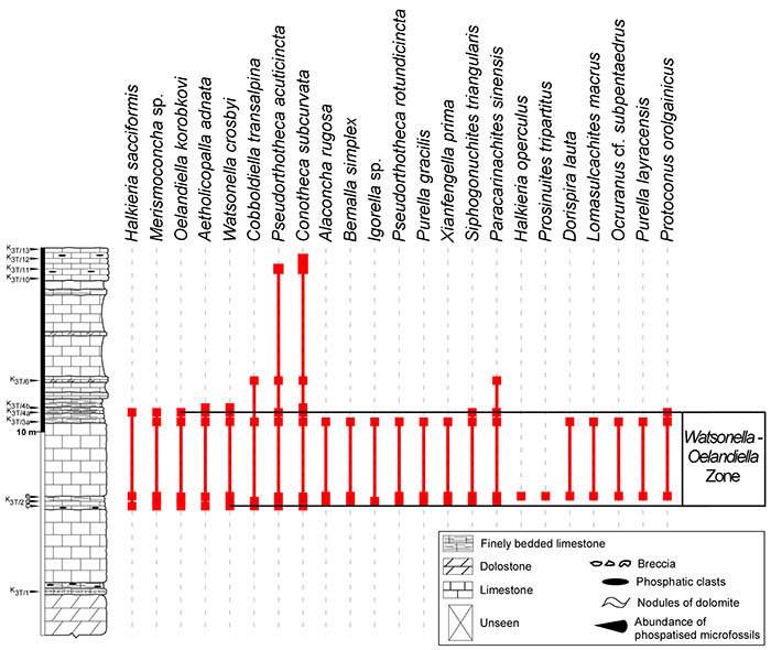
FIGURE 32. Stratigraphic range of taxa identified in section K4 (Saint Geniès de Varensal).
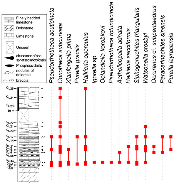
FIGURE 33. Stratigraphic range of taxa identified in section K5 (Saint Geniès de Varensal) and new biozone reported.
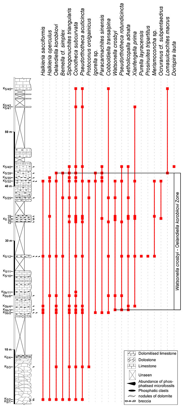
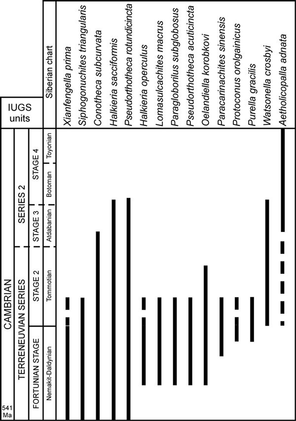
FIGURE 34. Composite ranges of widely distributed taxa present in the studied sections (for references, see the section 'other occurrences' for each of the taxa). The Siberian stratigraphic chart is reported here due to its most common use.