FIGURE 1. General view of Cambrian echinoderm types of preservation. 1.1. Stereom microstructure of a Cambrian echinoderm preserved as iron-oxide replacement (after Clausen and Smith, 2008). 1.2. Stereom microstructure of a Cambrian echinoderm preserved as silica coating (after Clausen and Smith, 2008). 1.3. Stereom of a recent echinoderm. 1.4. Specimen of Protocicnctus mansillaensis preserved in recrystallized calcite, as specimens presented in this study (after Rahman and Zamora, 2009). 1.5. Latex cast of the edrioasteroid Aragocystites belli preserving superficial traces of stereom on plates around central mouth (after Zamora, 2013). 1.6. Same specimen preserved as a natural mould. 1.7. Isolated echinoderm element obtained after acid etching (image courtesy of Olaf Elicki).
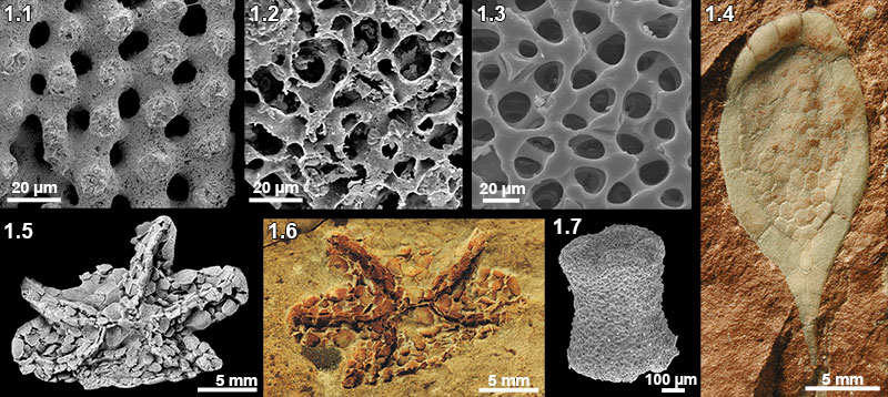
FIGURE 2. Microstructural organization and basic geochemical characteristics of the Middle Cambrian echinoderms: 2.1, Transverse section of the integument plate of Protocinctus in SEM; 2.2, transverse section of the integument plate of Protocinctus in SEM back-scattered electron (BSE) images; 2.3, geochemical analyses of the integument plate of Protocinctus (o) and the sediment (s); 2.4, CL-activated UV-VIS spectrum of the intense orange-red luminescent integument plate of Protocinctus showing Mn2+ emission maximum at ~620 nm; 2.5, Transverse section of the thecal plate of Stromatocystites in SEM; 2.6, transverse section of the thecal plate of Stromatocystites in BSE images; 2.7, geochemical analyses of the ossicle of thecal plate of Stromatocystites and the sediment; 2.8, CL-activated UVVIS spectrum of the intense orange-red luminescent thecal plate of Stromatocystites showing Mn2+ emission maximum at ~610 nm; 2.9, Axial section of the thecal plate of Dibrachicystidae in SEM; 2.10, transverse section of the thecal plate of Dibrachicystidae in BSE images; 2.11, geochemical analyses of the ossicle of thecal plate of Dibrachicystidae and the sediment; 2.12, CL-activated UV-VIS spectrum of the intense orange-red luminescent thecal plate of Dibrachicystidae showing Mn2+ emission maximum at ~610 nm. Scale bar equals 100µm.

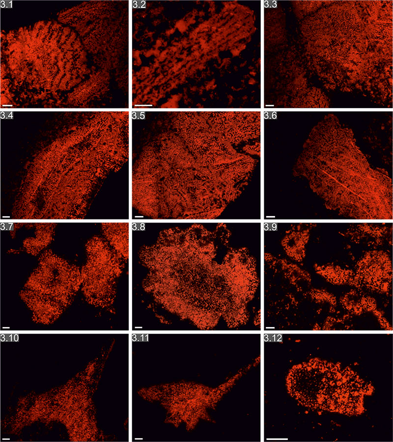
FIGURE 3. Cathodoluminescence photomicrographs of the Middle Cambrian echinoderms showing orange-red luminescent stereom and pore system filled with non-luminescent likely ferroan calcite: 3.1.-3.3, 3.5, Integument plates of Protocinctus; 3.4, 3.6, marginal plates of Protocinctus; 3.7, ambulacral plate of Stromatocystites; 3.8, thecal plate of Stromatocystites; 3.9, ambulacral plate of Stromatocystites; 3.10, 3.11, thecal plate of Dibrachicystidae; 3.12, columnal? plate of Dibrachicystidae. Scale bar equals 100µm.