Figure 1. (1) Location map of the Iharkút vertebrate locality (Upper Cretaceous [Santonian] Csehbánya Formation, Bakony Mts, western Hungary). (2) Schematic section of the open-pit Iharkút (after Ősi and Mindszenty, 2009). The black arrow indicates the position of the bone-yielding beds that, among other fossils, provided the fragmentary skull referred to Hungarosaurus sp. (in this work) and the associated Hungarosaurus skeletons. (3) The Iharkút locality from a bird's eye view. Asterisk marks the site where the fragmentary skull was found.
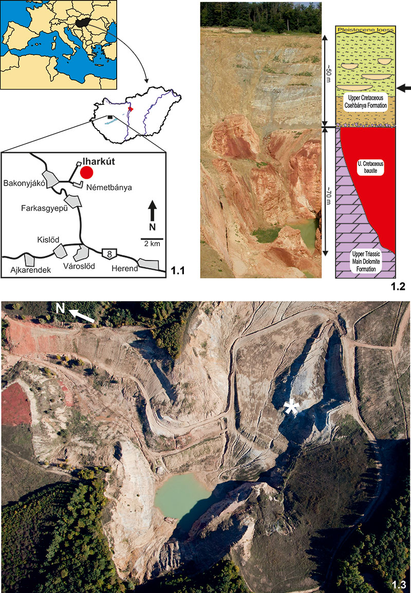
Figure 2. Fragmentary skull (PAL 2013.23.1) and its CT scan visualisation referred to Hungarosaurus sp. from the Upper Cretaceous (Santonian) Csehbánya Formation, Iharkút, western Hungary. (1–2) right lateral; (3–4) left lateral; (5–6) ventral view. Scale bar is 2 cm. For abbreviations see text.
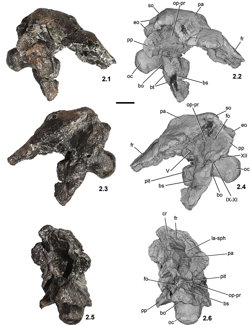
Figure 3. Fragmentary skull (PAL 2013.23.1) and its CT scan visualisation referred to Hungarosaurus sp. from the Upper Cretaceous (Santonian) Csehbánya Formation, Iharkút, western Hungary. (1–2) anterior; (3–4) posterior; (5–6) dorsal view. Scale bar is 2 cm. For abbreviations see text.
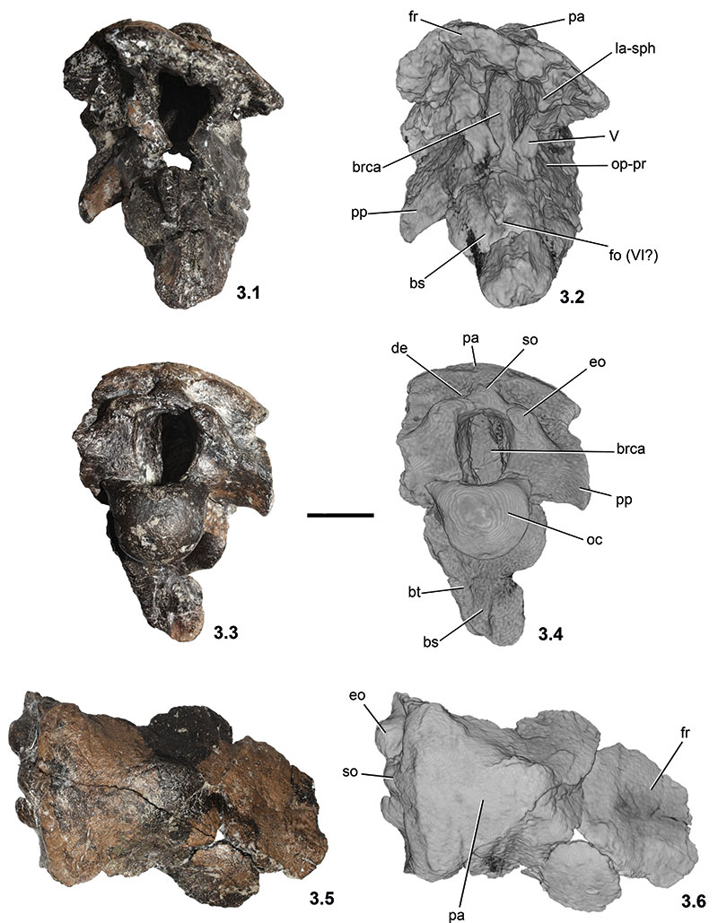
Figure 4. CT slices through the fragmentary skull (PAL 2013.23.1) referred to Hungarosaurus sp. from the Upper Cretaceous (Santonian) Csehbánya Formation, Iharkút, western Hungary. (1–2) axial slice through the basisphenoid; (3–4) parasagittal slice through the openings of the posterior cranial nerves.
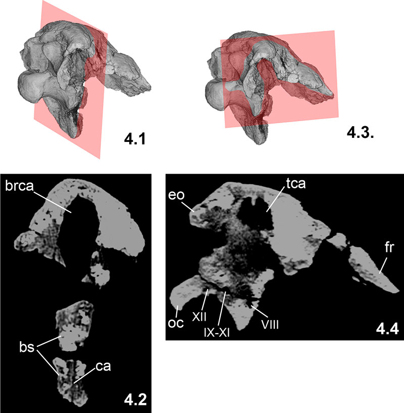
Figure 5. Topographic drawing of a silicone rubber mould of the endocranial cavity taken from the braincase of Hungarosaurus sp. (PAL 2013.23.1). (1) dorsal; (2) right lateral; (3) ventral view. Scale bar is 2 cm. For abbreviations see text.
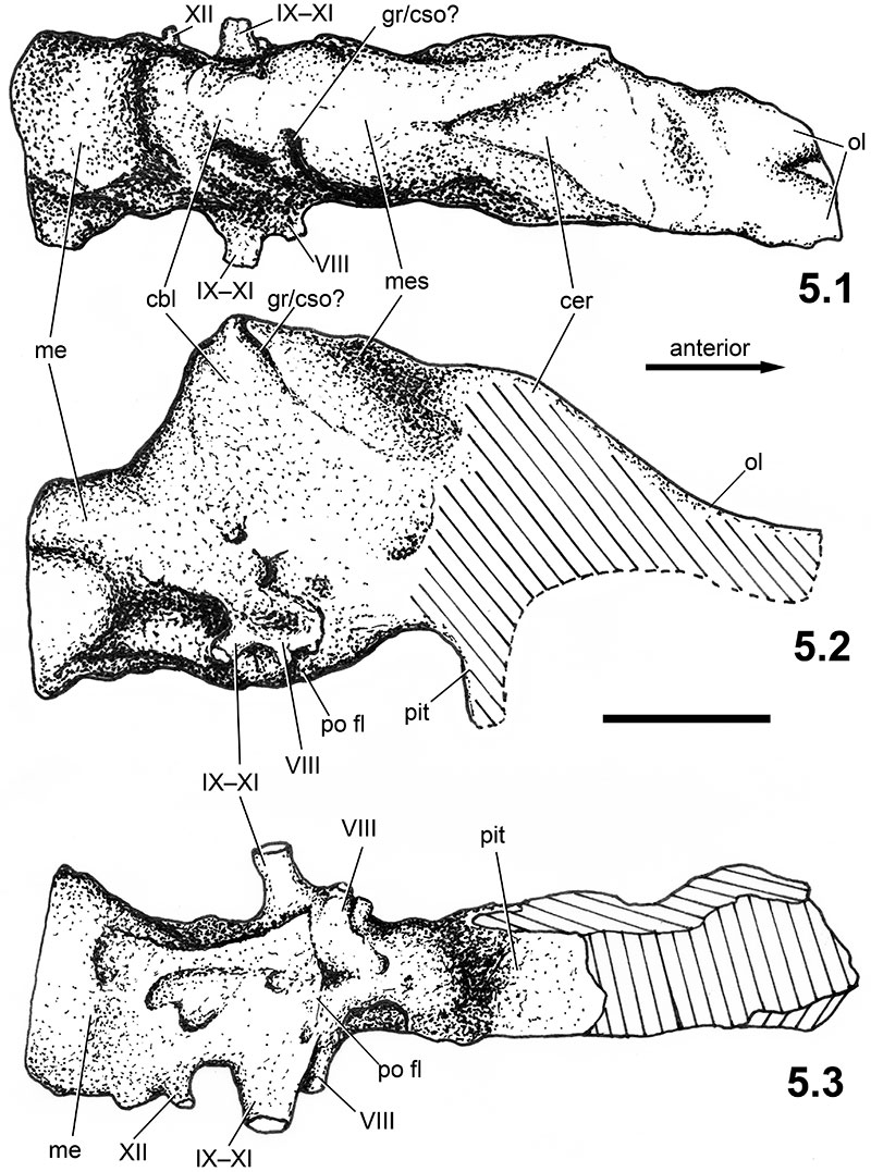
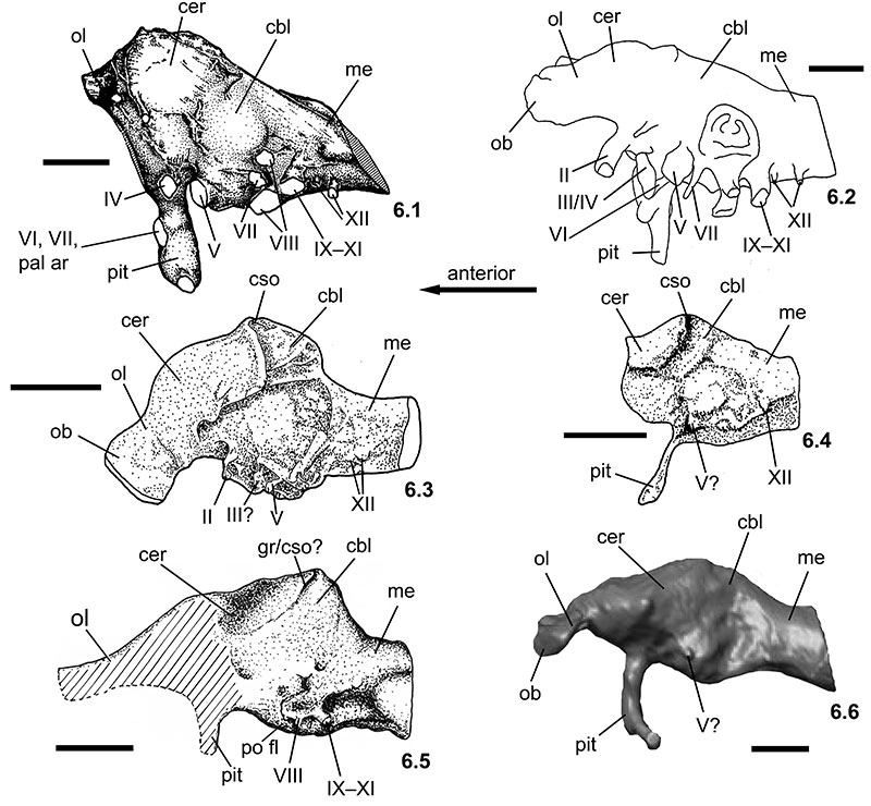
Figure 6. Comparison of ankylosaur endocasts in left lateral view. (1) cf. Polacanthus sp. (inverted image; redrawn from Norman and Faiers, 1996); (2) Euoplocephalus sp. (redrawn from Coombs, 1978a); (3) Struthiosaurus transylvanicus (redrawn from Pereda Suberbiola and Galton, 1994); (4) Struthiosaurus austriacus (redrawn from Pereda Suberbiola and Galton, 1994); (5) Hungarosaurus sp. (reversed image); (6) Panoplosaurus mirus (modified from Witmer and Ridgely, 2008). Note the hypertrophied cerebellum in Struthiosaurus transylvanicus and Hungarosaurus sp. Scale bars are 2 cm. For abbreviations see text.