FIGURE 1. Reconstructions of Bothriolepis canadensis: 1.1, redrawn from Patten (1904, figure 1); 1.2, redrawn from Stensiö (1948, text-figure 38); 1.3, redrawn from Vézina (1996, figure 1); 1.4, redrawn from Arsenault et al. (2004, figure 8C-8D); 1.5, new reconstruction based on 3D model. Alignment and scaling based on the dorsal thoracic armor length.
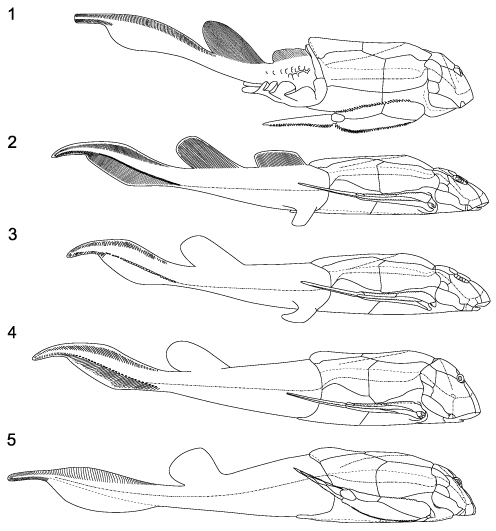
FIGURE 2. 3D reconstruction of Bothriolepis canadensis. Odd numbers without pectoral fins showing the brachial process, even numbers with pectoral fins; 1-2, dorsal view; 3-4 lateral view; 5-6, ventral view; 7-8, front view; 9-10, posterior view; 11, lateral view with posterior part of the body. Scale bar equals 1 cm. Use this figure as a target for the PaleoAR mobile application to get the 3D model.
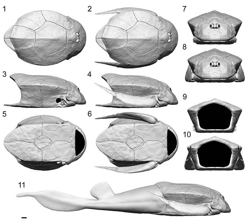
FIGURE 3. New reconstruction of Bothriolepis canadensis. Odd numbers without pectoral fins showing the brachial process, even numbers with pectoral fins; 1-2, dorsal view; 3-4 lateral view; 5-6, ventral view; 7-8, front view; 9-10, posterior view. Scale bar equals 1 cm.
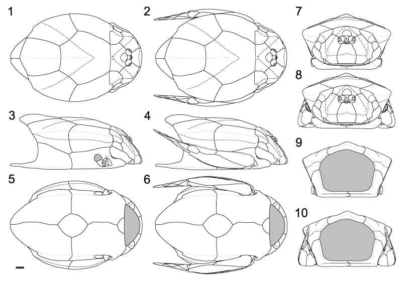
FIGURE 4. Gill opening of Bothriolepis canadensis. Head plates and pectoral fin in transparency to illustrate the relationships between the anterior ventrolateral plate (AVL) and the submarginal plate (SM).
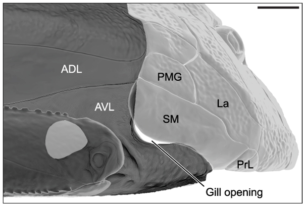
FIGURE 5. 3D reconstruction of the right pectoral fin of Bothriolepis canadensis: 1, lateral view; 2, medial view; 3, ventral view; 4, dorsal view. Scale bar equals 1 cm.
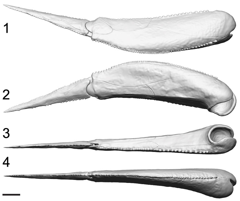
FIGURE 6. 3D reconstruction of the right pectoral fin segments of Bothriolepis canadensis: 1, lateral view of proximal segment; 2, medial view of proximal segment; 3, ventral view of proximal segment; 4, lateral view of distal segment; 5, medial view of distal segment. Scale bar equals 1 cm.
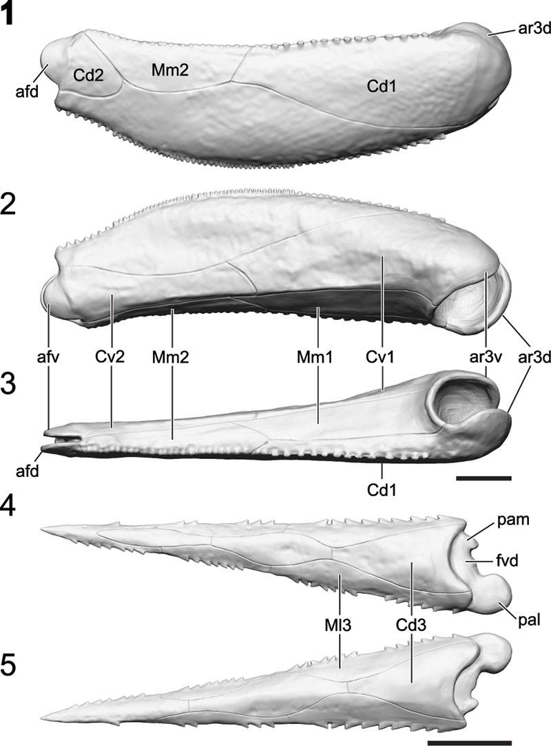
FIGURE 7. Range of movements for the pectoral fin of Bothriolepis canadensis: 1, rotation around the brachial process in fully retracted position; 2, fully protracted position; 3, fully protracted position front view; 4, latero-medially movement of the distal segment; 5, minimum angle of protraction for maximum mobility of the pectoral fin; 6, rotation around the brachial process in a protracted angle of 16°; 7, up-and-down movement in a protracted angle of 16°. Use this figure as a target for the PaleoAR mobile application to get a video of pectoral fins possible movement range.
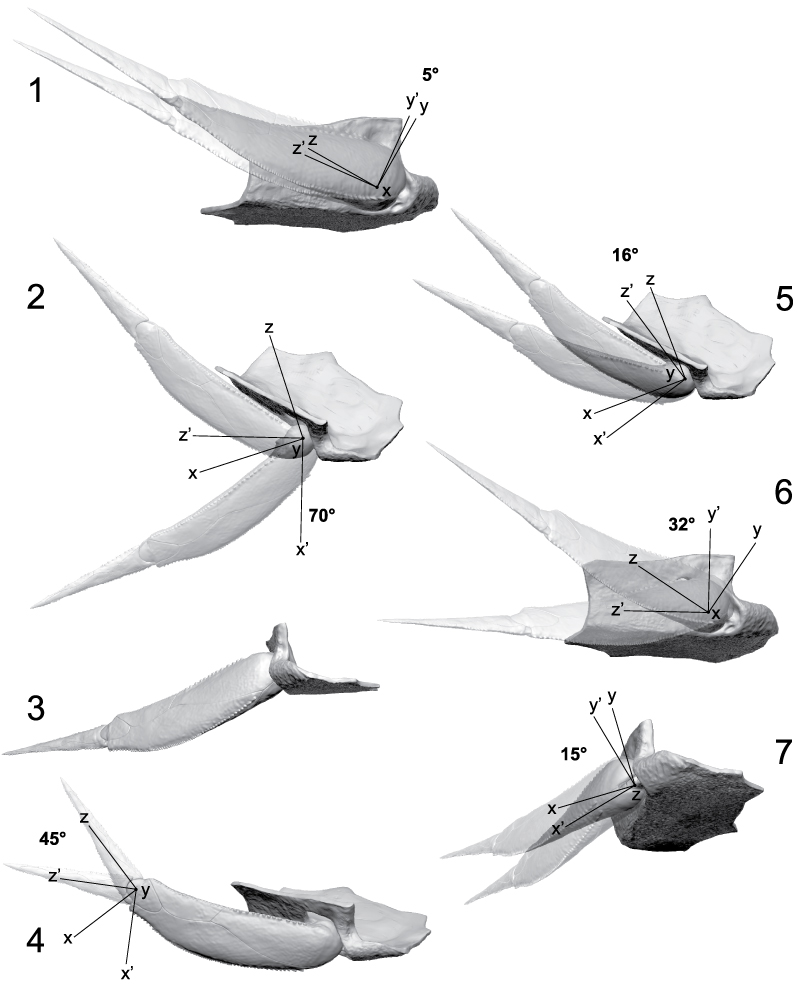
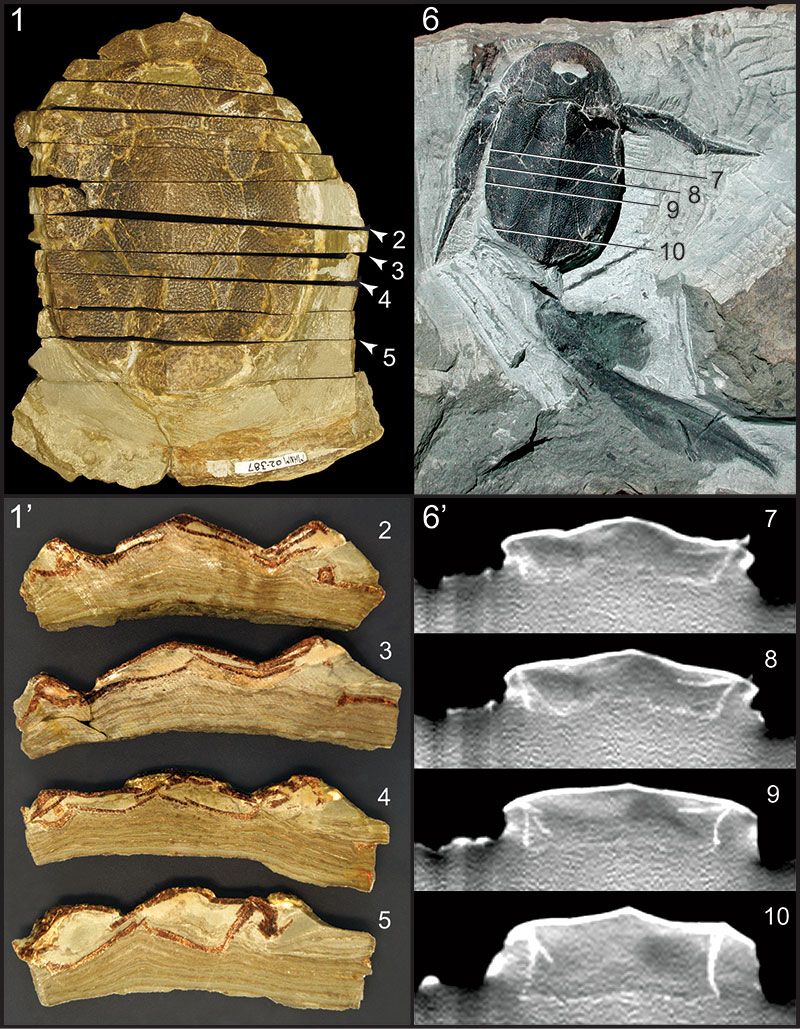
FIGURE 8. Taphonomic differences with reference to two types of sediment compaction for Bothriolepis canadensis: 1 and 1’, specimen MHNM 02-387 in dorsal view preserved in laminites with coronal sectioned with positioning of the sections illustrated in 2-5; 2-5, coronal sections showing the flattening and distortion of the specimen; 6 and 6’, specimen MHNM 02-2676 in dorsal view preserved in siltstone with positioning of the CT-scan coronal sections 7-10; 7-10, coronal sections showing the 3D condition of preservation and a weak lateral compaction.