APPENDIX 1
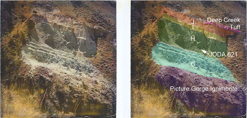
Photograph of the locality accompanying the field notes. Archive of the John Day National Park. Curtesy of Joshua X. Samuels, JODA.

APPENDIX 2
List of taxa in alphabetic order and character matrix used for the phylogenetic analysis. The characters are derived from own observations (partially from Stefen, 2005, Stefen and Moers, 2008) and the relevant literature (Barbour and Schultz, 1937; Korth and Emry, 1997; Korth, 2001; MacDonald, 1963; Martin, 1987; Mörs and Hulbert, 2010; Moore, 1890a, b; Peterson, 1905; Rybzynski et al., 2010; Schreuder, 1929; Stirton, 1934, 1935, 1965; Xu, 1996) (available in PDF format).
APPENDIX 3
List and explanation of characters used for the phylogenetic analysis.
1 dentition: 0 non-beaver pattern; 1 beaver pattern
2 hypsodonty: 0 teeth basically brachydont; 1 teeth subhypsodont; 2 clearly hypsodont, root is hardly ever formed
3 incisor inf. frontal face: 0 clearly convex; well rounded; 1 slightly convex; 1 clearly flat
4 incisor sup. Frontal face: 0 clearly convex; well rounded; 1 slightly convex; 1 clearly flat
5 incisor inf. enamel: 0 smooth; 1 clearly striated; 2 faintly striated
6 incisor sup enamel: 0 smooth; 1 clearly striated; 2 faintly striated
7 form and curvature of lower incisor tip: 0 chisel shaped; 1 pointed
8 form and curvature of upper incisor tip: 0 chisel shaped; 1 pointed
Characters of the skull
9 masseter arrangement: 0 non sciuromorph masseter arrangement ; 1 sciuromorph masseter arrangement
10 skull shape measured: 0 narrow (length/width approximately 1.5) ; 1 intermediate (length/width approximately 1.3) ; 2 broad (length/Width approximately 1.2 ) ; 3 broadest (length/Width approximately 1.0)
11 skull shape appearance: 0 skull longer than wide; 1 skull approximately square, nearly as long as wide (width at zygomatic arch); 2 skull wider than long
12 position of infraorbital foramen: 0 low, in ventral third of snout; 1 approximately in the middle of the snout height; 2 high, in the upper third of the snout height
13 form of infraorbital foramen in frontal view: 0 oval, geometrical; 1 tearshaped; 2 slitlike (markedly straight sides); 3 other form
14 course of infroaorbital canal: 0 – straight; 1 – not straight
15 position of masseter superfacials process: 0 – ventral to slightly posterior to infraorbital foramen; 1 anterior to infraorbital foramen; 2 not applicable
16 course of crista facialis (masseter ridge) in lateral view: 0 straight; 1 slightly curved; 2 "s"-shaped;
17 relative distance of premaxillary-maxillary suture to crista facialis masseter: 0 close; 1 well anterior
18 course of premaxillary-maxillary: 0 more or less straight dorso-ventrally; 1 in a marked angle not relatively straight dorso-ventrally
19 infraorbital in relation to root of zygomatic arch: 0 posterior (still level to part of zygomatic root though; 1 ventral to it; 2 anterior to it (to the most antrior part of the zyg root)
20 connection between jugal and lacrimal: 0 yes; 1 no
21 jugal extension on zygomatic arch: 0 jugal extending upwards up to dorsal rim of orbita, meeting fronto-maxillar suture; 1 jugal only extending to about half way up the maxillar component of the zygomatic arch; 2 jugal extending upwards to less than one third of the maxillar component of the zygomatic arch
22 dorsal component of lacrimal: 0 yes; 1 no
23 position of orbital constricion in relation to length of skull: 1 clearly in anterior part of skull length; 0 approx. in the middle of skull length; 2 clearly in posterior skull length
24 angle of occipital plane to a plane extended from the base of the skull: 0 about 90°; 1 less than 90 ° (exoccipital plane bending anteriorly); 2 more than 90 ° (exoccipital plane bending posteriorly)
25 fossa occipitalis: 0 absent; 1 present
26 rugosities on parietale/interparietale: 0 no (or very few); 1 yes, marked
27 groove dorsal to incisor in lateral view: 0 absent; 1 partial; 2 complete
28 divergence of tooth rows, ratio of distance between M3s to distance between P4s: 0 parallel; 1 slightly diverging (up to 1.7); 2 diverging more strongly 1.7 – 2.499; 3 larger than 2.5; 4 larger than 4
29 distal palatal termination: 1 at level of M3s; 2 at level of M2s; 0 at level of post M3s
30 Development of crest at posterior sagittal-lambdoidal area: 0 undeveloped; 1 well developed
31 Form of fronto-parietal crest: 0 frontal crest meet anterior of interorbital constriction and form 1 crest; 1 frontal crest meet at interorbital constriction to form 1 crest; 2 frontal crest meet posterior of interorbital constriction to form 1 crest; 3 frontal crest never meet
32 Formation of fronto-parietal crest: 0 – crest form one narrow crest when they meet; 1 crest remains broad when they meet
33 form of nasals: 0 maximum length/width greater than 2.5; 1 maximum length/width smaller than 2.0; 2 between 2.0 and 2.5
34 nasal form: 0 broader anterior than posterior; 1 distal broader than anterior (<); 2 - broadest in approximately the middle of the nasal; 3 – of about equal width throughout
35 position of caudal end of nasals: 0 distal of complete root of zygomatic arch; 1 approximately above root of zygomatic arch; 2 anterior to root of zygomatic arch
36 interpremaxillary foramen: 0 absent; 1 present
37 distal ending of the lower incisor: 0 lingual, at least no blub on labial side of mandible; 1 a slight blub on labial side of mandible; 2 a marked blub on labial side of mandible
38 curvature of premaxillary-maxillary in the diastema: 0 little, nearly straight; 1 curved
2 very strongly curved
39 ratio diastema to tooth row: 0: 0 > 1.1; 1 0.91-1.09; 2 < 0.9
40 intersection of premaxillary-maxillary suture and incisive foramen: 0 posterior to foramen; 1 really intersecting foramen
41 relation of depth of skull at bulla and at tooth row: 0 bulla and tooth row about same ventral depth; 1 bulla extending further ventral than tooth row; 2 tooth row more ventral than bulla
42 ratio of maximum width of nasals versus maximum width of snout: 0 greater than 0.85; 1 between 0.80-0.65; 2 smaller than 0.55
43 relation of length of incisive foramen to diastema: 0  0,1; 1 0,11-0,4; 2 0,41-0,6; 3 >0,61
44 position of the incisive foramen: 0 about in the middle of the diastema; 1 position more caudally; 2 position more anterior
45 presence of maxillar/palatal grooves: 0 none; 1 from incisive foramen to anterior palatal foramen; 2 only partial grooves directly posterior to incisive f; 3 only in front of anterior palatal foramen
46 Location of anterior palatal foramina: 0 within palatal bone; 1 not completely in palatal bone; 2 completely in maxillary bone
47 tip of incisor in relation to tooth row: 0 line extending from occlusal surface of cheek teeth approximately level with tip of incisor; 1 tip of incisor well above this line; 2 tip of incisor well below this line
48 presence of dp3/dP3 known: 0 yes; 1 no
49 extension of postglenoid constriction: 0 about equal in width to snout; 1 broader than snout; 2 at least twice as broad than snout; 3 postglenoid constriction narrower than snout
50 appearance of the bullae: 0 unconspicious; 1 well rounded and globose; 2 extremely small in relation to the skull size
51 presence of temporal foramen: 0 absent; 1 single; 2 multiple
52 auditory tube: 0 absent; 1 present
53 direction of the external auditory meat: 0 lateral; 1 lateral and dorsal; lateral and ventral
54 presence of interparietal: 0 present; 1 absent or fused
55 foramen ovale: 0 confluent with foramen lacerum; 1 separate
56 spehnopalatine foramen: 0 small; 1 mittel; 2 large;
57 masticatory and buccinator foramen: 0 separate; 1 conjunct;2 absent
58 amxillary alisphenoid contact (Rybc 18): 0 absent; 1 posterior to M3; 2 dorsal to M3; 3 dorsal to M2/M3
59 lateral pterygoid plate (proportion of alisphenoid in lateral view of skull): 0 not clear, small; 1 clear like Cator; 2 enlarged
60 Hamulus pertygoideus (or internal pt. process): 0 not visible; 1 small; 2 large (like in Paramys)
61 alisphenoid part of pterygoid fossa: 0 small to non marked; 1 up to half length to bulla
2 extended to bulla (like in Joda specimen)
62 a angle of interorbital constriction (angle between frontale and parietale at interorbital constriction): 0 about 110-145°; 1 about 150-180°; 2 ca 90-110°
63 form of interorbital constriction: 0 hourglass, cranium (parietale in the orbit) well rounded; 1 gross concave (like in recent Castor); 2 nearly, straight, only little interorbital constriction
Mandible
64 chin process or mandible digastric eminence: 0 chin process absent; 1 chin process clearly present
65 course of angular shelf (ventral rim of angular process): 0 extending in horizontal elongation of mandibular ramus; 1 extending upwards in an angle to the ventral rim of the mandibular ramus; 2 extending downwards in extension to the ventral rim of the mandibular ramus
66 course of ventral rim of mandibular ramus : 0 about in horizontal line; 1 clearly bending upwards; 2 clearly bending downwards
67 orientation of angular shelf: 0 flat, main surface only visible in ventral view; 1 tilting laterally (part of ventral surface is visible in lateral view)
68 posterior view of mandible: 0 condyle, coronid and angular process aligned; 1 condyle, coronid and angular process alternating – usually coronoid and angular procesi in line; 2 condyle, coronoid and angular aligned
69 position of mental foramen: 0 anterior to p4; 1 ventral to anterior rim of p4; 2 posterior to anterior rim of p4; 3 none
70 tip of mandibular incisor in relation to tooth row: 0 line extending from occlusal surface of cheek teeth approximately level with tip of incisor; 1 tip of incisor well above this line; 2 tip of incisor well below this line
71 course of mandibular tooth row: 0 about horizontal and parallel to horizontal course of mandibular ramus; 1 tilted to horizontal course of mandibular ramus
72 proximal end of mandibular incisor: 0 about level with tooth row; 1 above tooth row; 2 below tooth row
73 bulb at posterior end of incisor: 0 no; 1 yes
74 anterior rim of mandibular ramus: 0 crossing m3; 1 crossing m2; 2 crossing m1 or p4; 3 distal to m3
75 ratio mandibular diastema to tooth row: 0: 0 > 1.1; 1 = 0.91-1.09; 2 < 0.9
76 groove between ventral rim of mandible and angular process: 0 yes; 1 no

