FIGURE 1. Indocetus ramani. 1-2, IITR-SB 2986 in lingual and labial view, left mandible with fragment of d3, complete d4 and m1, and unerupted m2. This specimen preserves the mandibular foramen (mandib. for.). 3-4, IITR-SB 4001 in palatal and right lateral view, palate and orbit with right P3-M1, showing supraorbital process (supraorb. proc.).
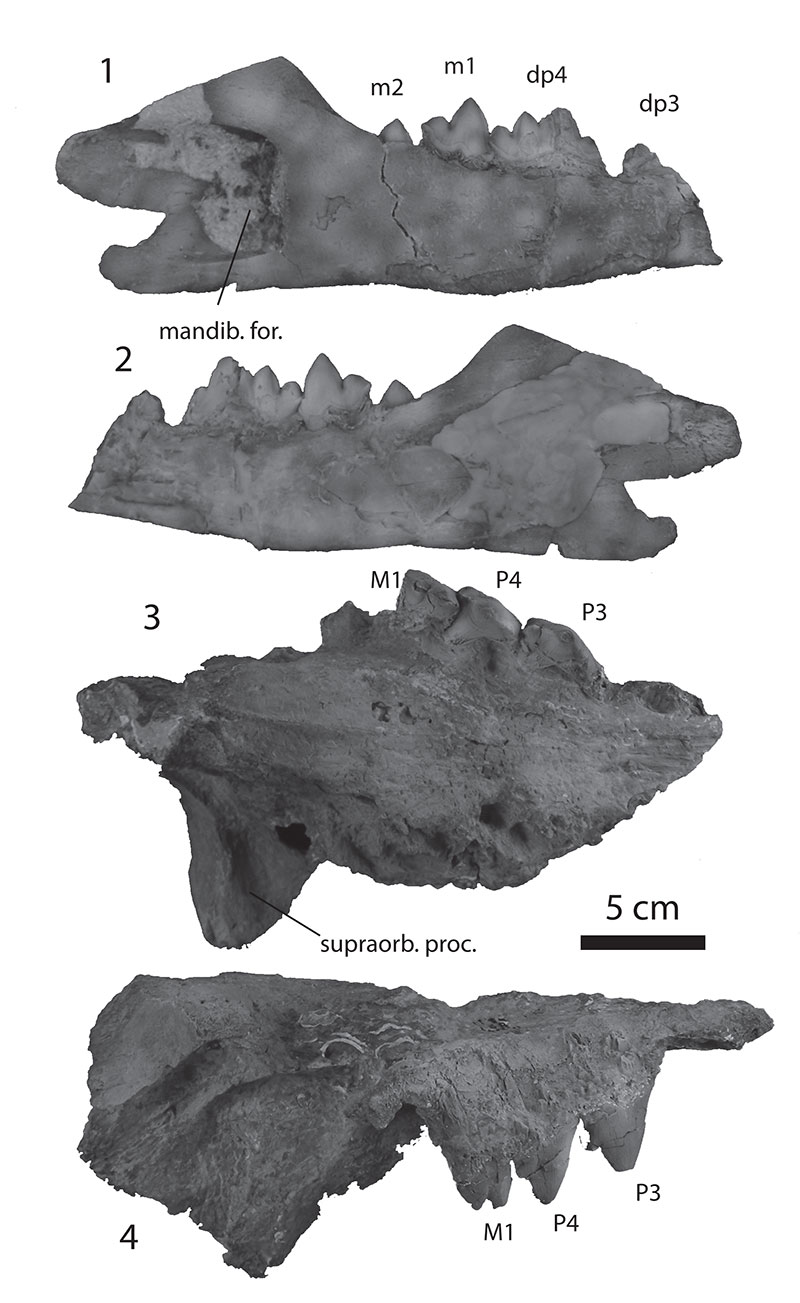
FIGURE 2. Length of M1 plotted against length of P4 for some protocetids, black squares are Indo-Pakistani species, white squares are from other continents. Tooth size data in this figure are based on casts or values given in Gingerich et al. (1995b, 2001b, 2005), Hulbert et al. (1998), and Bianucci and Gingerich (2011).
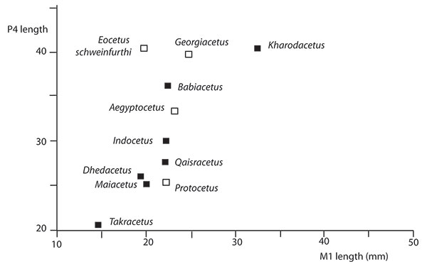
FIGURE 3. Kharodacetus sahnii. 1-5, IITR-SB 3189, rostrum in occlusal, right lateral, and dorsal view ( 1-3 ), and mandibles in left lateral and occlusal view ( 4-5 ).
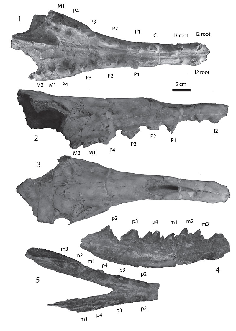
FIGURE 4. Kharodacetus sahnii. 1-4, IITR 3189, left upper dentition in labial and occlusal view ( 1-2 ), and left lower dentition in occlusal and labial view ( 3-4 ).
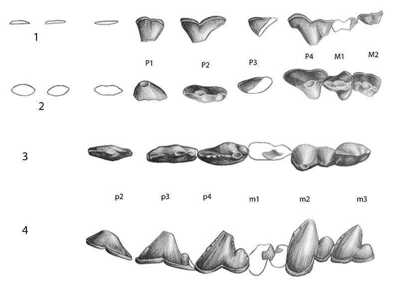
FIGURE 5. Dhedacetus hyaeni. IITR-SB 2870, holotype skull. 1-2, right lateral view and its outline drawing. 3-4, ventral view and its outline drawing. 5, detail of caudal view of right maxilla with M2 roots, lateral to left, ventral to top. Abbreviations: alv. edge, edge of alveolar process of M2; ant. root M2, anterolabial root of M2; bul., bulla; ext. aud., external auditory meatus; lat. max., broken edge of maxilla lateral to molar roots; nuc. cr., nuchal crest; occ. con., occipital condyle; pal., palate, medial to teeth; pos. orb., postorbital process; post. root M2, posterolabial root of M2. Abbreviations for teeth (I1, C, P4, M2) refer to wns or roots preserved. Scale bar pertains to 1 through 4.
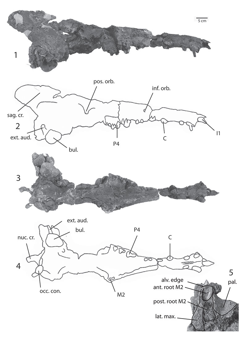
FIGURE 6. Dhedacetus hyaeni, IITR-SB 2870, upper teeth of holotype in occlusal ( 1 ) and labial ( 2 ) view. Crowns were in place when specimen was found, but were detached from skull in collecting and have not been reattached to the skull because of their fragility.
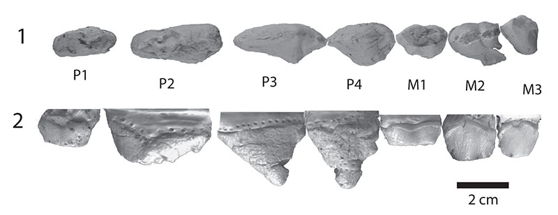
FIGURE 7. Dhedacetus hyaeni, IITR-SB 2870, holotype. 1, mid-thoracic vertebra in anterior view (2870.8). 2-3, mid-thoracic vertebra in anterior view and lateral view (2870.3). 4-5, anterior caudal vertebrae in lateral view (2870.II and IV), 6-7, anterior caudal vertebra in anterior and ventral view (2870.V). 8-9, anterior caudal vertebra in dorsal and lateral view (2870.VIII and VII).
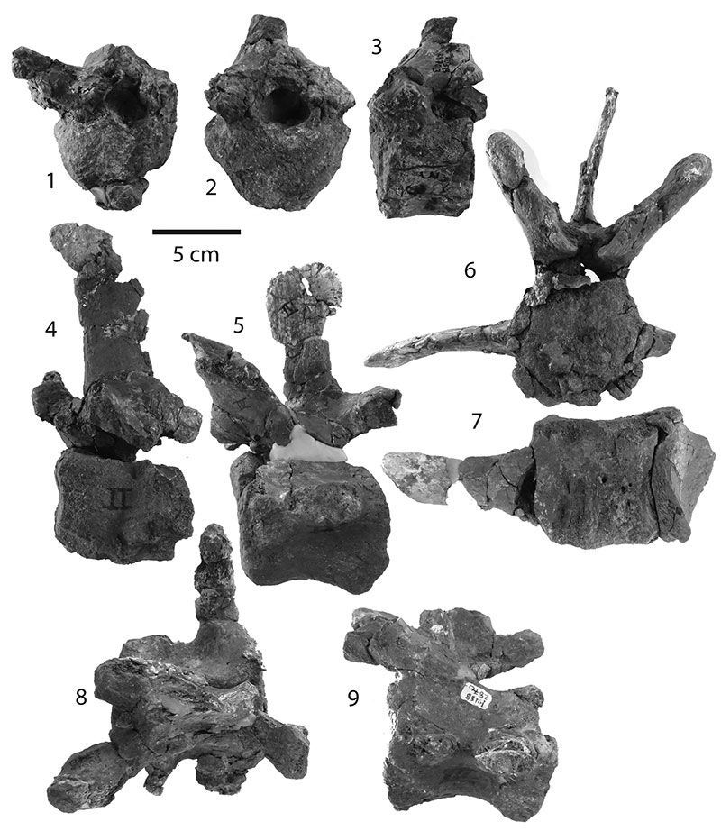
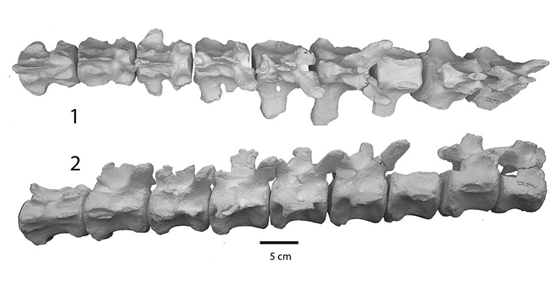
FIGURE 8. Dhedacetus hyaeni. 1-2, articulated caudal vertebrae of referred specimen (IITR-SB 2625) in left labial and dorsal view, respectively. Rostral is to right.