FIGURE 1. Scanning electron micrographs of right humerus (reversed) type 1 (Scraeva hatherwoodensis), M61009, Mammal Bed Hordle, Hampshire, in anterior (1), posterior (2) and medial (3) views. Scale bar equals 1 mm.
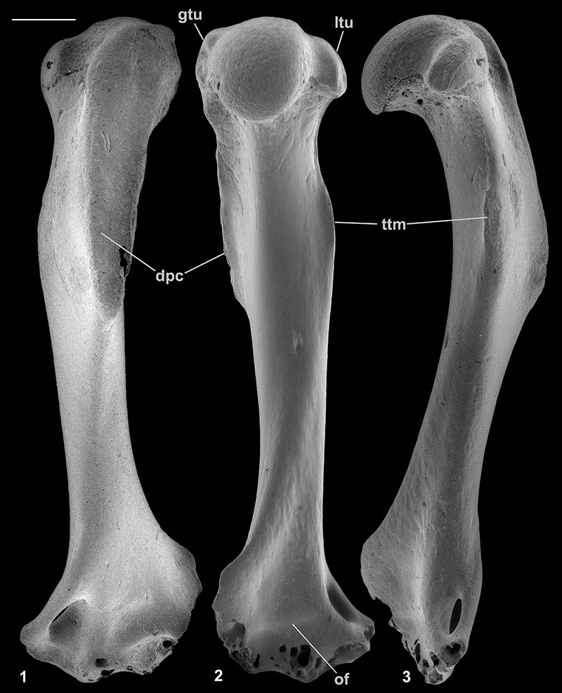
FIGURE 2. Scanning electron micrographs of humeri of Nyctitheriidae. 1, right humerus (reversed) type 1 (Scraeva hatherwoodensis), M61009; 2, distal left humerus type 2 (Euronyctia grisollensis), M60810; 3, distal right humerus (reversed) type 4 (Paradoxonycteris sp. 1?), M61422; 4, distal right humerus (reversed) type 3 (Cryptotopos woodi), M95898. Views are proximal (1), anterior (2.1, 3, 4.4), distal (2.2, 4.2), posterior (2.3, 4.5), medial (2.4, 4.3) and lateral (2.5, 4.1). 3 is from the How Ledge Limestone, Headon Hill, Isle of Wight, the rest are from the Mammal Bed, Hordle, Hampshire. Scale bar equals 1 mm.
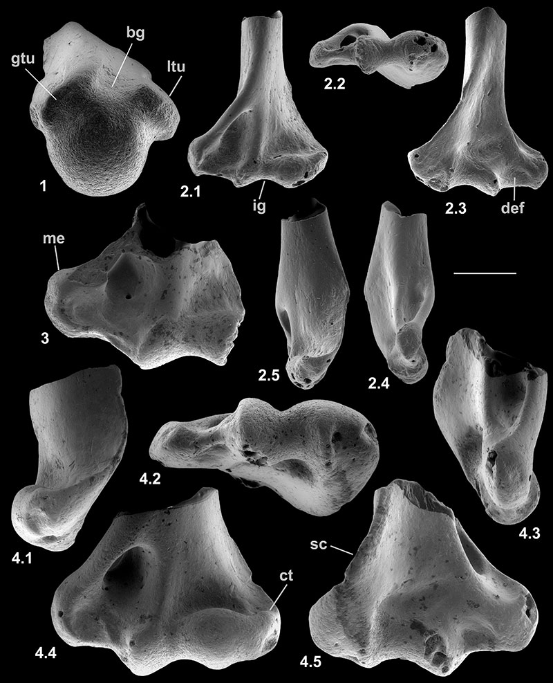
FIGURE 3. Scanning electron micrographs of right radii (reversed) of Nyctitheriidae. 1, Nearly complete radius lacking end, M95899; 2, proximal fragment, M60817; 3, proximal fragment, HZM.324.28063. 1-2 are attributed to Scraeva hatherwoodensis, 3 to Paradoxonycteris sp. 1? The extent of the ulnar facet (uf) in 3.1 is shown by arrows. Views are: anterior (1.1, 2.1, 3.2), lateral (1.2, 2.2, 3.3), posterior (2.3, 3.4), medial (2.4, 3.5) and proximal (3.1). 1-2 are from the Mammal Bed, Hordle, Hampshire; 3 is from the Rodent Bed, Hordle. Scale bar equals 1 mm.
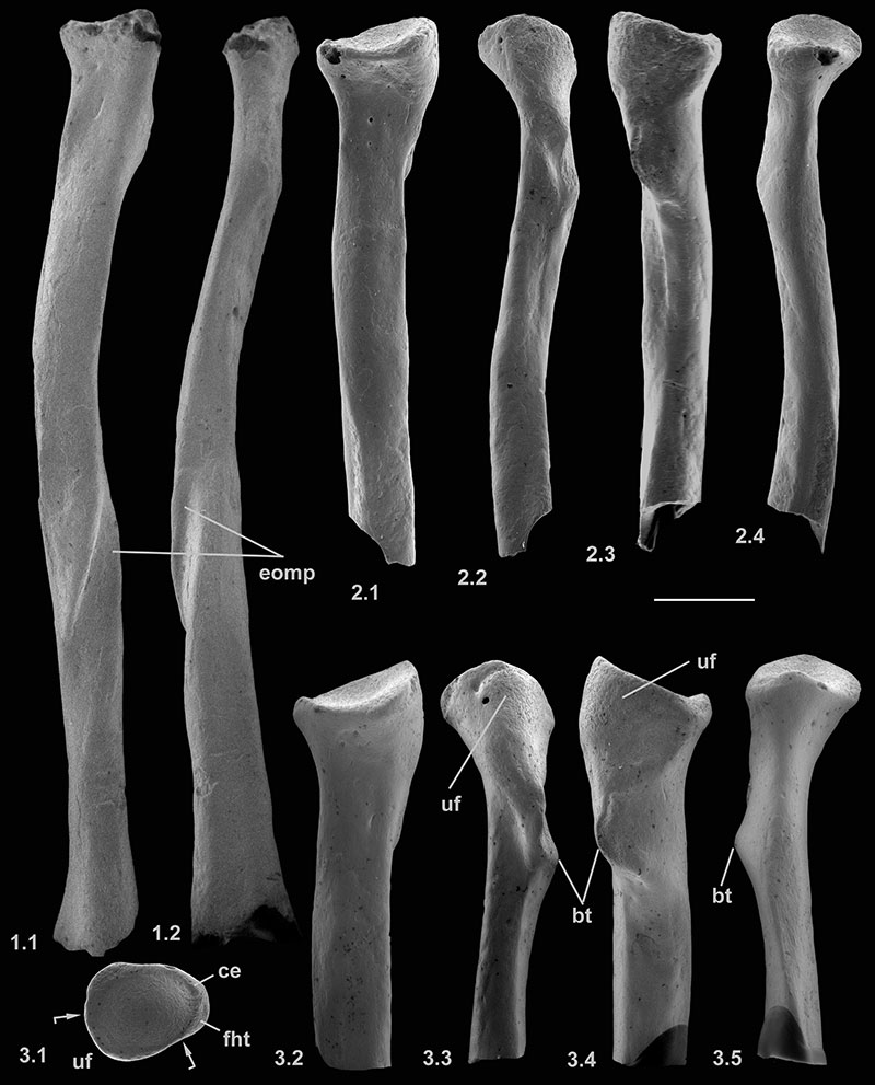
FIGURE 4. Scanning electron micrographs of right femora (reversed) of Nyctitheriidae. 1, nearly complete femur type 1 (Scraeva hatherwoodensis ?), lacking distal end, M60701; 2, proximal fragment of femur type 2 (Cryptotopos woodi ?), M95900; 3, left metacarpal II, HZM.266.27528. Views are: anterior (1.1, 3.1), posterior (1.2, 2) and proximal (3.2). All from the Mammal Bed, Hordle, Hampshire. Scale equals 1 mm.
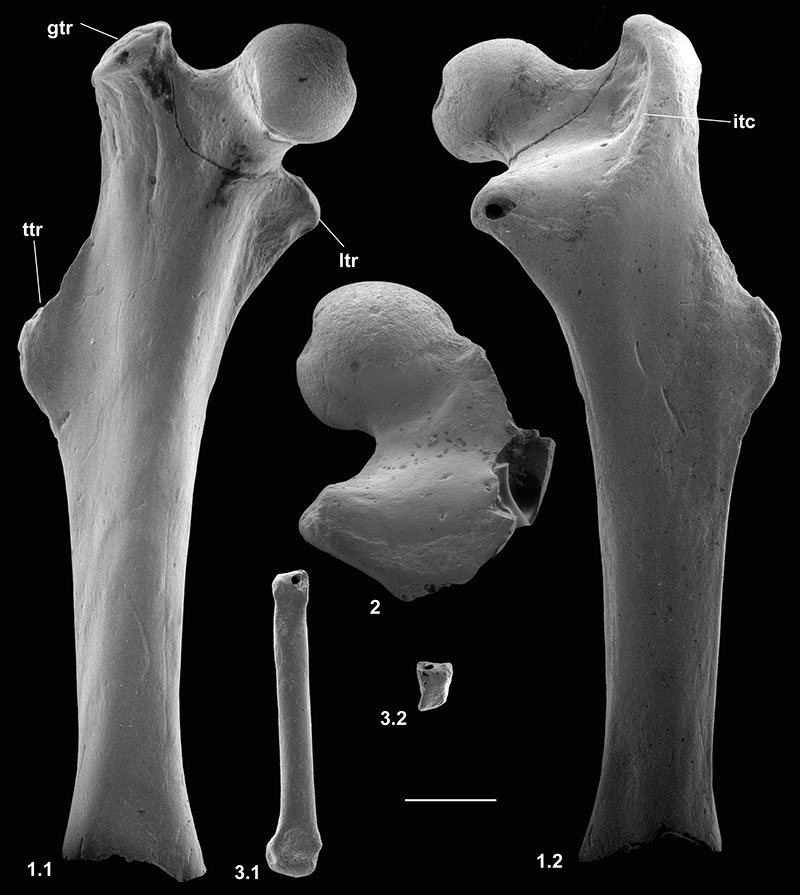
FIGURE 5. Scanning electron micrographs of right femora (reversed) of Nyctitheriidae. 1, nearly complete femur type 1 (Scraeva hatherwoodensis ?), lacking distal end, M60701; 2, proximal fragment of femur type 2 (Cryptotopos woodi ?), M95900. Views are: medial (1.1), proximal (1.2, 2) and lateral (1.3). Both from the Mammal Bed, Hordle, Hampshire. Scale equals 1 mm.
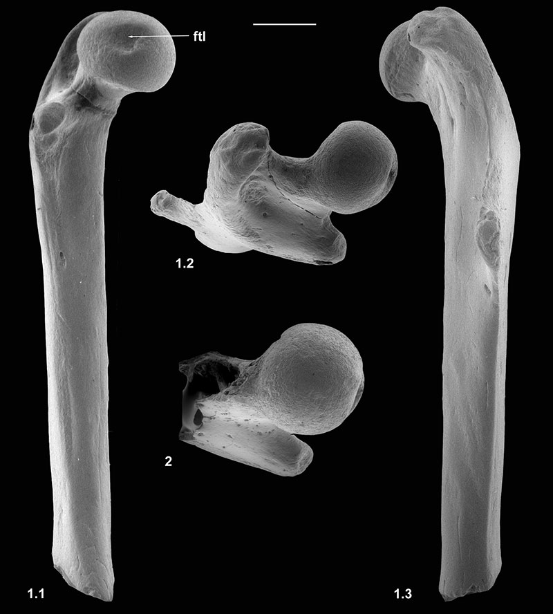
FIGURE 6. Scanning electron micrographs of distal tibiae, shown as from left side, of Nyctitheriidae. 1.1, right tibia (reversed) type 1 (Cryptotopos woodi), M60823; 1.2, left tibia type 1 (Cryptotopos woodi), M60822; 2, left tibia type 2 (Scraeva hatherwoodensis), M61087; 3, right tibia (reversed) type 3 (Paradoxonycteris sp. 1), M95949; 4, left tibia type 4 (Euronyctia grisollensis), M95951. Views are: distal (1.1, 2.1, 3.1, 4.1) and posterior (1.2, 2.2, 3.2, 4.2). 1-2 from the Mammal Bed, Hordle, Hampshire; 3-4 from the How Ledge Limestone, Headon Hill, Isle of Wight. Scale equals 1 mm.
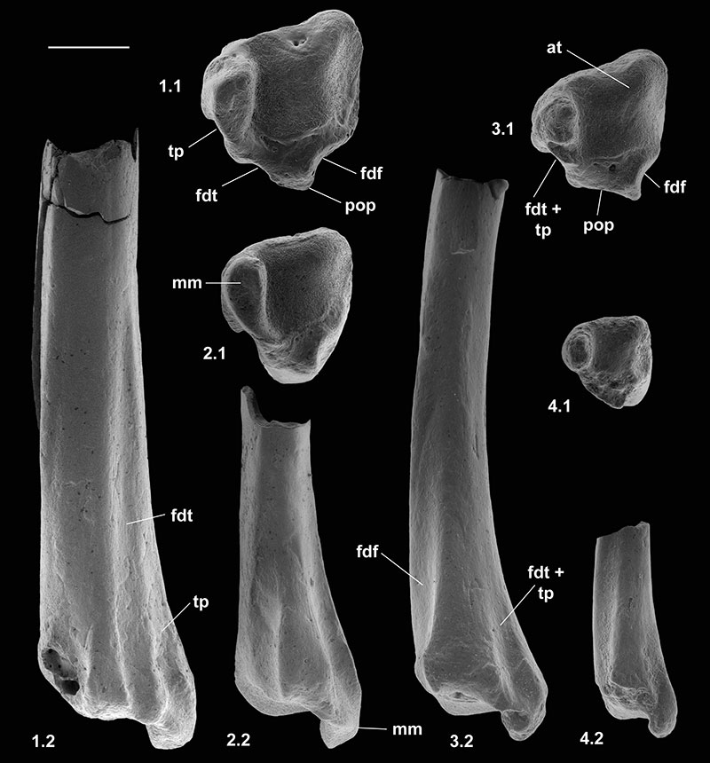
FIGURE 7. Scanning electron micrographs of right distal tibia (reversed) type 1 (Cryptotopos woodi), M95901, Mammal Bed, Hordle, Hampshire. Views are: anterior (1), medial (2) and lateral (3). Scale equals 1 mm.
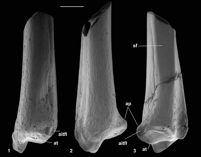
FIGURE 8. Scanning electron micrographs of right distal tibia (reversed) type 3 (Paradoxonycteris sp. 1), M95949, How Ledge Limestone, Headon Hill, Isle of Wight. Views are: anterior (1), medial (2) and lateral (3). Scale equals 1 mm.
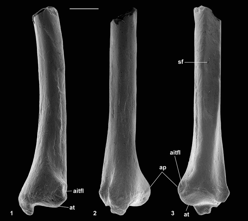
FIGURE 9. Scanning electron micrographs of left astragali of Nyctitheriidae. 1, type 2 (Paradoxonycteris sp. 1), M95952; 2, type 3 (Scraeva hatherwoodensis), M60101; 3, type 4 (Euronyctia grisollensis), HZM.108.25595. Views are: dorsal (1.1, 2.1, 3.1), ventral (1.4, 2.2, 3.2), lateral (1.2), medial (1.3), proximal (1.5) and distal (1.6). 3 is from the Rodent Bed, Hordle, Hampshire; the others from the Mammal Bed, Hordle. Scale equals 1 mm.
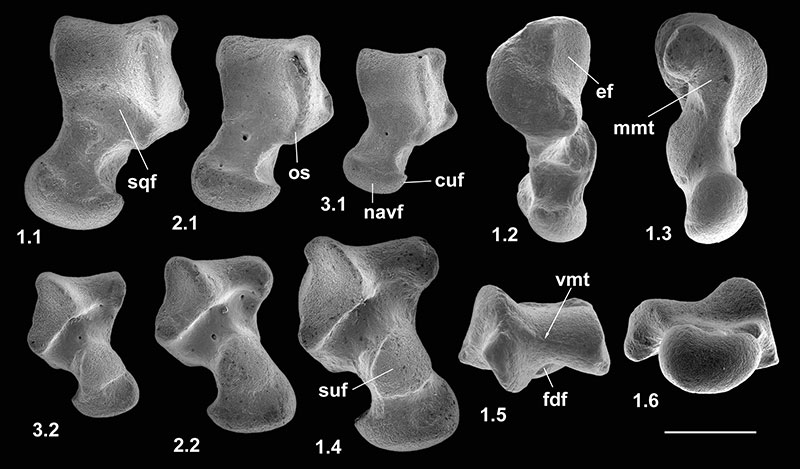
FIGURE 10. Logarithmic linear regressions of tooth versus astragalar and calcaneal dimensions in millimetres, following the methodology of Cifelli (1983) and Coillot et al. (2013). 1, M 2 area versus astragalar length of a range of mammals of known tooth and tarsal association; measurements of the numbered taxa are from Table 2; regression slope 1.4264, intercept -0.18646. 2, M 2 area versus calcaneal length as for 1; regression slope1.4686, intercept -0.52107. 3, M 2 area of named nyctitheres versus lengths of astragalar (A) and calcaneal (C) numbered types, using the regression lines from 10.1 and 10.2; M 2 areas of Saturninia gracilis and Euronyctia grisollensis (How Ledge Limestone, Headon Hill) are nearly identical; those of Cryptotopos woodi and Paradoxonycteris sp. 1, although similar, are from the Mammal Bed, Hordle, and How Ledge Limestone, Headon Hill, respectively, removing doubt of attribution of type1 and 2 tarsals from the same locations; calcaneal plots are closer to the regression line than are those of the astragali. 4, M 1 distal width versus lengths of astragalar (A) and calcaneal (C) numbered types; here the substantial difference in M 1 distal width between E. grisollensis and S. gracilis supports the referral of calcaneum type 5 to the latter; Scraeva hatherwoodensis is from the Mammal Bed, Hordle. Measurements in 10.3 and 10.4 are derived from Table 3.
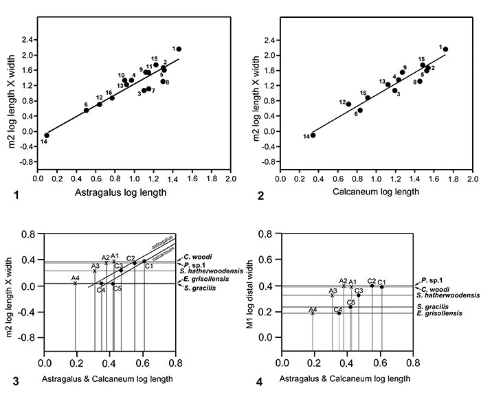
FIGURE 11. Scanning electron micrographs of calcanea, shown as left, of Nyctitheriidae. 1, left calcaneum type 2 (Paradoxonycteris sp. 1), M95960; 2, right calcaneum (reversed) type 3 (Scraeva hatherwoodensis), M95911; 3, left calcaneum type 4 (Euronyctia grisollensis), M95959; 4, left calcaneum type 5 (Saturninia gracilis ?), M95965. Views are: dorsal (1.1, 2.1, 3.1, 4.1), ventral (1.2, 2.2, 3.2, 4.2) and lateral (1.3, 2.3, 3.3, 4.3) . 1-2 are from the Mammal Bed, Hordle, Hampshire; 3-4 are from the How Ledge Limestone, Headon Hill, Isle of Wight. Scale equals 1 mm.
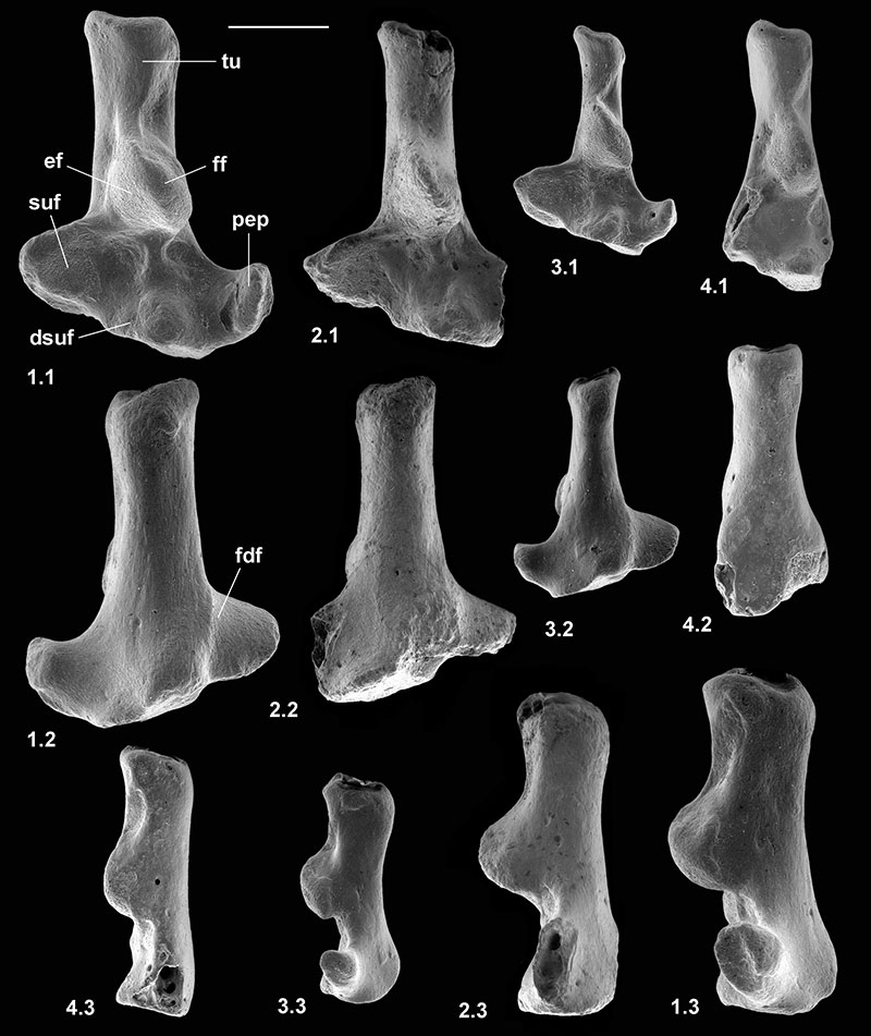
FIGURE 12. Scanning electron micrographs of calcanea, shown as left, of Nyctitheriidae. 1, left calcaneum type 2 (Paradoxonycteris sp. 1), M95960; 2, right calcaneum (reversed) type 3 (Scraeva hatherwoodensis), M95911; 3, left calcaneum type 4 (Euronyctia grisollensis), M95959; 4, left calcaneum type 5 (Saturninia gracilis ?), M95965. Views are: medial (1.1, 2.1, 3.1, 4.1) and distal (1.2, 2.2, 3.2, 4.2) . 1-2 are from the Mammal Bed, Hordle, Hampshire; 3-4 are from the How Ledge Limestone, Headon Hill, Isle of Wight. Scale equals 1 mm.
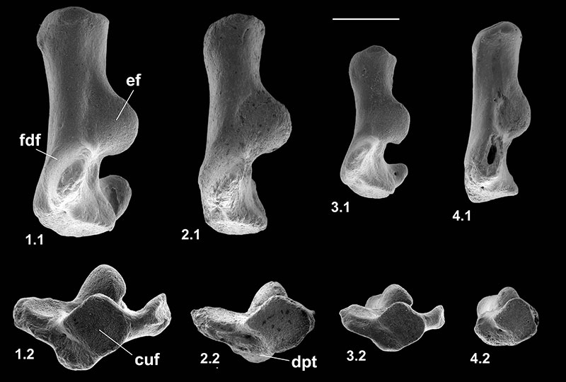
FIGURE 13. Scanning electron micrographs of naviculars, cuboids and ectocuneiform, shown as left, of Nyctitheriidae. 1, left navicular type 1 (Cryptotopos woodi), HZM.204.27205; 2, right navicular (reversed) type 2 ( Paradoxonycteris sp.1?), M95967; 3, right cuboid (reversed) type 1 (Cryptotopos woodi), M60970; 4, right cuboid (reversed) type 2 (Scraeva hatherwoodensis), M95918; 5, right cuboid (reversed) type 3 (Euronyctia grisollensis), M95969; 6, left ectocuneiform (Scraeva hatherwoodensis ?), M95919. Views are: dorsal (1.1, 3.1, 4, 5.1, 6.1), distal (1.2, 2.1, 3.5, 6.5), proximal (1.3, 2.2, 3.6, 5.4, 6.4), ventral (1.4, 3.3, 5.3), medial (1.5, 3.4, 6.3), lateral (1.6, 2.3, 3.2, 5.2, 6.2) . 1, 3, 4, 6 are from the Mammal Bed, Hordle, Hampshire; 2, 5 are from the How Ledge Limestone, Headon Hill, Isle of Wight. Scale equals 1 mm.
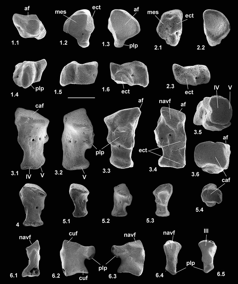
FIGURE 14. Scanning electron micrographs of composite reconstructed right ankle region (reversed), inverted at astragalo-calcaneal joint, of Cryptotopos woodi (type 1), Mammal Bed, Hordle, Hampshire. Distal tibia, M95901; astragalus, M95905; calcaneum, M95910; navicular, M95913; cuboid, M60970. Tibia is omitted from 6-7. Ankle dorsiflexed (1, 4), plantarflexed (2, 3, 5) . Views are: medial (1-2), posterior (3), anterior (4-5), dorsal (6), ventral (7) . Scale equals 2 mm.
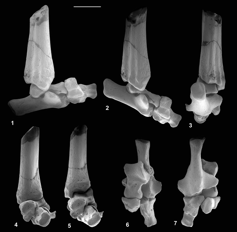
FIGURE 15. Scanning electron micrographs of metatarsals and phalanges of Nyctitheriidae. 1, right metatarsal IV (reversed), M95976; 2, left metatarsal III, M95970; 3, right metatarsal II (reversed), M95975; 4-5, 7-8, first phalanges; 6, second phalanx. 5 is manual?, 4, 7-8 are pedal?. 4, M60843; 5, M61114; 6, M60989; 7, HZM.588.30474; 8, M60790. Views are: proximal (1.1, 2.1, 3.1, 4.5, 5.3, 6.1, 8.1), distal (1.2, 4.4), dorsal (1.3, 2.2, 3.2, 4.1, 5.1, 6.2, 7.1, 8.2), medial (1.4), ventral (4.3, 6.4) and side (4.2, 5.2, 6.3, 7.2, 8.3) . 2, Paradoxonycteris sp. 1?; 4-6, Cryptotopos woodi ?; 7, Euronyctia grisollensis or Saturninia gracilis ; 8, Scraeva hatherwoodensis ? 1, lower Hamstead Member, Bouldnor, Isle of Wight; 2, How Ledge Limestone, Headon Hill, Isle of Wight; 3, Fishbourne Member, Woodside, Isle of Wight; 7, Rodent Bed, Hordle, Hampshire; 4-6, 8, Mammal Bed, Hordle. Scale equals 1 mm.
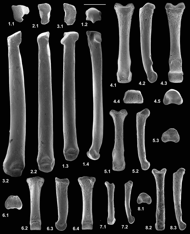
FIGURE 16. Scattergram of length against proximal width of first phalanges of Nyctitheriidae.
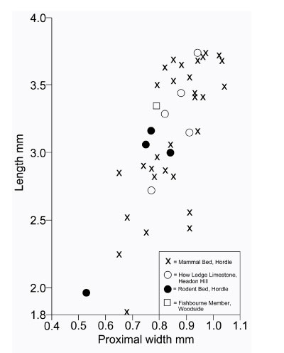
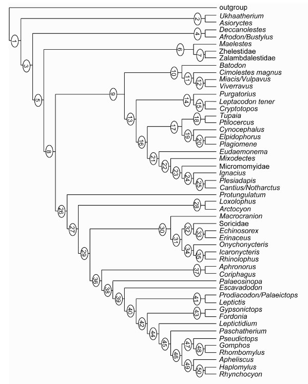
FIGURE 17. Strict consensus of four maximum parsimony trees generated by PAUP 4.0b10 from the character-taxon matrix in Appendix 2. Node numbers are Bremer decay indices. Groups shown as monophyletic are labelled. Abbreviations: Anagal, Anagalida; Carniv, Carnivora; Chiropt, Chiroptera; Dermopt, Dermoptera; Erin, Erinaceidae; Lept, Leptictidae; Lipo, Lipotyphla; Macro, Macroscelidea; Pholid, Pholidota; Plag, Plagiomenidae; Prim, Primates; Scand, Scandentia;
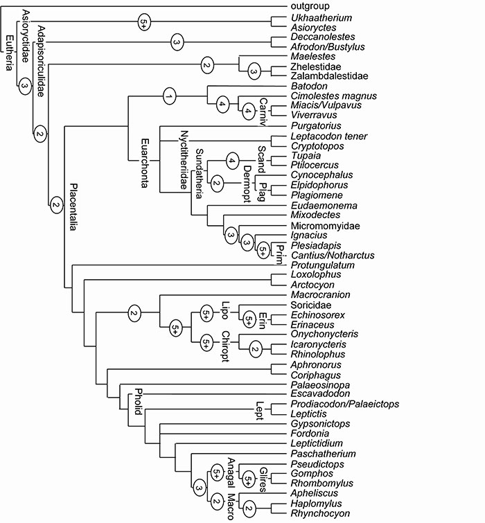
FIGURE 18. One of four maximum parsimony trees of 2704 steps generated by PAUP 4.0b10 from the character-taxon matrix in Appendix 2. Character states at numbered nodes are listed in Appendix 3.