FIGURE 1. Map showing the location of the Kaseki-Kabe fossil site, Ishikawa Prefecture.
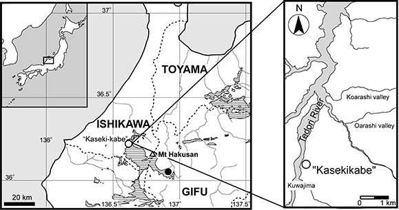
FIGURE 2. Kuroyuriella mikikoi gen. et sp. nov., holotype, SBEI 1510. 1, the four blocks making up the specimen; 2-3, the main association; 4-5, circumorbital bones, pterygoid and premaxilla; 6-7, frontal region in ventral view.
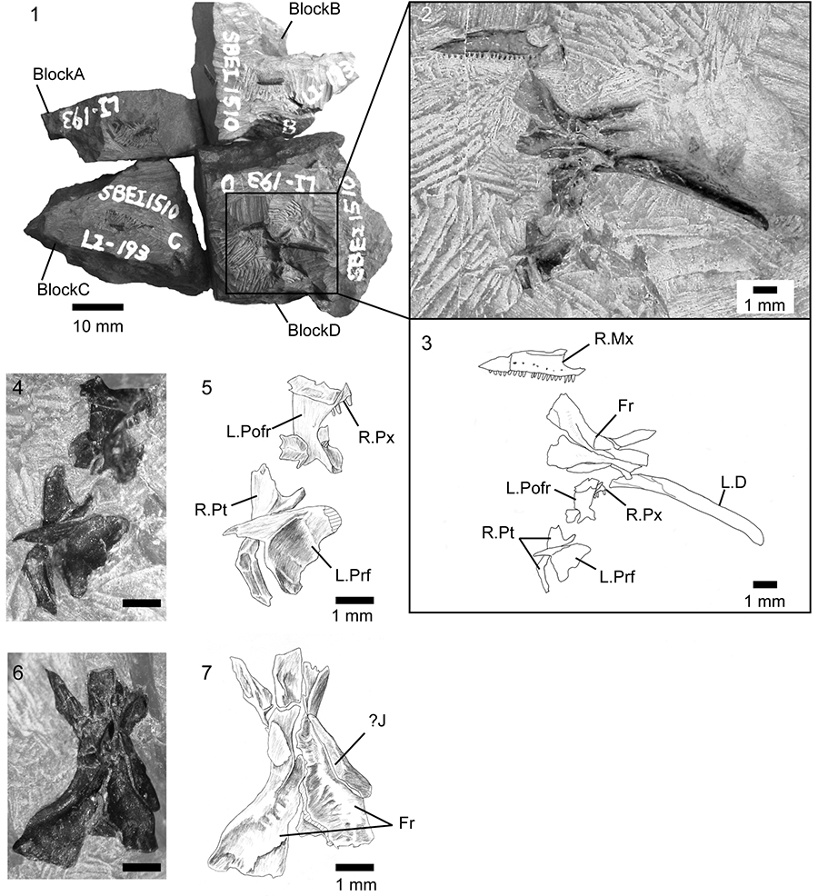
FIGURE 3. Kuroyuriella mikikoi gen. et sp. nov., referred specimen, SBEI 1608. 1-2, main association; 3-4, offset association; 5-6, detail of parietal and frontal; 7-8, detail of quadrate; 9, detail of postdentary bones. For abbreviations, see Material and Methods.
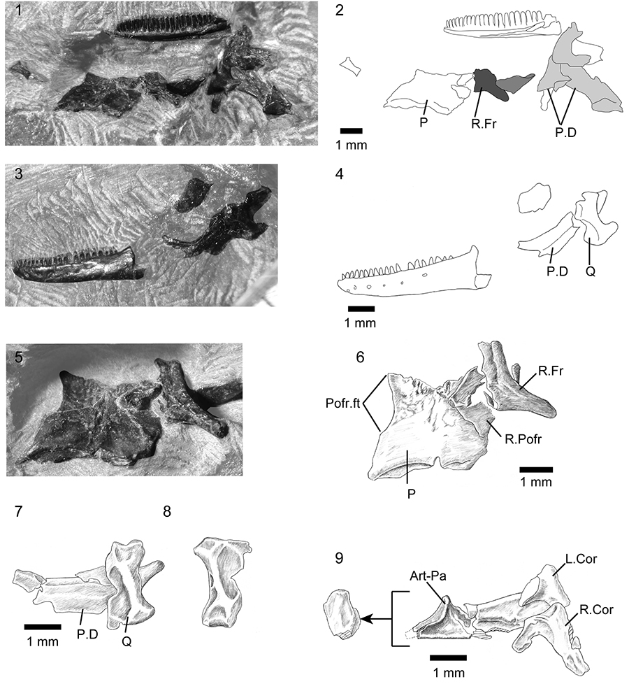
FIGURE 4. Kuroyuriella mikikoi gen. et sp. nov., jaw elements. 1-2, SBEI 1510, right maxilla in labial view; 3-4, SBEI 1608, left dentary in labial view; 5-6, SBEI 1608, right dentary in lingual view; 7-10, SBEI 1510, right mandible in 7-8, labial view, and 9-10, ventrolateral view. For abbreviations, see Material and Methods.
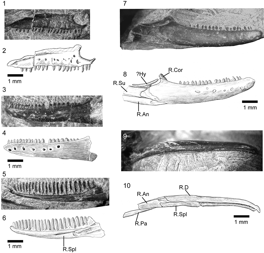
FIGURE 5. Phylogenetic position of Kuroyuriella mikikoi gen. et sp. nov. and Asagaolacerta tricuspidens gen. et sp. nov. in squamate trees, using the morphological data matrix of Gauthier et al. (2012), extended by Longrich et al. (2012). 1, Strict consensus of 1000 trees using unordered equally weighted characters, and run using TNT with 'minisearch.run'. Major clades are condensed. 1Note that Iguania was not monophyletic and comprised four smaller clades whose position was unresolved in relation to Borioteiioidea. Support values at nodes are Bremer/Jacknife/Symmetric resampling; 2, one of three trees (identical at this level) with clades condensed, run using the same matrix as in Figure 5.1 and analysed with TNT (with sectorial search, ratchet [20 iterations], and tree fusion all activated), but with the molecular tree of Wiens et al. (2010) and Pyron et al. (2013) providing the backbone constraint.
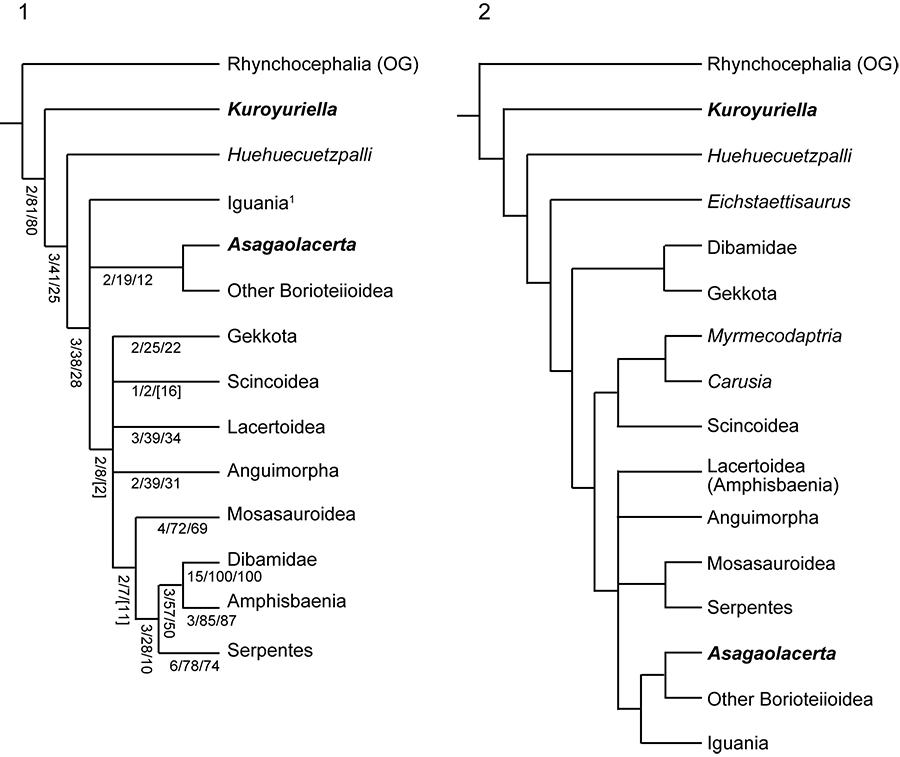
FIGURE 6. The phylogenetic position of Kuroyuriella mikikoi gen. et sp. nov. as recovered by analyses run with Implied Weighting (k=7), and character ordering. 1, no backbone constraint; 2, backbone constraint tree based on the molecular trees of Wiens et al. (2010) and Pyron et al. (2013). Only the scincoid section of each tree is shown as in all analyses with equally weighted taxa, Kuroyuriella lies in the stem-squamate position shown in Figure 5.
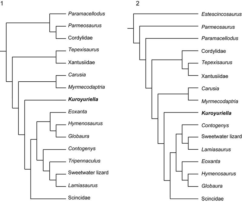
FIGURE 7. Asagaolacerta tricuspidens gen. et sp. nov., holotype, SBEI 1566. 1, the main association with the offset second bone group in the smaller image; 2, explanatory drawing of the same. For abbreviations, see Material and Methods.
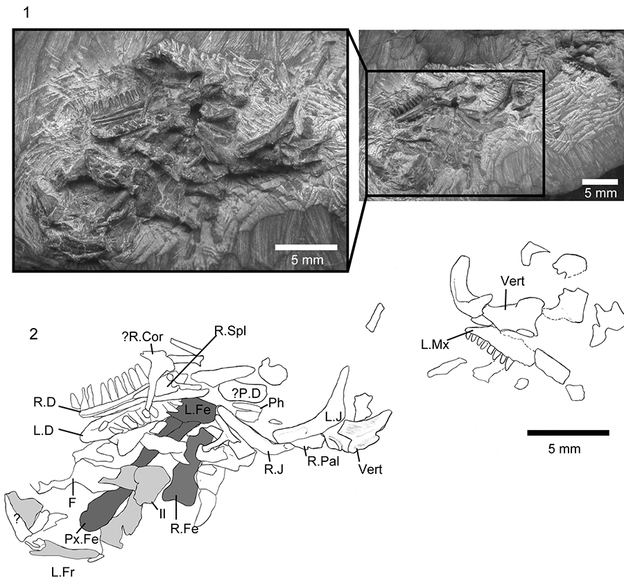
FIGURE 8. Asagaolacerta tricuspidens gen. et sp. nov., holotype, SBEI 1566. 1, Left partial maxilla in labial view; 2, left dentary in lingual view; 3, right dentary in lingual view; 4, details of teeth on the right dentary (enlarged from 3). For abbreviations, see Material and Methods.
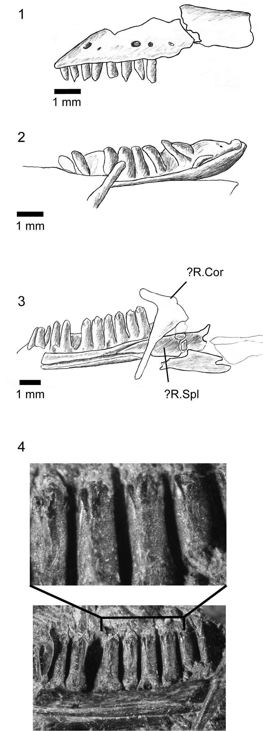
FIGURE 9. Asagaolacerta tricuspidens gen. et sp. nov., holotype, SBEI 1566, details of cranial bones. 1, left and right jugals; 2-3, explanatory drawings of right and left jugal respectively; 4-5, right quadrate in 4, lateral, and 5, anterior views; 6, biradiate bone, possibly coronoid. For abbreviations, see Material and Methods.
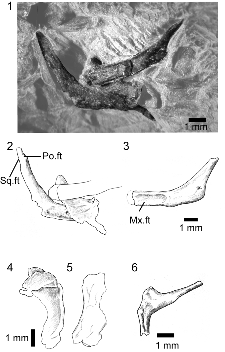
FIGURE 10. Asagaolacerta tricuspidens gen. et sp. nov., holotype, SBEI 1566, postcranial elements. 1, presacral vertebrae; 2, left ilium and distal head of right femur. For abbreviations, see Material and Methods.
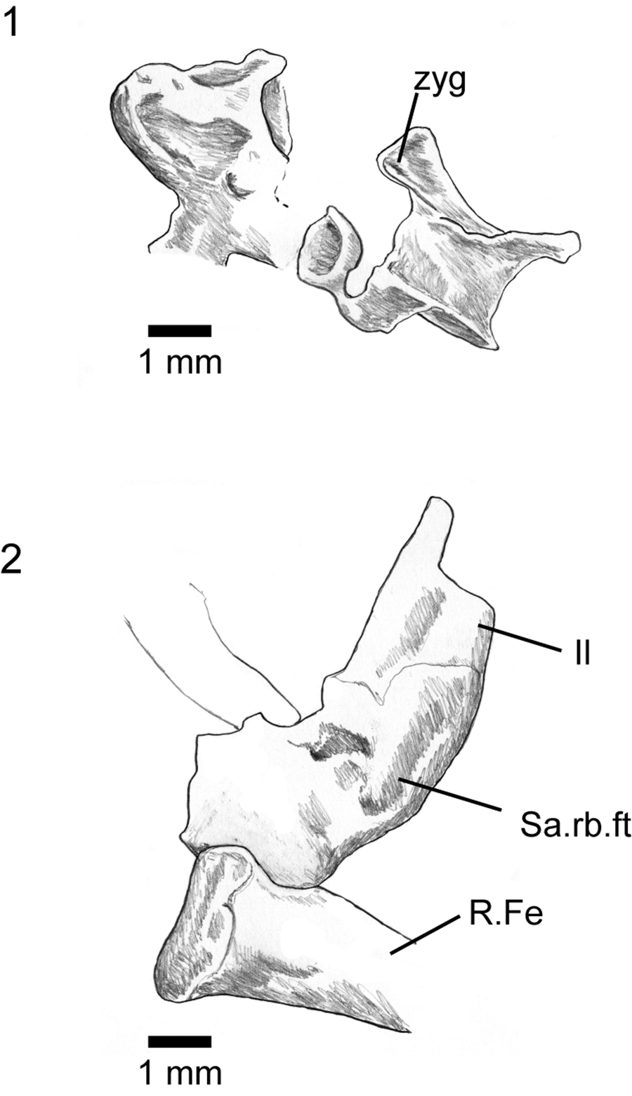
FIGURE 11. Associated postcranial specimens that may be referable to Asagaolacerta gen. nov. 1-2, SBEI 190; 3-4, SBEI 193. For abbreviations, see Material and Methods.
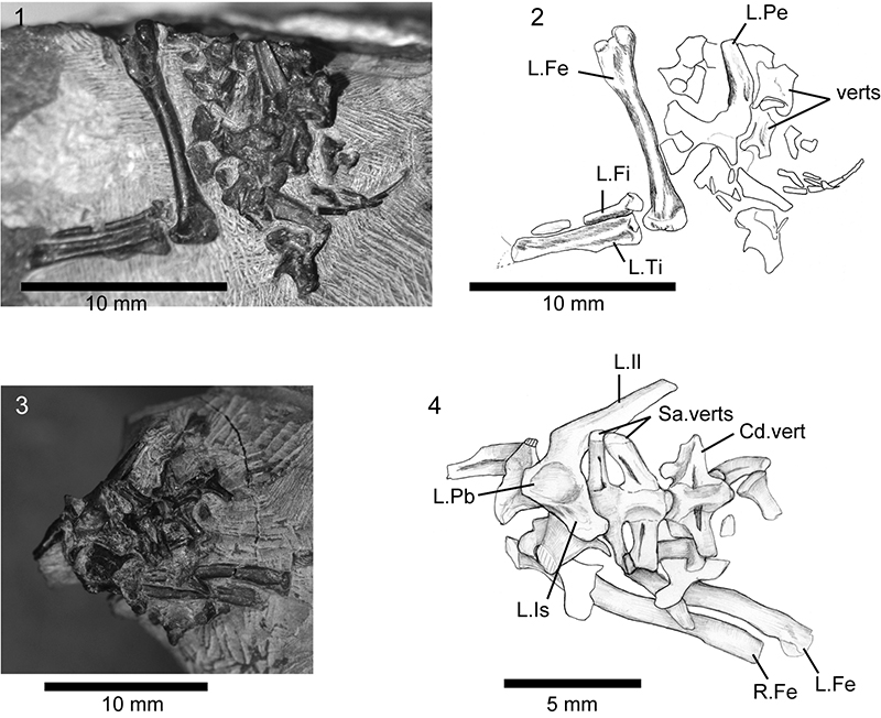
FIGURE 12. The phylogenetic position of Asagaolacerta tricuspidens gen. et sp. nov. tested using different analytical protocols within TNT. 1, detail of Strict Consensus of 1000 trees using the protocol that yielded the tree in Figure 5.1, node support values Bremer/Jacknife/Symmetric sampling; 2, one of three trees resulting from an analysis using with the molecular backbone constraint tree, but no character ordering or weighting; 3, one of 58 trees from an analysis run as in (2), but with character ordering as per Gauthier et al. (2012) and Longrich et al. (2012), and Implied Weighting (k=7); 4, one of 34 trees from an analysis run as in (3), but without the molecular backbone constraint; 5, 70% MRT of 19 trees resulting from an analysis (characters ordered but equally weighted, no constraints) run using only the boreoteiioid taxa, with Gekko gecko as the outgroup taxon. The 70%MRT is presented rather than the unresolved Strict Consensus to show that Asagaolacerta tricuspidens is usually (88% of trees) placed in the basal position (see text for further discussion). Abbreviation: poly, polyglyphanodont (as used in Longrich et al., 2012).
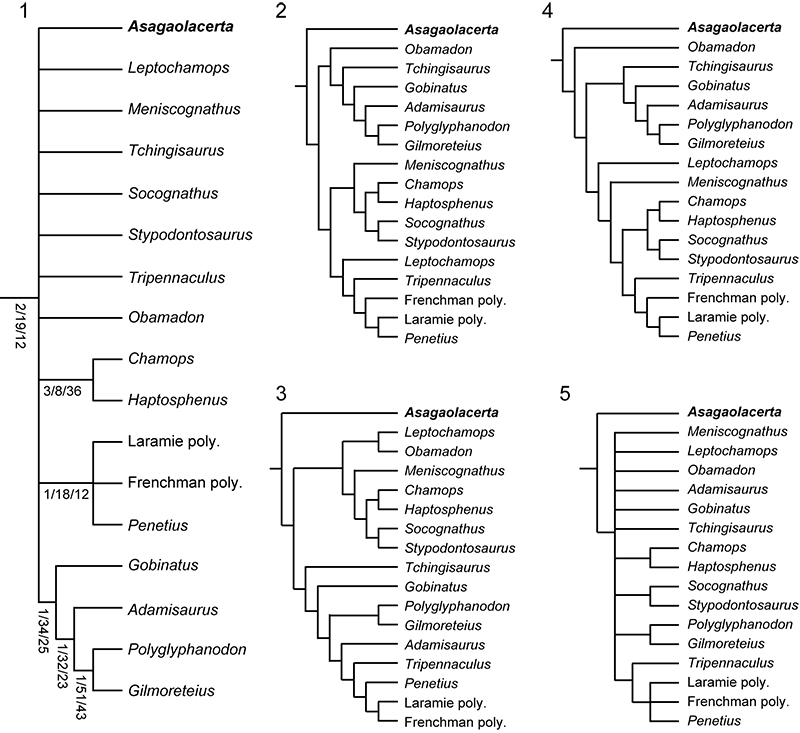
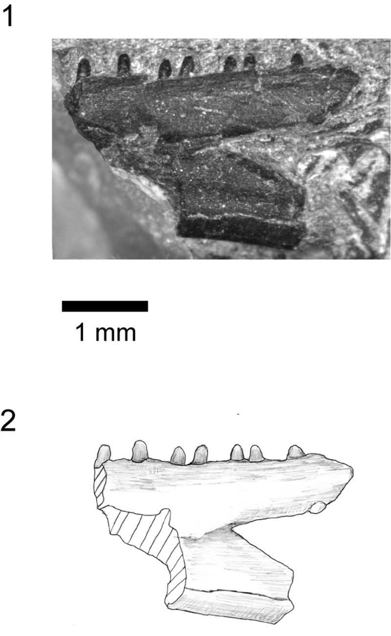
FIGURE 13. Hakuseps imberis gen. et sp. nov., holotype left dentary, SBEI 2086. 1-2, labial view; 3-4, lingual view; 5-6, occlusal view; and 7, ventral view. For abbreviations, see Material and Methods.
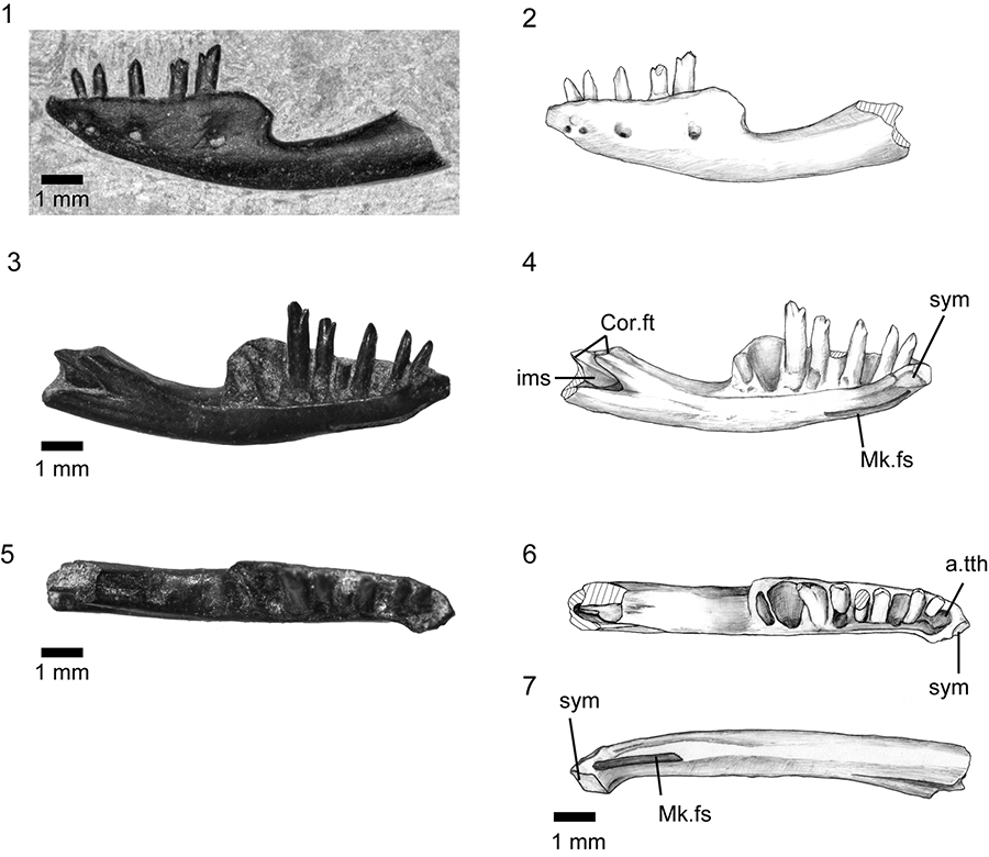
FIGURE 14. Comparison of Pachygenys thlastesa (Gao and Cheng, 1999) and Hakuseps imberis gen. et sp. nov. Labial and lingual views respectively of 1-2, Pachygenys thlastesa (redrawn from Gao and Cheng, 1999), 3-4, Hakuseps imberis gen. et sp. nov.
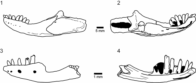
FIGURE 15. Shiramine Morphotype A, bicuspid dentition. 1-2, left maxilla, SBEI 1525 in labial view; 3-4, right maxilla, SBEI 1501, in labial view; 5-10, left dentary, SBEI 808, in two parts, in labial view, with 5-6, symphysial region, 7-8, posterior dentary, and 9-10, detail of bicuspid teeth.
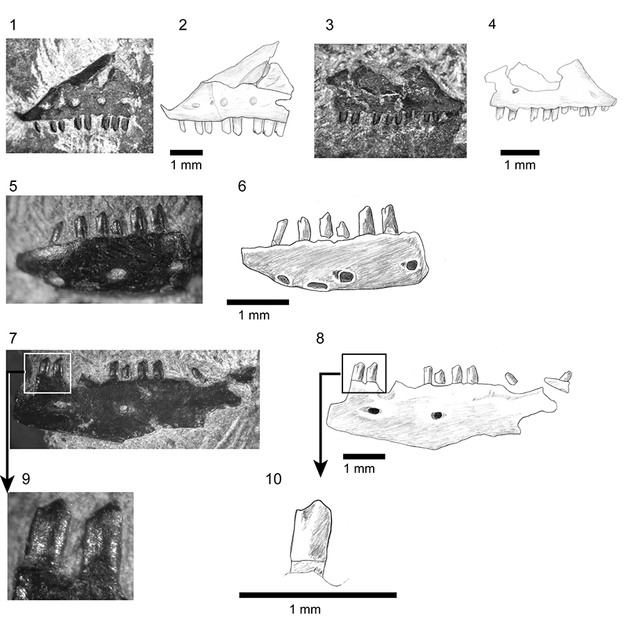
FIGURE 16. Shiramine Morphotype B, right mandible SBEI 827. 1-2, dentary in labial view; 3-4, dentary and associated splenial in lingual view; 5-11, postdentary bones in 5-6 dorsomedial view; 7, ventral view; 8, dorsal view; 9, medial view; and 10, posterior view of articular surface. For abbreviations, see Material and Methods.
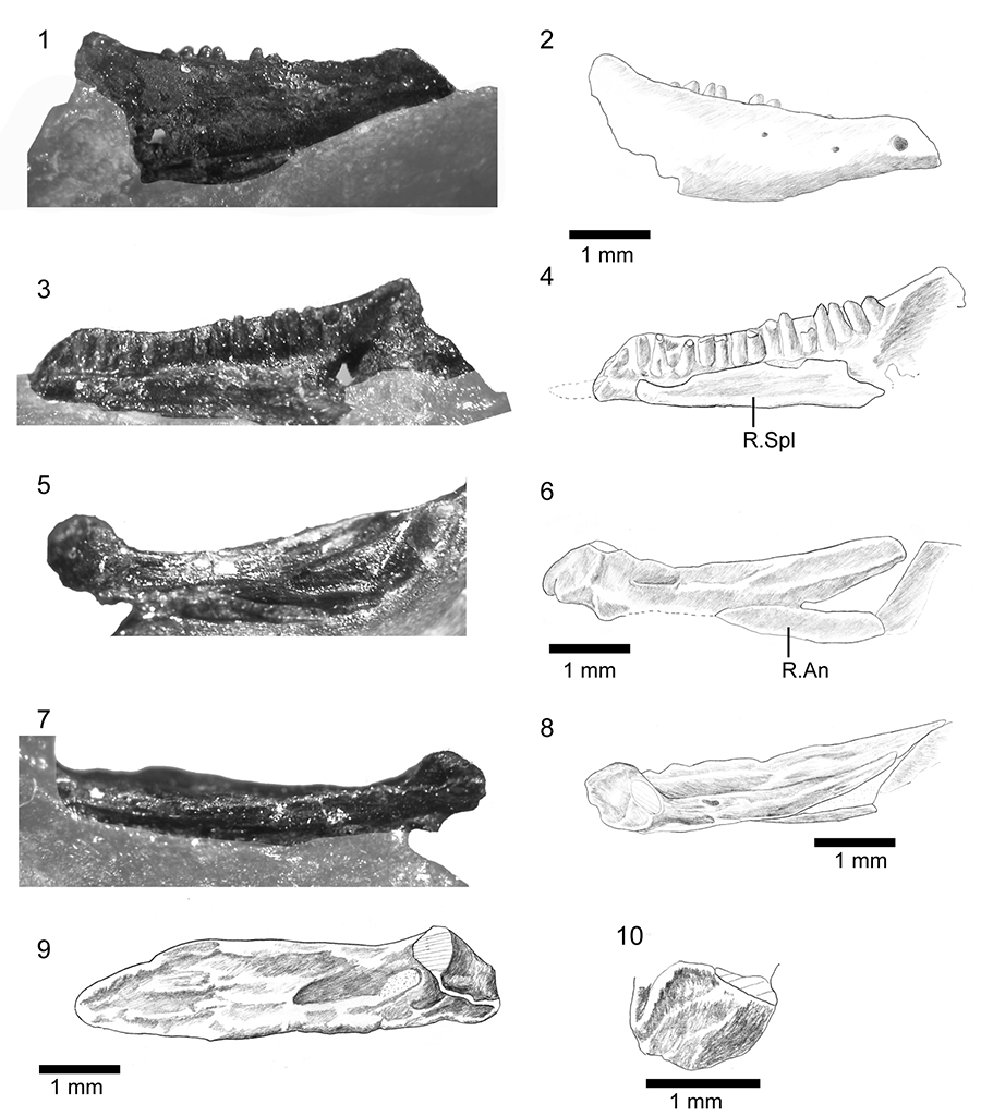
FIGURE 17. Comparison of Shiramine Morphotype B and Myrmecodaptria microphagosa (Gao and Norell, 2000) from the Late Cretaceous of Mongolia. 1-2, left lateral and dorsal views respectively of the skull of Myrmecodaptria microphagosa, redrawn from Gao and Norell (2000). Scale bar equals 5mm; 3, dentary of morphotype B, SBEI 827, labial view; 4, isolated left maxilla, SBEI 2407, in labial view; 5, isolated median frontal, SBEI 1803, in dorsal view.
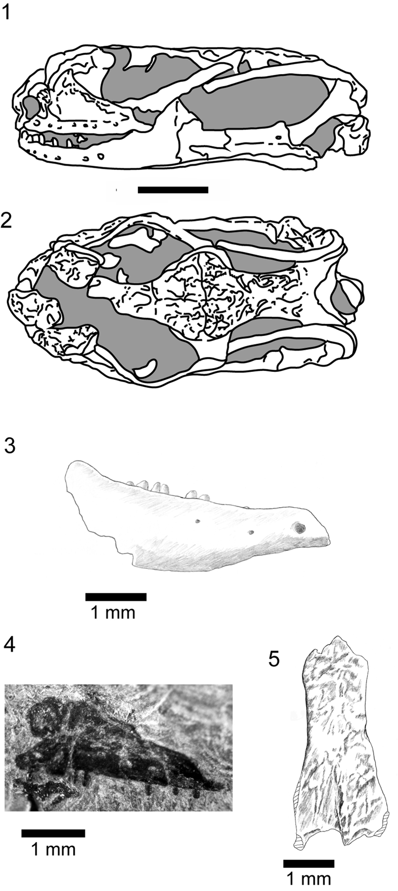
FIGURE 18. Shiramine Morphotype C, SBEI 1277, posterior region of a left dentary in 1-2, labial view.