FIGURE 1. Type specimens of Scottish Middle Devonian diplacanthids: 1, Diplacanthus crassisimus (Duff, 1842) holotype NMS G.1891.92.333, from Tynet Burn, Moray; 2, Rhadinacanthus longispinus (Agassiz, 1844) holotype NMS G.1953.4.3, from Cromarty, Ross and Cromarty; 3, Homacanthus borealis holotype spine NMS G.1892.91.1, from Lybster, Caithness; 4, Diplacanthus tenuistriatus (Traquair, 1894) syntype NMS G.1891.92.340, from Gamrie, Banffshire. Scale bars equal 1 cm.
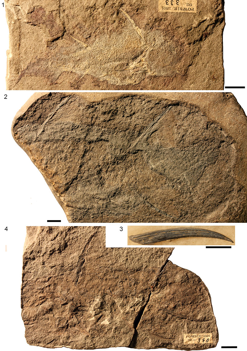
FIGURE 2. 1, Site map indicating the most important localities with Middle Devonian diplacanthids on northern Scotland; 2, biostratigraphic table of diplacanthid occurrences in the Orcadian Basin of northern Scotland.
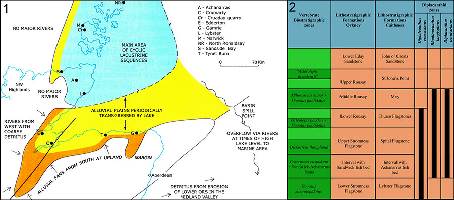
FIGURE 3. Diplacanthus crassisimuss, NMS G.2014.4.29, from Edderton, Ross and Cromarty, showing surface morphology of spines: 1, complete specimen; 2, line drawing identifying various elements; 3, circumorbital bone; 4, occlusal bones; 5, procoracoid with ventral plate attached. Scale bars equal: 1 cm for 1, 2; 1 mm for 3-5. Abbreviations: a.f=anal fin web; a.fs=anal fin spine; ad.f=anterior dorsal finweb; ad.fs=anterior dorsal fin spine; adm.s=admedian spine; co.p=circumorbital plate; lt.sc=left scapula; lt.pelv.fs=left pelvic fin spine; oc.p=occlusal plate; pd.f=posterior dorsal finweb; pd.fs-=posterior dorsal fin spine; pelv.f=pelvic finweb; prepelv.s=prepelvic spine; proc=procoracoid; rt.p.fs=right pectoral fin spine; rt.pelv.fs=right pelvic fin spine; rt.sc=right scapula; v.p=ventral plate. Arrow indicates anterior.
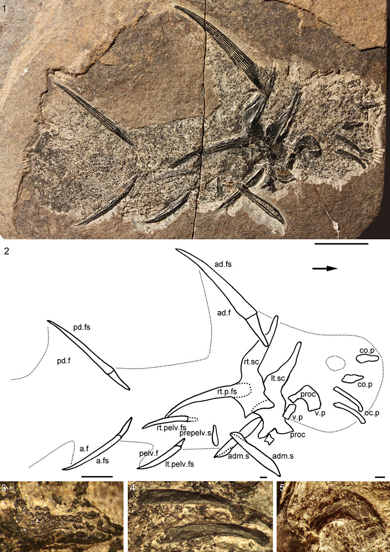
FIGURE 4. Diplacanthus crassisimus, articulated specimens: 1-3, NMS G.2014.44.3 from Edderton, Ross and Cromarty: 1, complete specimen; 2, right occlusal plate; 3, left circumorbital and postorbital plates; 4,NMS G.2014.15.21, a ventrodorsally flattened specimen from Cruaday Quarry, Orkney, showing prepelvic spines; 5-7, NMS G.1891.99.10 from Tynet Burn, Moray: 5, head region; 6, impression of a displaced cheek, rotated 90° counterclockwise; 7, postorbital plate; 8-10, NHM OR.36582 from Tynet Burn, Moray: 8, complete specimen with head region ventrodorsally flattened, with articulated, displaced branchial region; 9, branchial region; 10, sketch of branchial region elements, rotated 90° clockwise. Scale bars equal: 1 cm for 1, 5, 8 and 5 mm for 2-4, 6, 7, 9. Abbreviations: ad.fs=anterior dorsal fin spine; bb=basibranchial; cb=ceratobranchials; ck.p=cheek plate; co.p=circumorbital plate; eb=epibranchials; oc=occlusal plate; pb=pharyngobranchials; prepelv.s=prepelvic spine; proc=procoracoid; scap=scapulocoracoid.
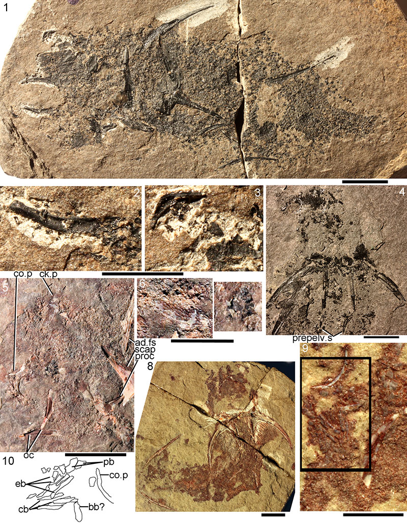
FIGURE 5. Diplacanthus crassisimus spine morphology and histology: 1, 2, NMS G.2014.4.29 from Blackpark, Ross and Cromarty; 1, anterior dorsal fin spine; 2, posterior dorsal fin spine; 3, pectoral spine of NMS G.1878.5.349 from Springpark, Caithness; 4, pectoral spine NMS G.2014.44.7 from west side of Sandside Bay, Caithness; 5, pectoral spine impression, Snaky Noust, Westray, Orkney (HY501.405); 6, longitudinal section of pelvic spine NMS G.2014.33.10 from Corbie Den, Banffshire; 7, transverse section of anterior dorsal fin spine NMS G.2014.33.9 from Corbie Den, Banffshire; 8, 9, transverse section of anterior dorsal fin spine NMS G.2014.33.11 from the south of East Murkle Bay, Caithness, with magnified image of same specimen; 10, transverse section NMS G.2014.44.2 of pectoral fin spine and bone of scapulocoracoid from Corbie Den, Banffshire. Scale bars equal 1 cm for 1-6 (1, 2 at same scale); 1 mm for 8, 10; 0.1 mm for 7, 9.
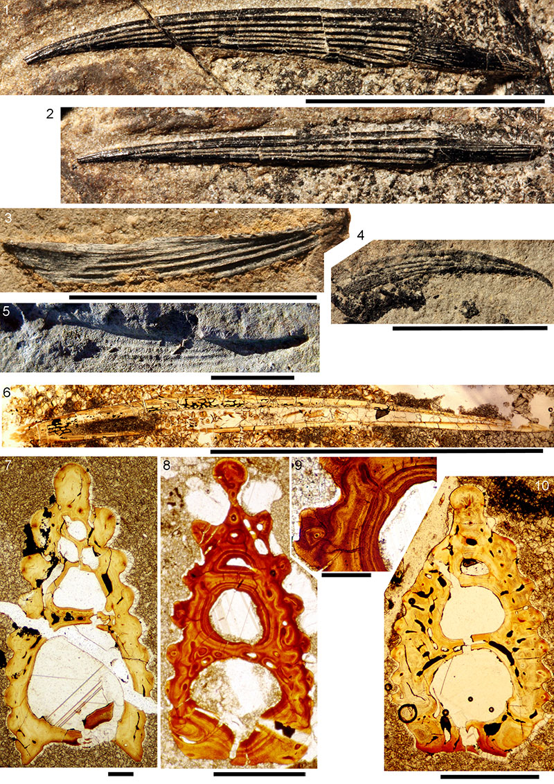
FIGURE 6. Drawings of serial transverse sections of the spines of Diplacanthus crassisimus. 1, sketch of NMS G.2014.15.7, from Edderton, Ross and Cromarty, showing the position of sections in 2-6; the sections run from the distal end to the base of each spine. 2, anterior dorsal fin spine (ad.fs); 3, anal fin spine (a.fs); 4, right pelvic fin spine (pelv.fs); 5, left pelvic fin spine; 6, left pectoral fin spine (p.fs). 7, Sollas sections of a prepelvic spine (prepelv.s) on NMS G.2014.44.4. Scale bars equal 1 mm.
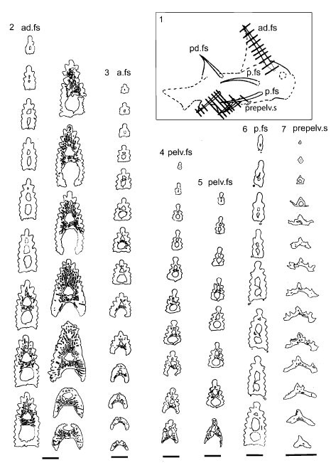
FIGURE 7. Diplacanthus crassisimus shoulder girdles. 1, NMS G.Canon Kyle no. 2 from Tynet Burn, Moray, showing ventral view of the shoulder girdle complexes; 2, NHM P.1357a from Tynet Burn, Moray, pectoral girdle region; 3, NHM P.22198 from Achanarras Quarry, Caithness, complete specimen; 4, NMS G.2014.44.3 from the Edderton, Ross and Cromarty; 5, 6, left shoulder girdle complex of NMS G.2014.4.30 from Cruaday Quarry, Orkney; 7, NMS G.2014.44.4 from the Edderton, Ross and Cromarty, natural transverse section through articulated uncompressed shoulder girdle region (prepelvic spines subsequently Sollas sectioned). Scale bars equal 1 cm.
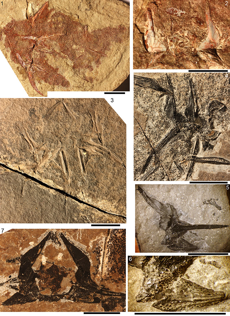
FIGURE 8. Diplacanthus crassisimus shoulder girdle thin section images. 1, 2, NMS G.2014.33.8 from west of Castletown Harbour, Caithness: 1, section through pectoral spine, pinnal plate, ?procoracoid, and scapulocoracoid; 2, transverse section through base of pectoral fin spine enclosed by pinnal plate and remnants of the endoskeletal coracoid plate; 3-5, section through admedian spine NMS G.2015.11.2 from Gamrie, Banffshire: 3, proximal end of admedian spine, inner bone base layer is the coracoid; 4, line drawing of reconstructed section; 5, scale-like ornament towards edge of admedian spine (area box in 4). Scale bars equal 1 mm in 1, 2, 4; 0.1 mm in 5.
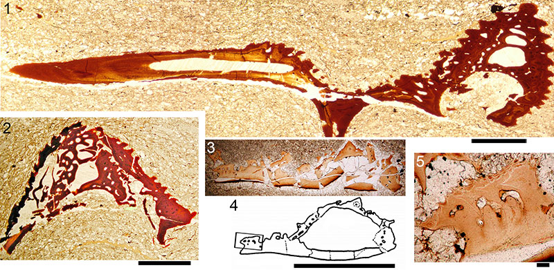
FIGURE 9. Line drawings of serial transverse sections through pectoral girdle elements of Diplacanthus crassisimus; sketch of whole specimen NMS G.2014.4.30 used to denote position of the sections shown in 2, 3 (mirror image sketch in latter). 1, serial destructive Sollas sections through admedian spine from west of Castletown, Caithness. 2, thin sections A-L through an isolated pectoral girdle complex NMS G.2014.33.8, from west of Castletown harbour. 3, Sollas sections 1-15 through an isolated juvenile pectoral girdle complex (without pectoral spine) from west of Castletown, Caithness. Abbreviations: adm.s=admedian spine; p.fs=pectoral fin spine; scap=scapula.
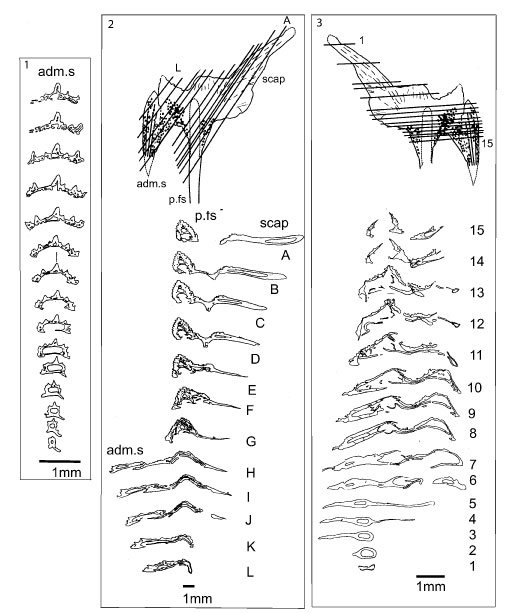
FIGURE 10. Reconstruction of the pectoral girdle of Diplacanthus crassisimus. 1, lateral view. 2, ventral view showing Miles' (1973a) interpretation on the left hand side of the fish and the authors’ revised version on the right hand side of the fish. Dark grey fill indicates dermal elements, light grey fill indicates endoskeletal elements.
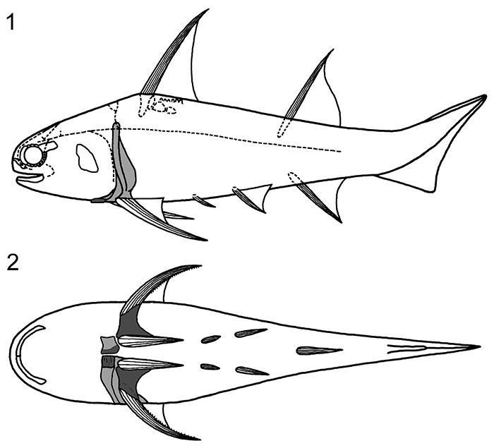
FIGURE 11. Diplacanthus crassisimus from Gamrie, Banffshire, squamation. 1-5, NMS G.1882.60.17, articulated specimen showing scale morphologies: 1, the complete specimen, with A-C denoting areas of squamation shown in Figure parts 2-5: 2, 3, area A; 4, area B; 5, area C. 6, NMS G.1892.8.5, mid-body squamation. 7-11, scales slide NMS G.1882.60.17.1, Scanning Electron Micrographs (SEMs): 7, 8, crown and anterior views; 9, lateral view of scale pair; 10, basal view; 11, undersurface of crown (neck and base broken off). 12-15, scales slide NMS G.2015.11.3.1, SEMs: 12, 13, crown and posterior views; 14, 15, laterocrown and lateral views. Scale bars equal 1 cm for 1; 1 mm for 2-5; 0.2 mm for 6-11, 14, 15; 0.1 mm for 12, 13. Arrows directed anteriorly.
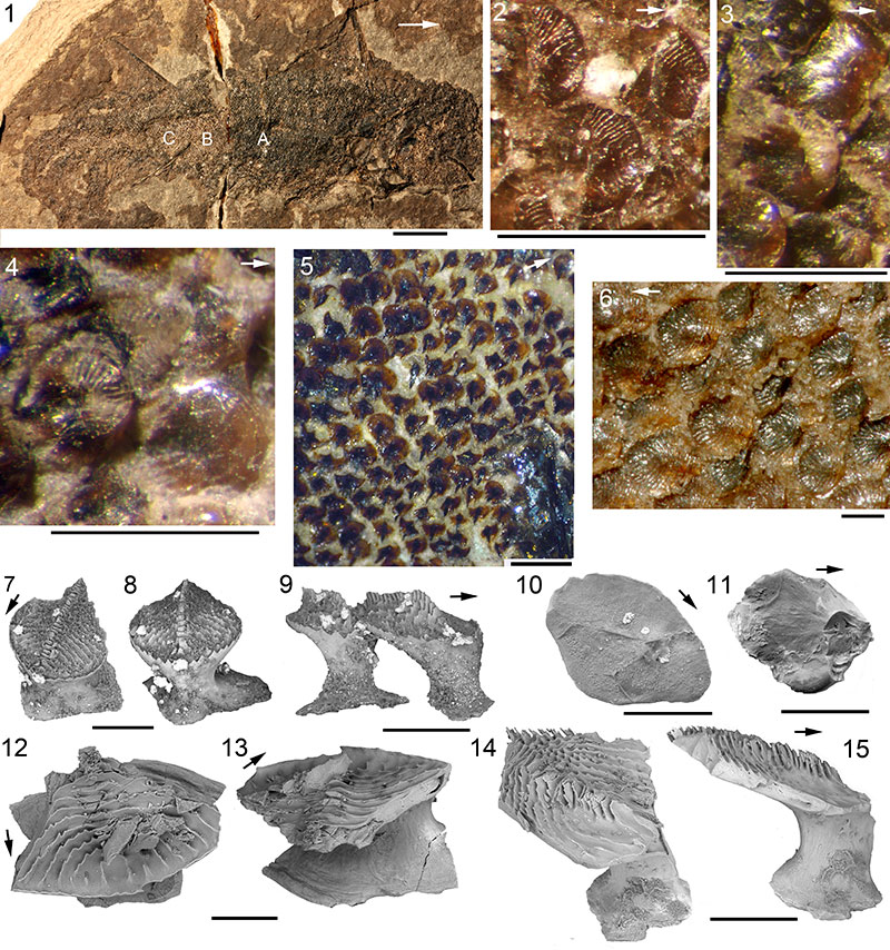
FIGURE 12. Diplacanthus crassisimus scale histology. 1-7, NMS G. 2014.33.10 from Corbie Den, Banffshire, thin sections of a patch of midflank scales: 1, group of scales sectioned in various orientations; 2, vertical longitudinal section; 3, crown horizontal section; 4, horizontal section through the base; 5, 6, vertical longitudinal sections through articulated scales; 7, crown horizontal section. 8, 9, NMS G.2015.11.3 from Gamrie, Banffshire, isolated midflank scales: 8, crown horizontal section; 9, vertical section through the base and lower neck, imaged with cross nicols. 10, 11, NMS G.2015.11.2 from Gamrie, Banffshire: 10, vertical longitudinal section; 11, vertical longitudinal section. Scale bars equal 0.1 mm. Abbreviations: cgz=crown growth zones; dt=dentine tubule; p=canal opening; pc=pore canal; spc=spiral canal. Arrows indicate anterior direction, where relevant.
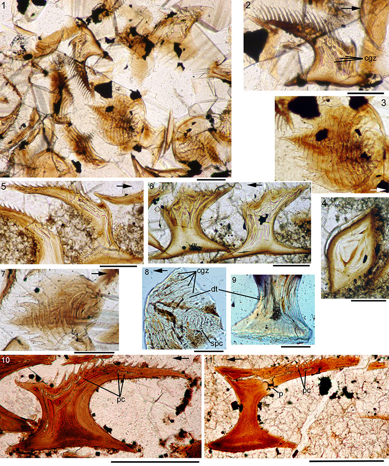
FIGURE 13. Diplacanthus tenuistriatus morphology. 1, NMS G.1897.55.2, counterpart of a headless large specimen from Achanarras Quarry, Caithness; 2, 3, NMS G.1897.55.1 (the part of NMS G.1897.55.2): 2, caudal fin, anal fin spine and fin web and right pelvic fin spine and fin web; 3, posterior dorsal fin spine and fin web; 4, NMS G.1870.14.144 from Tynet Burn, Moray, head and pectoral region. Scale bars equal 1 cm. Abbreviations: ad.fs=anterior dorsal fin spine; adm.s=admedian spine; ck.p=cheek plate; co.p=circumorbital plate; cor=coracoid; lt.sc=left scapula; lt.p.fs= left pectoral fin spine; oc=occlusal plate; p.p=pinnal plate; prepelv.s=prepelvic spines; rt.sc=right scapula.
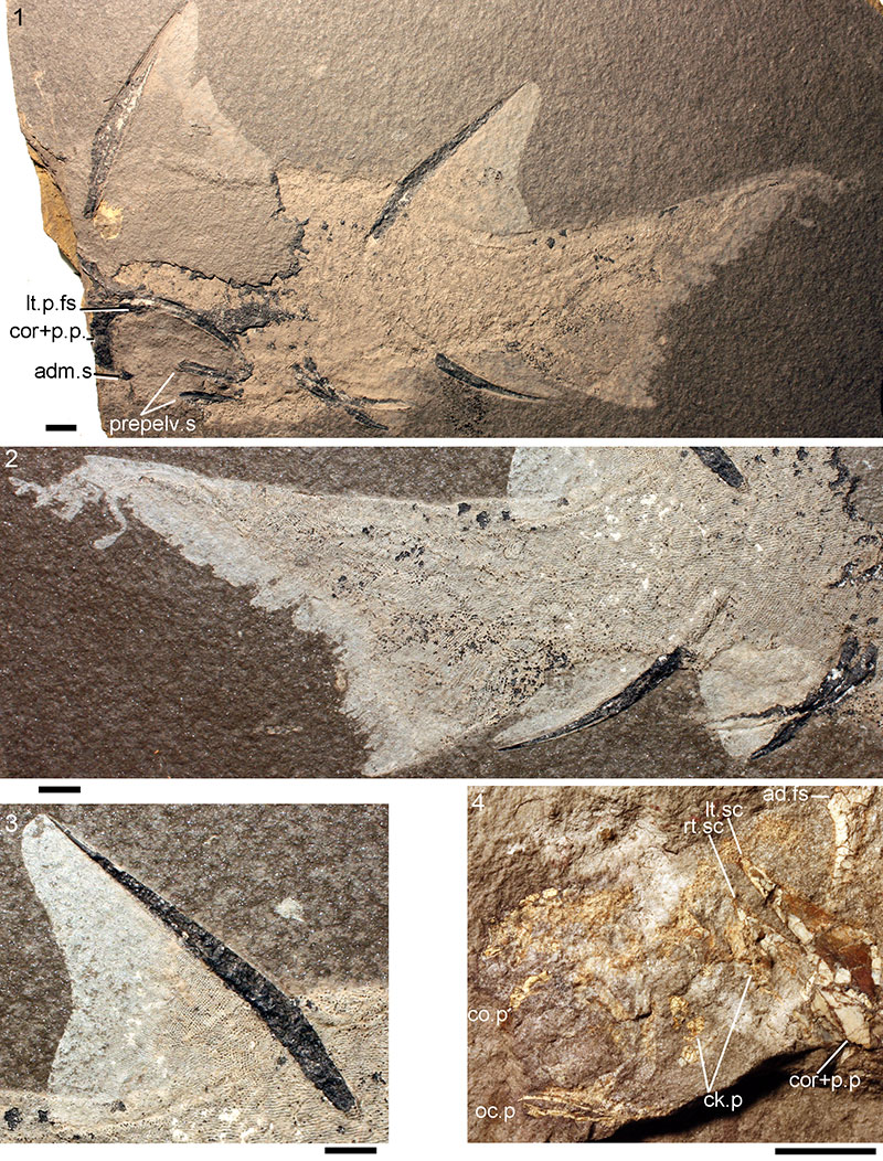
FIGURE 14. Diplacanthus tenuistriatus pectoral fin spines. 1-4, spine QM F58024 from Marwick, Orkney: 1, complete spine, dorsal surface; 2, ventral surface of spine; 3, recurved denticles on posterior edges of spine; 4, transverse natural fracture surface of distal part of spine, at point arrowed in (2); 5, NMS G.1901.153.1 from Appat Hill, Caithness; 6, NMS G.1898.163.2 from Birsay, Orkney; 7, NMS G.2014.4.18, from Marwick, Orkney, spine attached to pinnal plate. Scale bars equal 1 cm for 1, 2, 5-7; 1 mm for 3, 4.
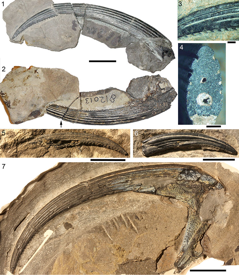
FIGURE 15. Diplacanthus tenuistriatus anterior dorsal fin spines. 1, NMS G.2014.7.35 from Marwick, Orkney. 2-4, NMS G.2014.44.5 from Marwick, Orkney: 2, complete specimen in matrix; 3, magnified view of proximal exserted part; 4, magnified view of the same area. Scale bars equal 1 cm in 1-3; 1 mm in 4.
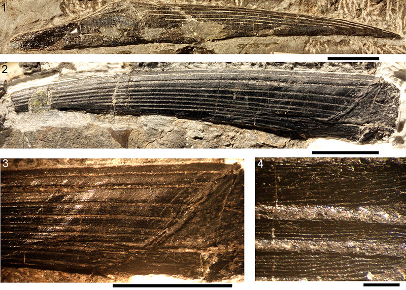
FIGURE 16. Diplacanthus tenuistriatus fin spine histology. 1-4, pectoral spine NMS G.2014.7.36 from Marwick, Orkney; 1-4, transverse section through the pectoral spine and the posterior end of the pinnal plate; 5, 6, anterior dorsal spine NMS G.2014.7.35 from Marwick Orkney: midspine transverse section. 7, NMS G.2014.33.1, midspine transverse section of anterior dorsal spine from North Ronaldsay, Orkney. Scale bars equal 1 mm in 1, 2, 5, 7; 0.1 mm in 3, 4, 6.
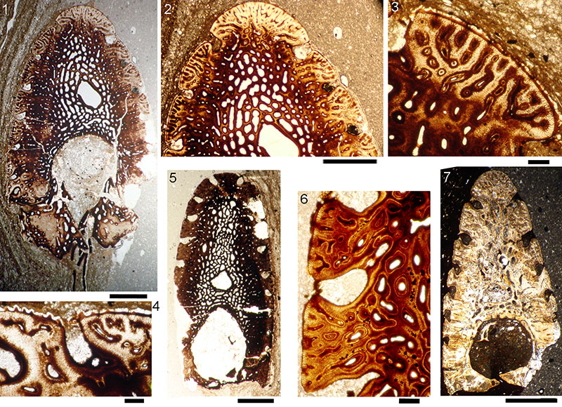
FIGURE 17. Drawings of transverse sections through Diplacanthus tenuistriatus pectoral and anterior dorsal fin spines. 1, 2, pectoral fin spine NMS G.2014.7.36 from Marwick, Orkney: 1, through pectoral spine and posterior end of pinnal plate; 2, slightly distal to the latter section; 3, 4, anterior dorsal fin spine NMS G.2014.7.35 from Marwick, Orkney; 3, midspine; 4, slightly proximal to latter section. Scale bars equal 1 mm. Abbreviations: cc=central pulp canal; e=enameloid; gl=growth line; ost=osteodentine; p.p=pinnal plate; uc=upper longitudinal canal. Arrows point to junction between pectoral fin spine and pinnal plate.
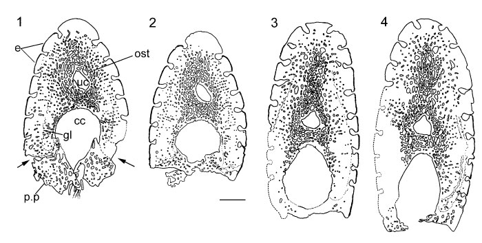
FIGURE 18. Diplacanthus tenuistriatus isolated shoulder girdle complexes. 1-4, NMS G.2014.15.21 from Marwick, Orkney: 1, external surface of scapulocoracoid, pectoral fin spine, pinnal plate and admedian spine; 2, admedian spine; 3, 4, scale-like ornament on pinnal plate; 5, NMS G.2014.15.22 from Marwick, Orkney, internal surface; 6-9, transverse sections of NMS G.2014.4.20 from Marwick, Orkney: 6, pinnal plate; 7, multiple odontode on plate; 8, scale-like ornament; 9, transverse section through pinnal plate, pectoral fin spine base, scapulocoracoid and perichondral bone sheet; 10, 11, NMS G.2014.4.27 from Flashes, Hoy, Orkney, transverse section through base of admedian spine and inner perichondral bone sheet; 10, whole width; 11, scale-like ornament in groove. Scale bars equal 1 cm in 1, 5; 5.0 mm in 2; 1.0 mm in 3, 4, 6; 0.5 mm in 9, 10; 0.1 mm in 7, 8, 11; 0.05 mm in 4.
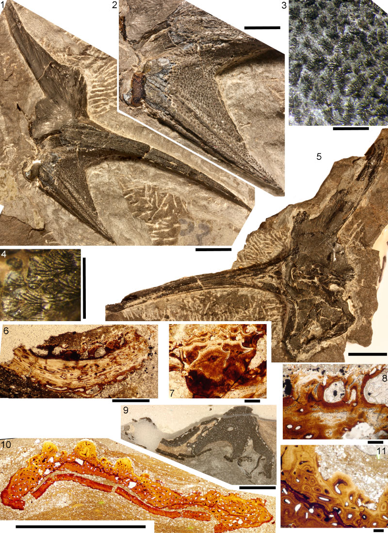
FIGURE 19. Drawings of serial sections throughDiplacanthus tenuistriatus shoulder girdle complexes from Marwick, Orkney. 1, schematic drawing of the external view of shoulder girdle complex NMS G.2014.4.21, showing levels at which transverse sections were made. 2, schematic drawing of the inner view of complex NMS G.2014.15.22. A2-F1 is the section series through the admedian spine on NMS G.2014.4.27; I1 is a section through the pectoral fin base enclosed by surrounding plates on NMS G.2014.4.27; I2, J2B, and J2A are sections through the admedian spine, pinnal plate, pectoral fin, base of scapulocoracoid and perichondral bone of the coracoid of NMS G.2014.4.20; 10A and 10B are sections through the girdle complex NMS G.2014.4.23; K2, L1, M2 and O1 are serial sections through the scapulocoracoid and associated structures of NMS G.2014.20.20. Scale bars equal 5 mm. Anatomical abbreviations: adm.s=admedian spine; pch.b=perichondral bone layer; p.fs=pectoral fin spine; p.p=pinnal plate; sc=scales; scap=scapula; scap.c=central pulp cavity of scapula shaft.
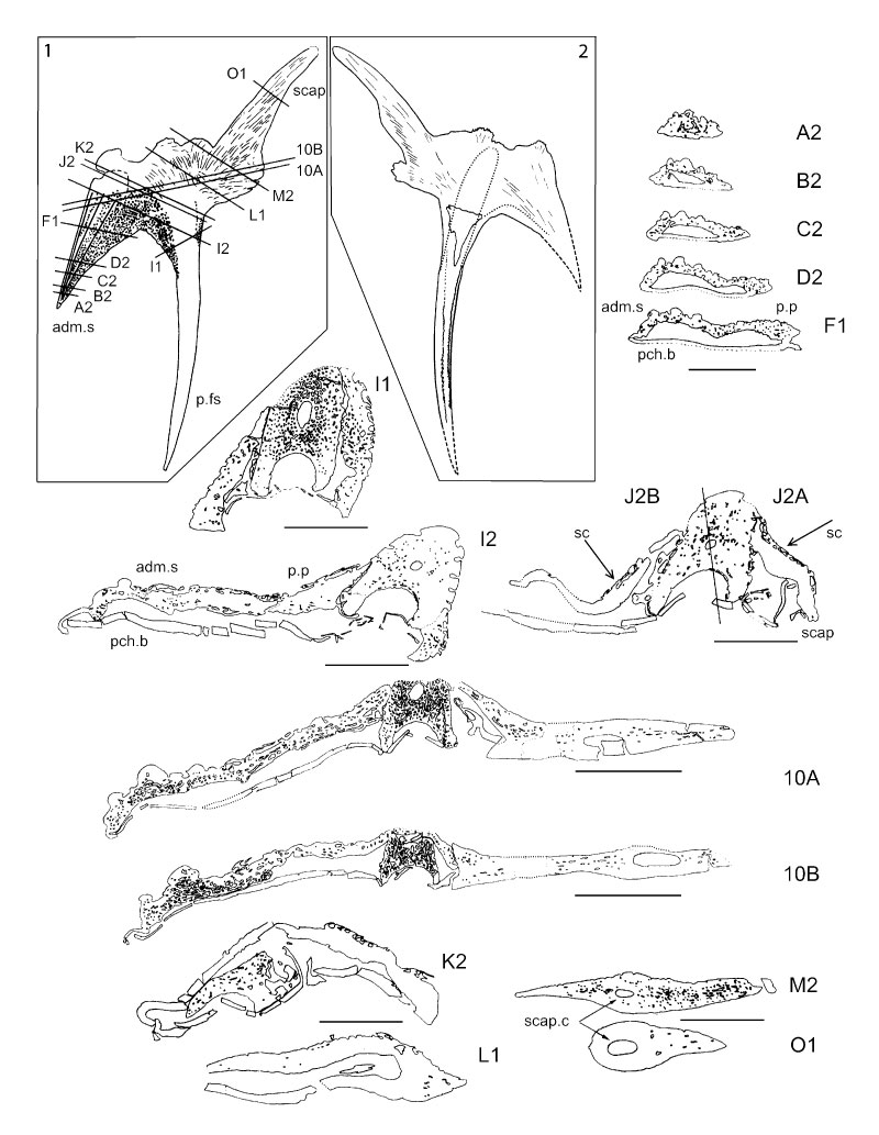
FIGURE 20. Reconstruction of the shoulder girdle region in Diplacanthus tenuistriatus. 1, lateral view. 2, ventral view. Dark grey fill indicates dermal elements, light grey fill indicates endoskeletal elements.
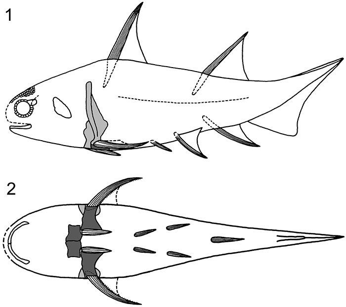
FIGURE 21. Diplacanthus tenuistriatus specimens from Achanarras Quarry, Caithness, squamation and scale histology. 1, NMS G.1897.55.2 (Figure 13.1), impression of squamation anterior to base of posterior dorsal fin spine; 2, 3, NMS G.1897.55.1 (Figure 13.2), squamation above the anal fin, scale crown detail partly obscured by matrix; 4-9, NMS G.1897.55.1, serial sections from patch of midflank squamation: 4, 5, vertical transverse section of scale; 6, 7, vertical longitudinal section of scale; 8, vertical longitudinal section of scale; 9, horizontal section near the level of the base/neck rim of scale. Scale bars equal 1 cm in 1; 1 mm in 2-3; 0.1 mm in 4-9. Arrows indicate anterior direction, where relevant.
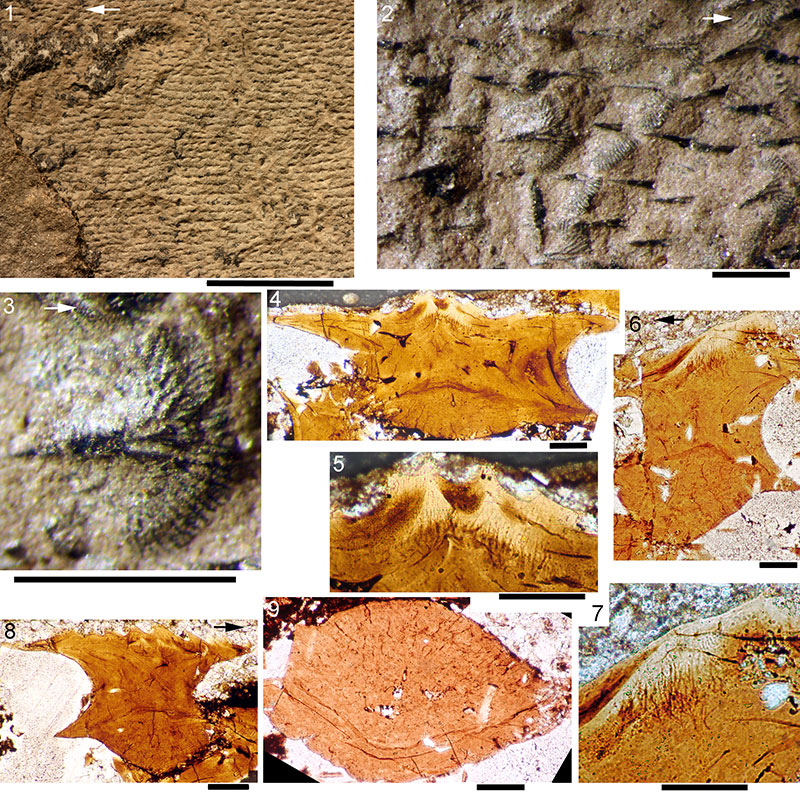
FIGURE 22. Diplacanthus tenuistriatus squamation and histology of scales from Tynet Burn and Gamrie. 1-8, NMS G.Canon Kyle no. 1 from Tynet Burn: 1, postpectoral-caudal peduncle region; 2, mid-body squamation; 3, scale crown; 4, vertical transverse section of scale from caudal region (area A); 5, vertical longitudinal section of scale from area A; 6, horizontal section through crown base, scale from posterior to ad.fs (area B); 7, mid-crown horizontal section, area B; 8, upper crown horizontal section, area B. 9-16, NMS G.1892.8.9 from Gamrie, Banffshire: 9, complete specimen; 10-16, sections of scales from upper flank area between ad.fs and pd.fs (area A) and lower flank in front of a.fs (area B); 10, calcified cartilage globules, posterior end of area B; 11, horizontal section through scale neck, posterior end of area B; 12, vertical longitudinal section, area A; 13, vertical transverse section, area A; 14, vertical longitudinal section, area A; 15, transverse section, area A; 16, vertical oblique section, area A. Scale bars equal 1 cm in 1, 9; 1 mm in 2-3; 0.1 mm in 4-8, 10-16. Abbreviations: a.fs=anal fin spine; pd.fs=posterior dorsal fin spine. Arrows indicate anterior direction, where relevant.
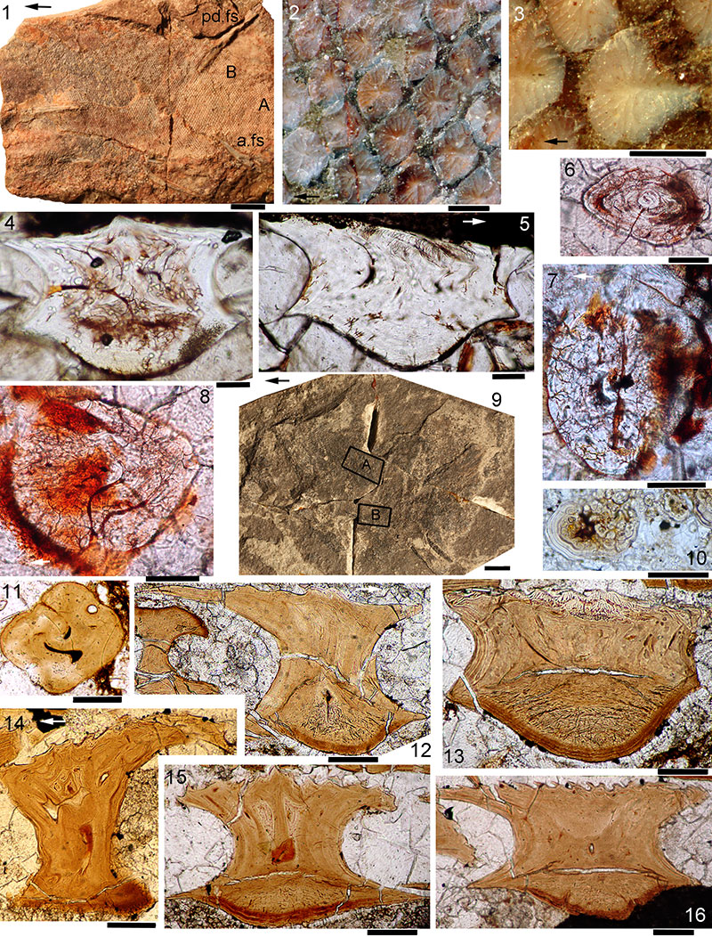
FIGURE 23. Scanning Electron Micrographs of Diplacanthus tenuistriatus scales. 1-4, NMS G.Canon Kyle no. 1 from Tynet Burn, Moray, scales slide NMS G.Canon Kyle no. 1.1, from dorsal side posterior to ad.fs, SEMs: 1, anterior view with broken crown; 2, 3, posterocrown and anterocrown views with end of posterior crown broken off; 4, posterior view. 5-13, NMS G.1892.8.9 from Gamrie, Banffshire, SEMs: 5, 6, 13-16, scales slide NMS G.1892.8.9.17: 5, crown view; 6, anterocrown view; 13, patch of scales in matrix, crown view; 14, crown view; 15, lateral view; 16, posterior view. 7-12, scales slide NMS G.1892.8.9.18: 7, crown view; 8, anterior view; 9, posterior view; 10, anterolateral view; 11, 12, posterobasal view and closeup of base surface. Scale bars equal 1 mm for 13; 0.3 mm for 1-11, 14-16; 0.1 mm for 12.
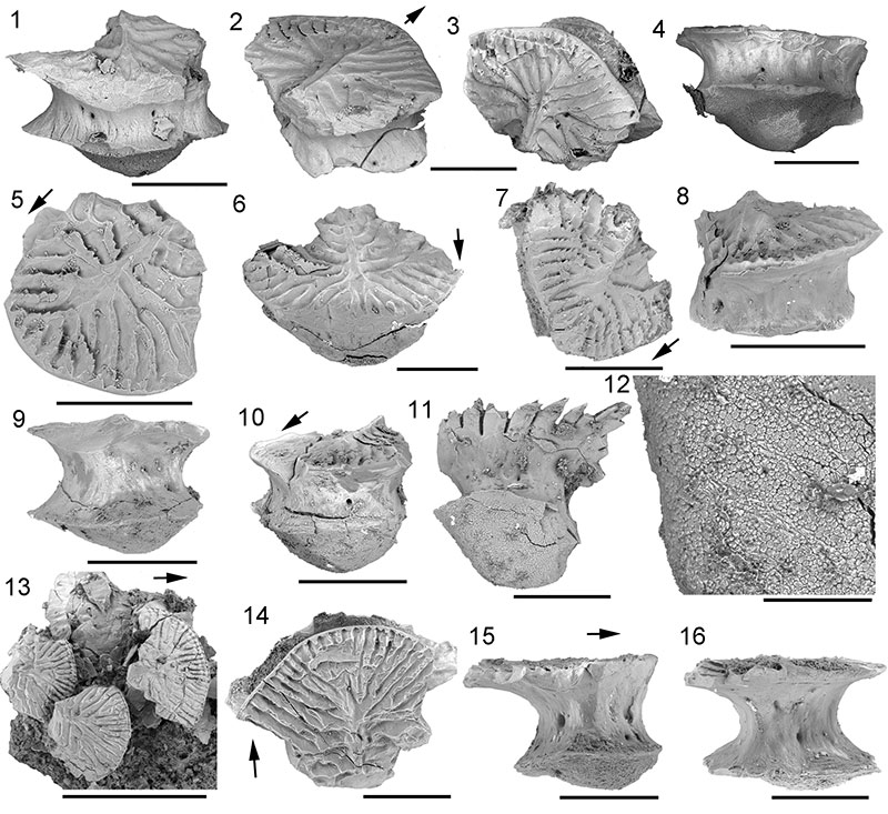
FIGURE 24. Rhadinacanthus longispinus from Gamrie, Banffshire, general morphology. 1, NMS G.1891.92.338; 2, 3, NHM P.4041 from Gamrie, part and counterpart respectively. Scale bars equal 1 cm.
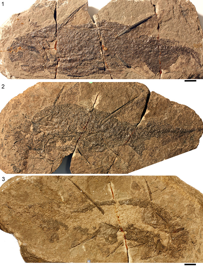
FIGURE 25. Rhadinacanthus longispinus head morphology. 1, 2, NMS G.1891.92.338 from Gamrie, Banffshire; 1, head region; 2, magnified image of sensory line scales; 3, NMS G.1892.8.15 from Gamrie, Banffshire, head region. 4, 5, NHM OR.43276 from Tynet Burn, Moray: 4, complete specimen, head to left; 5, internal surface of right cheek plate, long anterior circumorbital bone, polygonal tesserae on tectal region. 6, NMS G.1892.8.10 from Gamrie, head region. Scale bars equal 1 cm for 1, 3, 4, 5, 6; 1 mm for 2. Abbreviations: ck.p=cheek plate; co.p=circumorbital plate; e=eye stain; oc.p=occlusal plate; so.sl=supraorbital sensory line. Arrows indicates anterior direction.
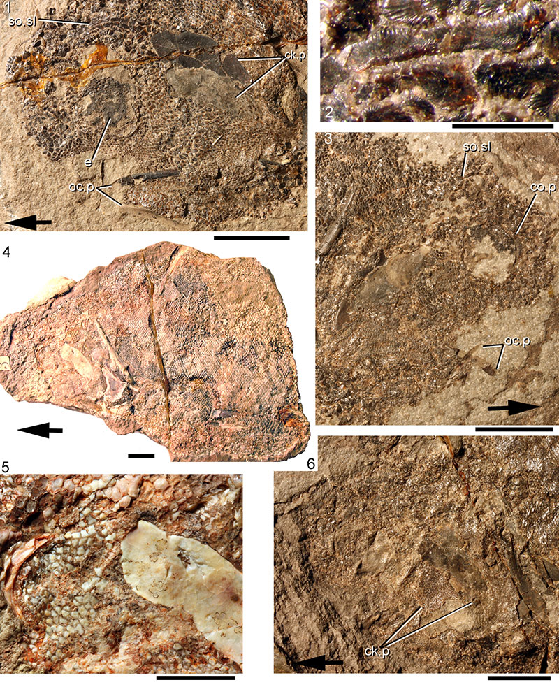
FIGURE 26. Rhadinacanthus longispinus spine morphologies. 1, 2, isolated anterior dorsal fin spine of NMS G.2014.33.4 from Marwick, Orkney: 1, whole spine; 2, long insertion zone showing fine parallel ribs; 3, posterior dorsal fin spine of NMS G.2014.33.7 from Marwick, Orkney; 4, anal fin spine of NMS G.1859.33.703 from Thurso, Caithness; 5, pectoral fin spine of NMS G.1968.5.2 from Taldale Quarry, Caithness; 6, denticulated posterior of pectoral fin spine on specimen NMS G.1870.14.143 from Gamrie, Banffshire. Scale bars equal 1 cm. Abbreviations: p.fs=pectoral fin spine; scap=scapula.
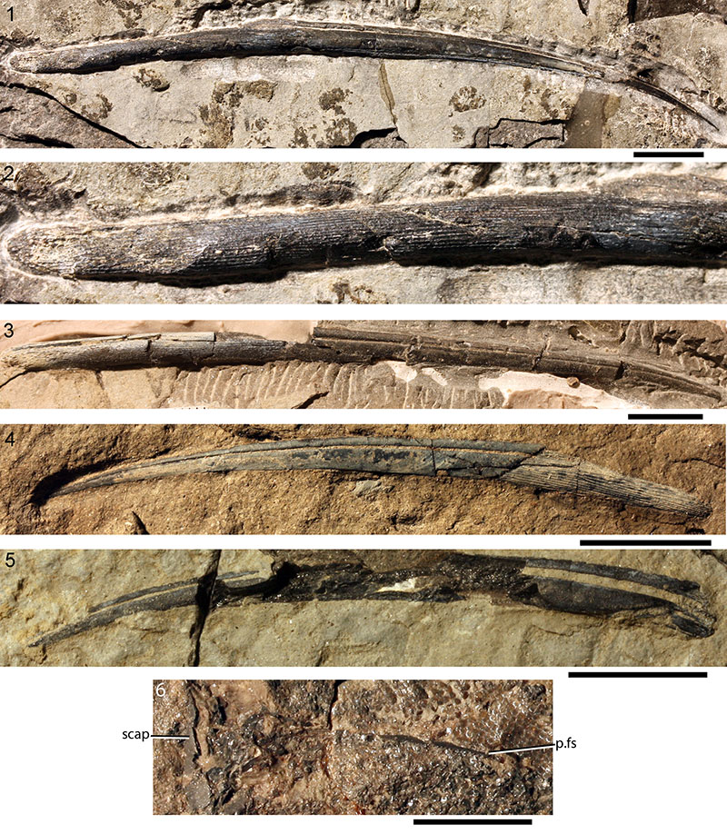
FIGURE 27. Rhadinacanthus longispinus dorsal spine histology. 1, 2, transverse section of anterior dorsal fin spine NMS G.2015.11.1 from Broad Taing, Orkney: 1, whole section indicated by white line; 2, magnified area; 3-5, transverse sections of posterior dorsal fin spine NMS G.2014.4.33 from the slates west of East Murkle Bay, Caithness (Mey subgroup): 3, section through insertion area; 4, 5, section through exserted part of spine; 6, transverse section of posterior dorsal spine NMS G.2014.33.2 from North Ronaldsay (Rousay Beds). Scale bars equal 1 mm in 1, 3, 4, 6; 0.1 mm in 2, 5.
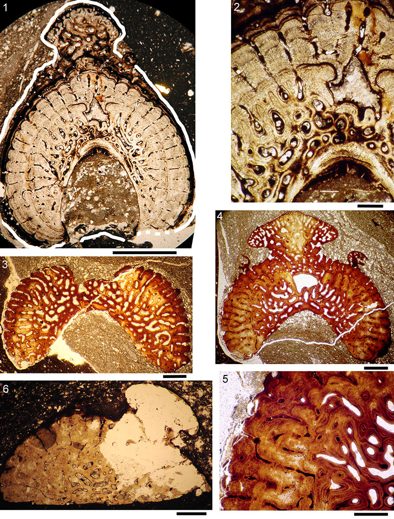
FIGURE 28. Rhadinacanthus longispinus paired and anal spines histology. 1-5, sections of spines on articulated fish NMS G.2014.4.32 from Achanarras; 1, transverse section of pectoral fin spine just distal to insertion area; 2, more distal transverse section of pectoral fin spine; 3, 4, longitudinal section of proximal half of anal fin spine; 5, longitudinal section of distal half of pelvic fin spine; 6, 7, NMS G.2014.44.1 from Cromarty, Ross and Cromarty, section through partial articulated specimen; 6, transverse section of pelvic fin spine; 7, transverse section of prepelvic spine. Scale bars equal 1 cm in 3; 0.5 mm in 1-2, 5-7; 0.1 mm in 4.
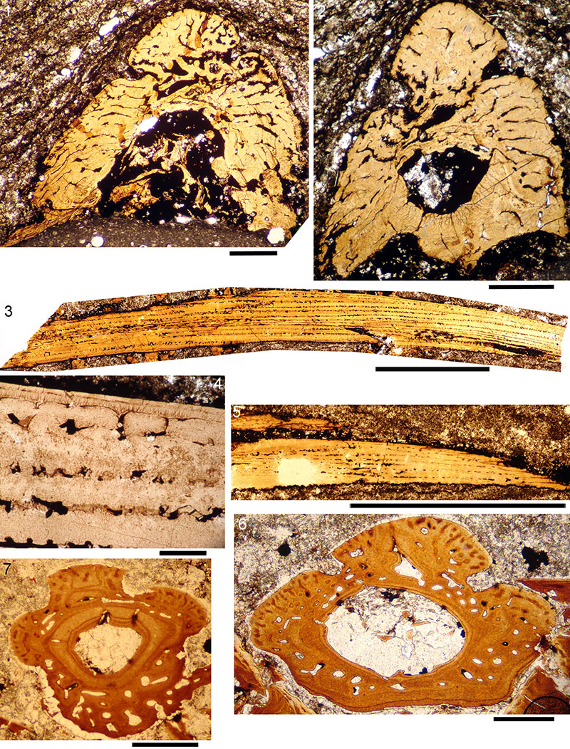
FIGURE 29. Rhadinacanthus longispinus, from a small bonebed (NMS G.2014.15.1 is a sample) at Clardon Haven, Caithness, spine histology. Drawings of Sollas serial sections of various spines from juvenile fish. Abbreviations: adm.s=admedian spine; ad.fs=anterior dorsal fin spine; a.fs=anal fin spine; pelv.fs=pelvic fin spine; p.fs=pectoral fin spine; prepelv.s=prepelvic spine. Length given for each spine.
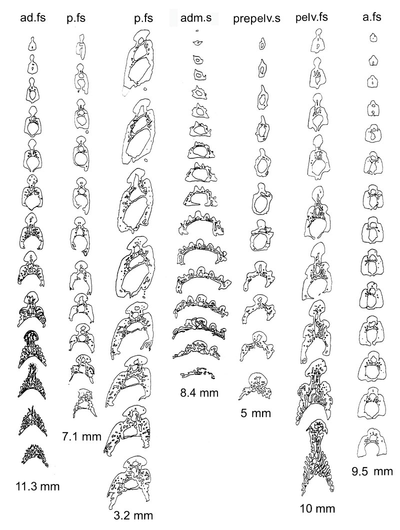
FIGURE 30. Rhadinacanthus longispinus median fin spines from mature fish, transverse section drawings. 1, 2, posterior dorsal spine NMS G.2014.4.33 from the slates west of East Murkle Bay, Caithness: 1, spine showing position of sections; 2, sections 1-8. 3, section of posterior dorsal spine NMS G.2014.33.2 from North Ronaldsay. 4, section of anterior dorsal spine NMS G.2014.11.1 from Broad Taing, Orkney. Scale bars equal 1 cm in 1; 5 mm in 2; 1 mm in 3, 4.
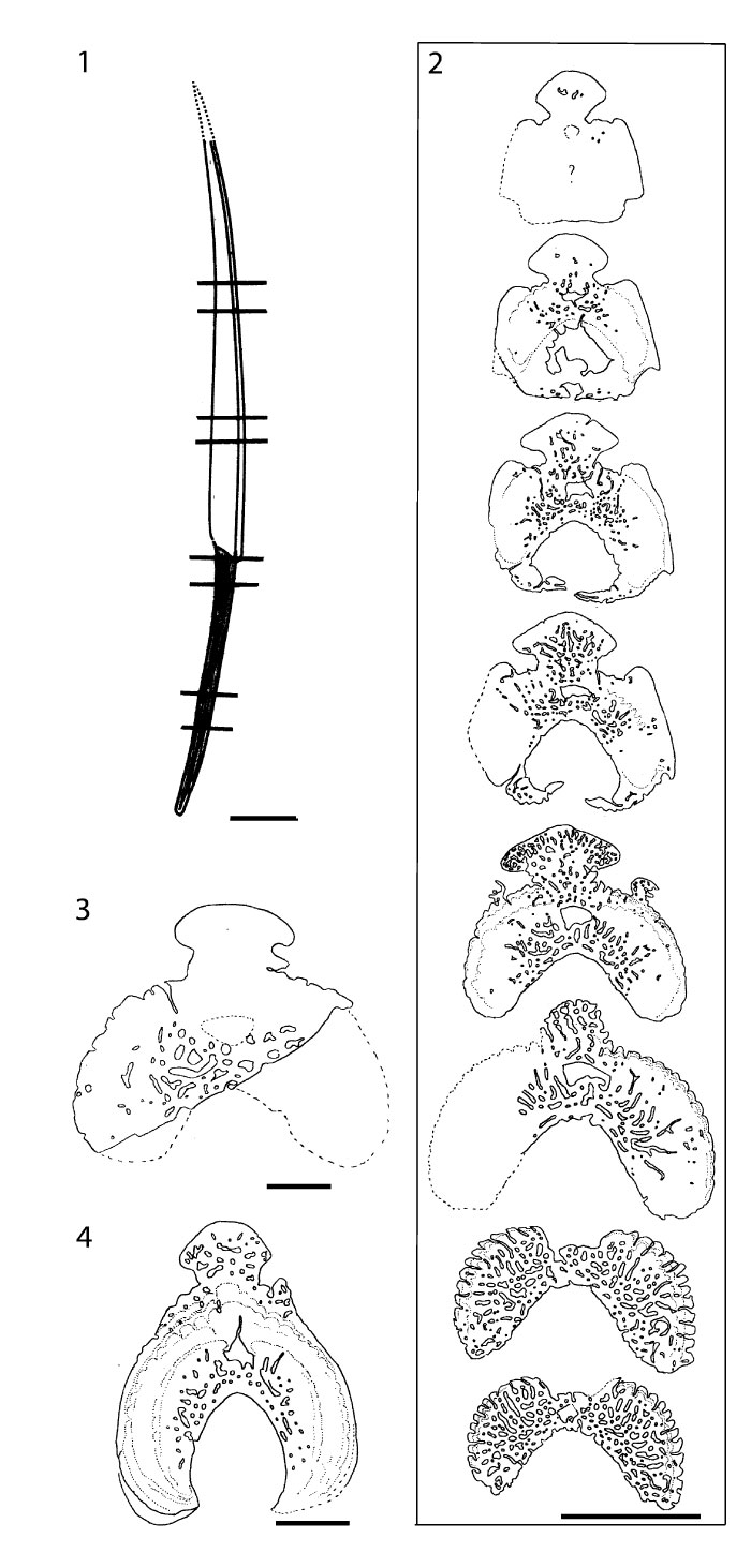
FIGURE 31. Rhadinacanthus longispinus NHM P.4041, from Gamrie, Banffshire, shoulder girdles; 1, 2, pectoral region and head on the part (anterior to right); 3, magnified view of the impression of the admedian spine; 4, 5, pectoral region and head on the counterpart (anterior to left). Scale bars equal 1 cm. Abbreviations: adm.s=admedian spine; ck.p=cheek plate; co.p=circumorbital plate; cor/adm.s=coracoid attached to base of admedian spine; lt.sc=left scapula; lt.p.fs= left pectoral fin spine; oc.p=occlusal plate; p.p=pinnal plate; rt.p.fs=right pectoral fin spine; rt.sc=right scapula; v.p=ventral plate.
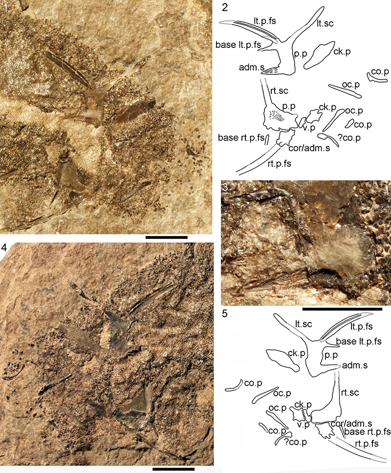
FIGURE 32. Rhadinacanthus longispinus. 1-2, NMS G.1966.49.9 from Orkney, pectoral region and head; 3-4, NMS G.1891.92.334 from Orkney, pectoral region and head; 5-7, isolated scapula NMS G.2014.15.2 from Marwick: 5, ?medial view; 6, ?lateral view, with 7, area with remnants of pinnal plate fused to scapula base. Scale bars equal 1 cm. Abbreviations: adm.s=admedian spine; lt.sc=left scapula; lt.p.fs= left pectoral fin spine; p.p=pinnal plate; rt.p.fs=right pectoral fin spine; rt.sc=right scapula.
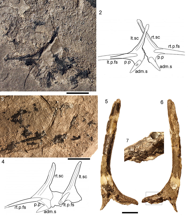
FIGURE 33. Reconstructions of Rhadinacanthus longispinus. 1, lateral view. 2, ventral view. Dark grey fill indicates dermal elements, light grey fill indicates endoskeletal elements.
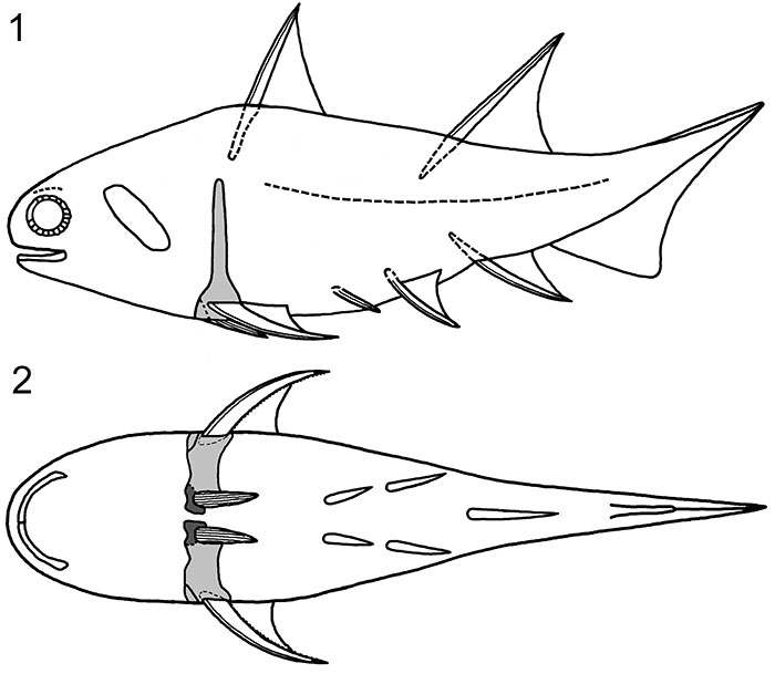
FIGURE 34. Rhadinacanthus longispinus squamation types on different body areas. 1-3, NHM OR.43276 from Tynet Burn, Moray; 1, surrounding scapula shaft; 2, anterior dorsal fin web; 3, mid-flank. 4, NMS G.1892.8.13 from Gamrie, Banffshire, mid-flank; 5, NMS G.2002.59.142pt from Cushnie Burn, Banffshire, mid-flank; 6-10, NHM P.11760 from Gamrie, Banffshire, 6, complete specimen with A, B, C areas magnified in the following images; 7, midflank with lateral line (area A); 8, below lateral line (area A); 9, tail (area B); 10, posterior dorsal fin web (area C). Scale bars equal 1 cm in 1-2, 6, 9-10; 1 mm in 3-5, 7, 8. Abbreviations: fw sc=fin web scales; ll sc=lateral line scales; sc=body scales; scap=scapula shaft, tess=head tesserae.
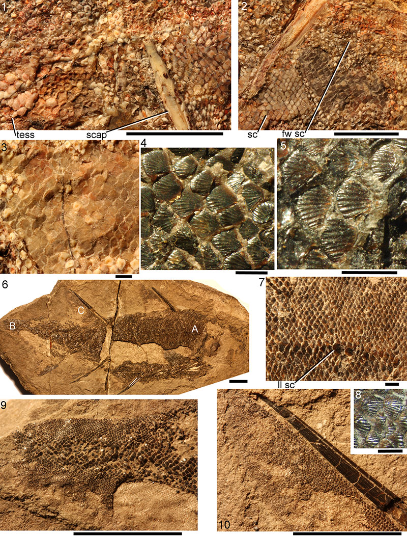
FIGURE 35. Rhadinacanthus longispinus, scanning electron micrographs of scales. 1-4, scales slide NMS G.1892.8.17.1 from Gamrie, Banffshire: 1, 2, crown view of patch of articulated scales, and closeup of scale; 3, lateral view; 4, anterior view. 5-7, scales slide NMS G.1870.14.143.1 from Gamrie, Banffshire: 5, laterocrown view; 6, lateral view; 7, scales from each side of body, stuck together base to base, in posterior view. 8-12, scales slide NMS G.1892.8.15.1 from Gamrie, Banffshire: 8, crown view; 9, crown view; 10, basal view; 11, lateral view; 12, anterior view. Scale bars equal 0.3 mm. White adhesions on scale bases are pyrite crystals. Arrows indicate anterior.
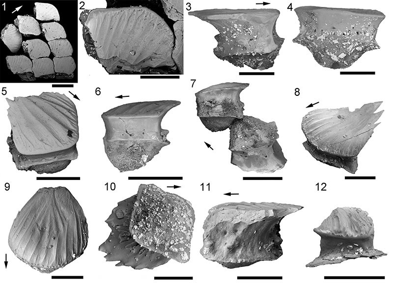
FIGURE 36. Rhadinacanthus longispinus NMS G.2014.4.32 from Achanarras Quarry, Caithness, scale histology. 1, articulated squamation from midflank area; 2-6, thin sections of midflank scales: 2, oblique vertical section; 3, magnified view of central crown of the same scale; 4, horizontal section of crown; 5, oblique vertical section; 6, oblique vertical section. Scale bars equal 0.1 mm. Abbreviations: ac=ascending canal; cW=canal of Williamson; gz=growth zones; hc=horizontal canal; w=neck swelling. Arrows indicate anterior direction, where relevant.
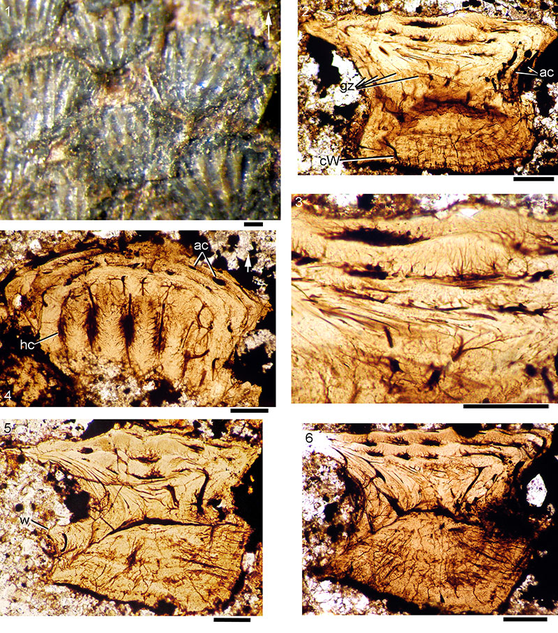
FIGURE 37. Rhadinacanthus longispinus NMS G.2014.44.1 from Cromarty, Ross and Cromarty, scale histology. 1, vertical transverse section; 2, vertical transverse section; 3, vertical longitudinal section; 4, oblique vertical section; 5, vertical section of double scale; 6, vertical longitudinal section. Scale bars equal 0.1 mm. Arrows indicate anterior direction, where relevant.
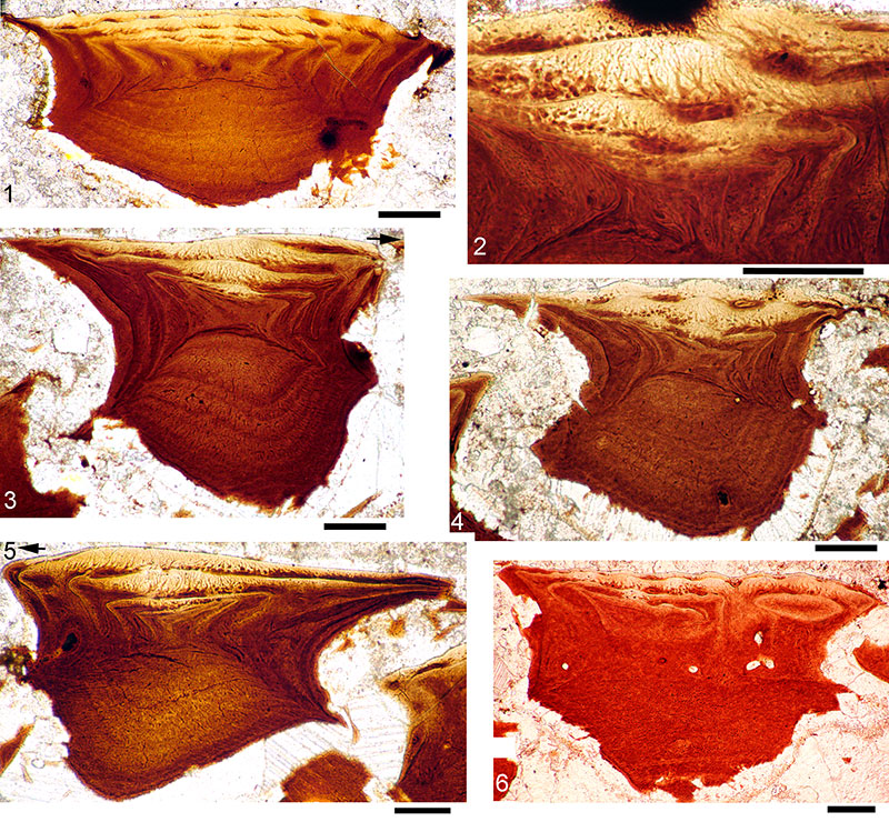
FIGURE 38. 'Life' reconstructions of: 1, Diplacanthus crassisimus, 2, Diplacanthus tenuistriatus, and 3, Rhadinacanthus longispinus.
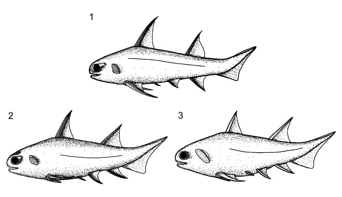
FIGURE 39. Relationship tree generated in phylogenetic analysis of selected early gnathostomes (77 taxa, 262 characters; Tree 1, 641 steps)
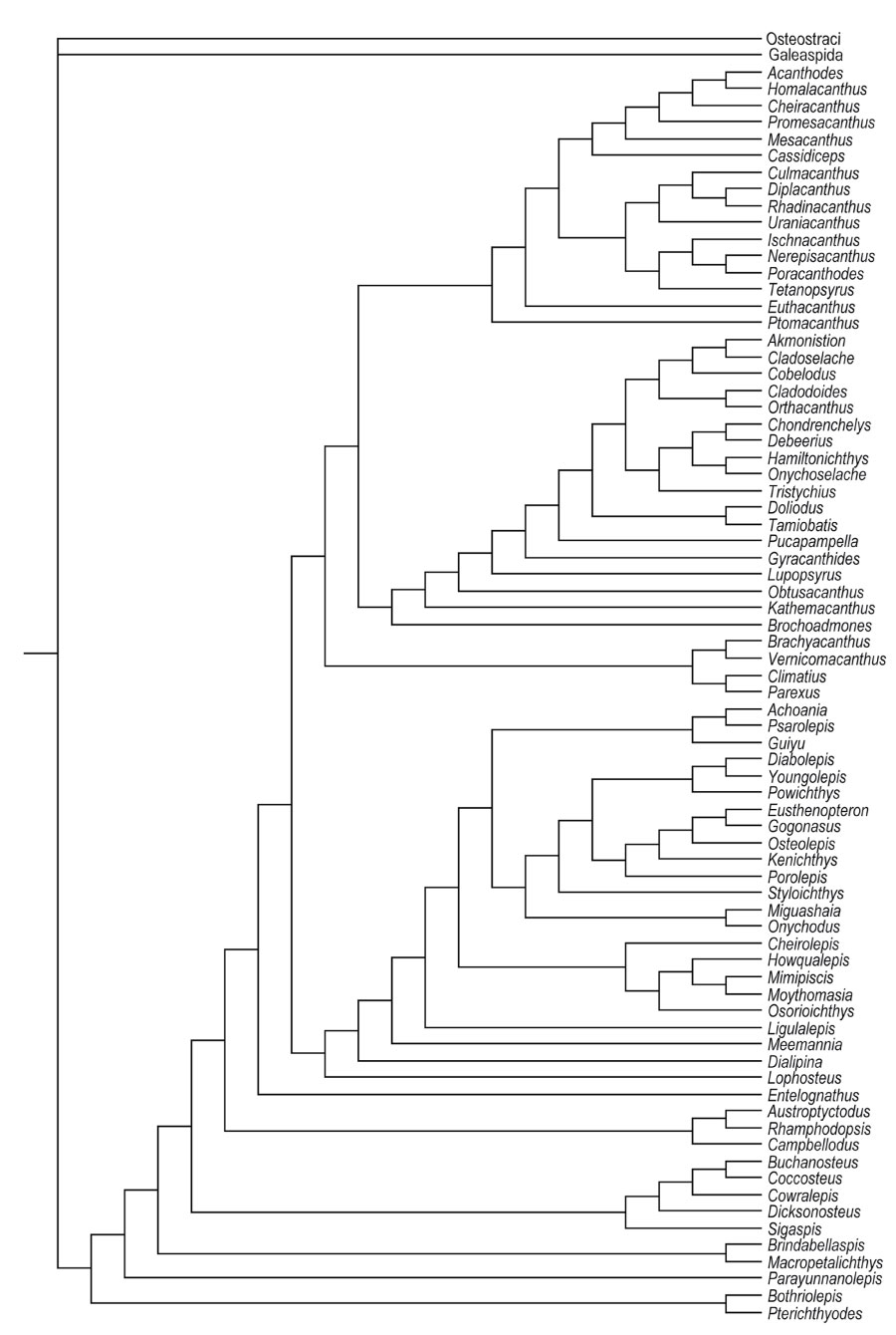
FIGURE 40. Strict consensus relationship tree generated in phylogenetic analysis of selected early gnathostomes (77 taxa, 262 characters).
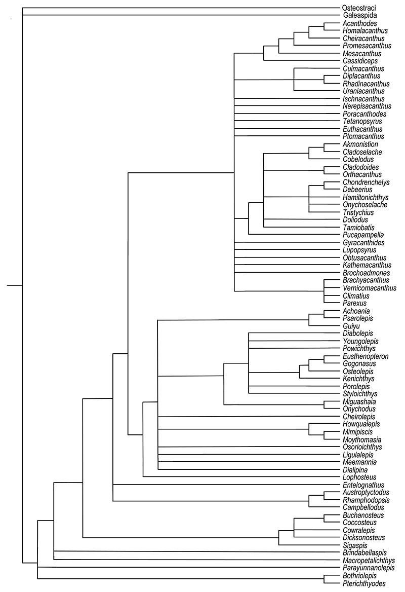
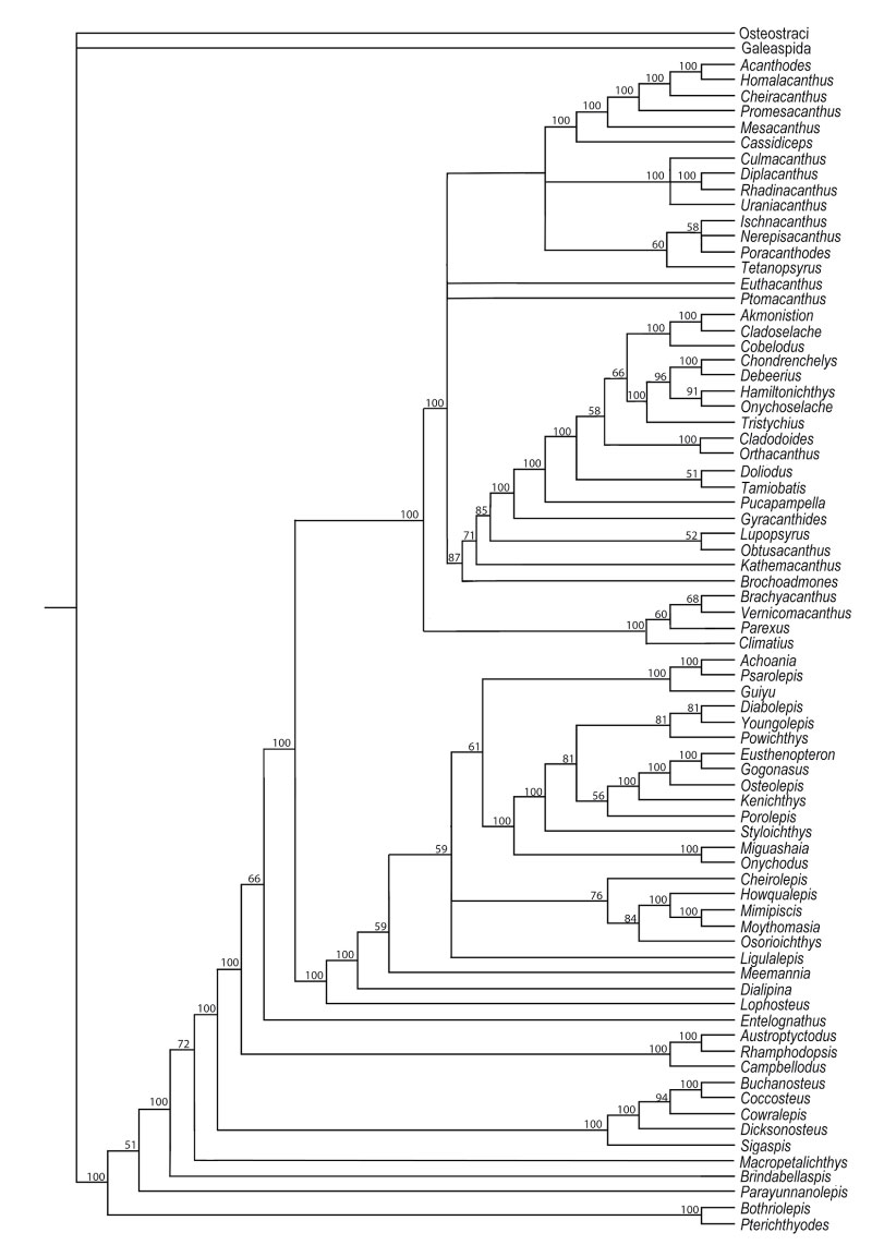
FIGURE 41. 50% majority rule consensus tree generated in phylogenetic analysis of selected early gnathostomes (77 taxa, 262 characters; numbers on branches show bootstrap support %).