FIGURE 1. Schematic geological map with location of the investigated section (1). The study section is located at Portella Colla (2) in the Madonie Mountains, Sicily (3).

FIGURE 2. Biostratigraphic interpretation of the investigated section. According to Benedetti (2010), the distribution of the main larger foraminifers in the displaced layers allows the recognition of four biozones in the Paleogene succession. The presence of SBZ 18 (late Bartonian) at the base of Paleogene succession is uncertain. The E/O boundary is here redefined by the study of poorly-preserved nannofossils occurring only in some levels, and following the biozonal schemes of Martini (1971), and Catanzariti et al. (1997).

FIGURE 3. Representative forms of foraminifers belonging to the six different functional morphogroups (redrawn after Jones and Charnock, 1985, figure 2).

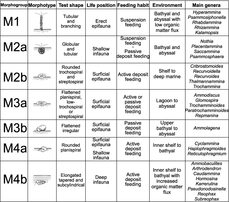
FIGURE 4. Summarizing scheme of the DWAF functional morphogroups and morphotypes described in this work (redrawn and modified after van den Akker et al., 2000, figure 6).

FIGURE 5. Results of the micropaleontological analysis along the investigated section cropping out at Portell Colla (Sicily). Vertical distribution of individuals counted per sample, number of taxa, Fisher’s Alpha diversity, species richness, evenness index, Simpson’s index, and percentage of CaCO 3 within the samples. The three boxes represent the relative abundances of agglutinated and calcareous hyaline taxa, the DWAF morphogroups, and the recognized assemblages.

FIGURE 6. Stratigraphic distribution (in percentage) of selected DWAF taxa (solid line). The abundances have been exaggerated (dashed lines) in order to show also the minimal fluctuations.

FIGURE 7. Stratigraphic distribution (in percentage) of selected DWAF taxa (solid line). The abundances have been exaggerated (dashed lines) in order to show also the minimal fluctuations.

FIGURE 8. Environmental interpretation of the investigated succession. Block diagrams schematically show the presence/absence of main taxa and the faunal density.

FIGURE 9. Schematic zonal schemes of late Eocene-lower Oligocene successions described from different regions (redrawn and modified after Kaminski and Gradstein, 2005). The most common DWAF taxa are added.

FIGURE 10. Depth distribution of some selected and frequent taxa according to data presented by van Morkhoven et al. (1986), Kaminski and Gradstein (2005) and Waśkowa (2014).

FIGURE 11. Location of the section FO; the reddish clay are interbedded by quartzose volcanic layers marked by the metallic labels.

FIGURE 12. Scanning electron micrographs of Astrorhizida ( 1-13) and Saccamminidae (14-15) foraminifers from the Caltavuturo Formation cropping out at Portella Colla. 1-2, Rhabdammina discreta Brady, 1884, PCs0 (1) and PC060618 (2). 3,Rhabdammina eocenica Cushman and Hanna, 1927, PC060601. 4-5,Psammosiphonella cylindrica (Glaessner, 1937), PC11 (4) and PCs0 (5). 6-7,Psammosiphonella linearis (Brady, 1879), PC060618 (6) and PC11 (7). 8,Bathysiphon sp., MM12. 9,Nothia excelsa (Grzybowski, 1898), PC060603. 10,Nothia robusta (Grzybowski, 1898) with Ammolagena clavata (Jones and Parker, 1860), PC060621. 11, Nothia cf. latissima (Grzybowski, 1898), PC060624. 12-13, Rhizammina indivisa Brady, 1884, PC060621. 14-15,Psammosphaera irregularis (Grzybowski, 1896), PC13 (14) and PC22 (15). 16,Placentammna placenta (Grzybowski, 1898), PCs0. 17,Saccammina grzybowskii (Schubert, 1902), PC060621. Scale bar equals 0.5 mm.

FIGURE 13. Scanning electron micrographs of Psammosphaeridae (1-4) and Ammodiscidae (5-27) from the Caltavuturo Formation cropping out at Portella Colla. 1,Psammosphaera cf. laevigata White, 1928, PCs0. 2,Psammosphaera sp. 1, PC13. 3, Psammosphaera sp. 2, PC060615. 4,Psammosphaera sp. 3, PC060625. 5,Ammodiscus cretaceus (Reuss, 1845), PC22. 6,Hyperammina sp., PC060625. 7-8,Ammodiscus incertus (d’Orbigny, 1839), PC060603 (7) and PC13 (8). 9-11,Ammodiscus tenuisimus (Gümbel, 1862), PC22 (9), PC060615 ( 10) and PC060615 (11). 12-13,Ammodiscus latus Grzybowski, 1898, PC11 (12) and PC11 (13). 14,Ammodiscus peruvianus Berry, 1928, PC060617. 15-16,Ammodiscus cf. latus Grzybowski, 1898, PC11. 17, Ammodiscus sp. 1, PC11. 18,Ammodiscus sp. 2, PC15. 19,Ammodiscus sp. 3, PC060603. 20,Annectina biedai Gradstein and Kaminski, 1997, PC060618. 21,Annectina cf. grzybowski (Jurkiewicz, 1960), PC060617. 22,Glomospira sp. 2, PC15. 23,Glomospira sp. 1, PC11. 24, Glomospira sp. 3, PC13. 25-26. Glomospira serpens (Grzybowski, 1898), PC060617 (25) and PC060618 (26). 27,Glomospira irregularis (Grzybowski, 1898), PC060618. Scale bar equals 0.5 mm.

FIGURE 14. Scanning electron micrographs of Hormosinelloidea from the Caltavuturo Formation cropping out at Portella Colla. 1, Ammolagena clavata (Jones and Parker, 1860) on a specimen of Psammosiphonella cylindrica (Glaessner, 1937), PCs0. 2,Ammolagena clavata (Jones and Parker, 1860), PC060607. 3-7,Caudammina gutta Benedetti and Pignatti, 2009, PC18. 8,Subreophax cf. guttifer (Brady, 1881), PC15. 9,Subreophax cf. pseudoscalaris (Samuel, 1977), PCs0. 10,Subreophax scalaris (Grzybowski, 1896), PC11. 11-12,Subreophax splendidus (Grzybowski, 1898), PC060624 (11) and PC11 ( 12). 13,Arthrodendron subnodosiformis (Grzybowski, 1898), PC060615. 14,Arthrodendron grandis (Grzybowski, 1898), PC050515. 15-19, Kalamopsis grzybowskii (Dylążanka, 1923), PC18 (15), PC11 (16), PC18 (17), PC060621 (18) and PC060621 (19). Scale bar equals 0.5 mm.

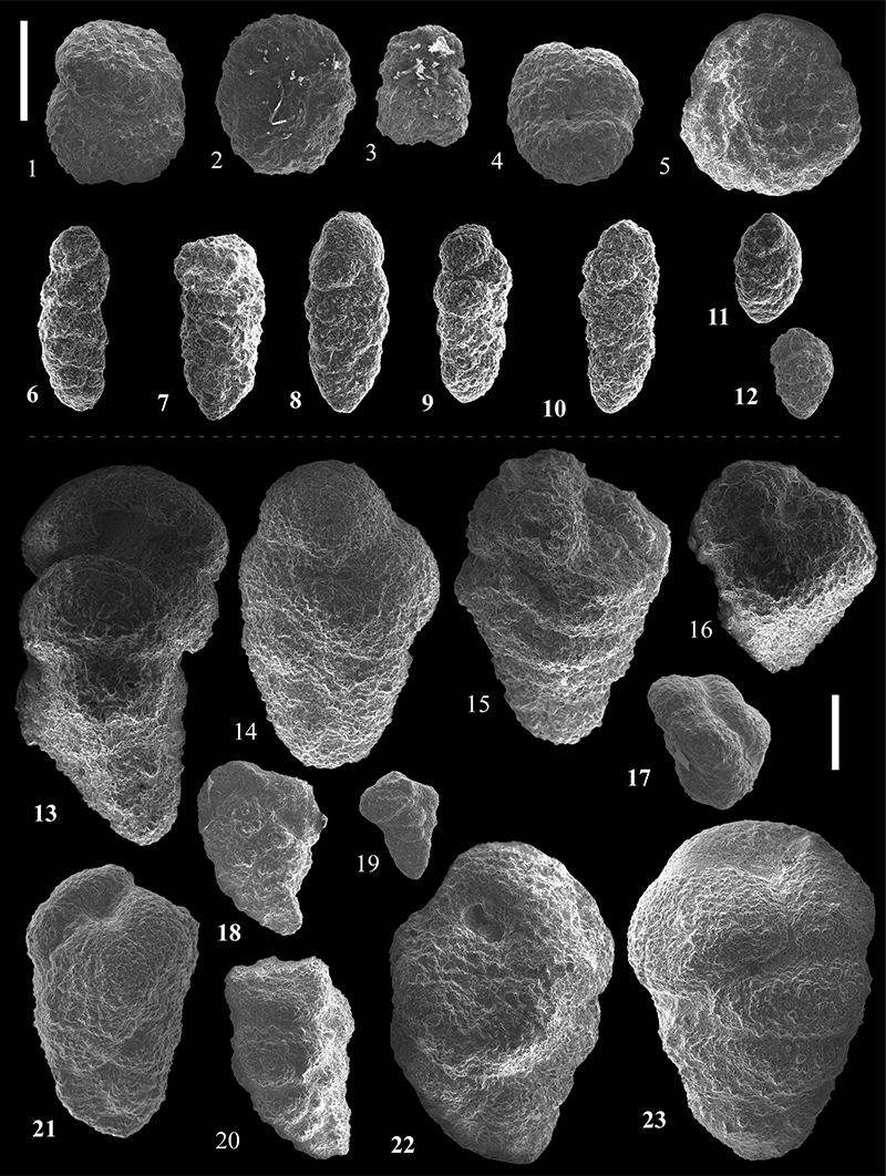
FIGURE 15. Scanning electron micrographs of Usbekistaniinae from the Caltavuturo Formation cropping out at Portella Colla. 1,Glomospira extendens Emiliani, 1954, PC11. 2,Glomospira gordialis (Jones and Parker, 1860), PC060604. 3-16,Repmanina charoides (Jones and Parker, 1860), PC060604 (3), PC060601 (4), PC060604 ( 5), PC060603 (6), PC060611 (7), PC060601 (8), PC060603 (9), PC060621 (10), PC060621 (11), PC060617 (12), PCs0 (13), PCs0 (14), PC060604 (15) and PCs0 (16). Scale bar equals 0.5 mm.

FIGURE 16. Scanning electron micrographs of Reophacidae (1-3), Hormosinidae (4-13), and Lituotubidae (14-20) from the Caltavuturo Formation cropping out at Portella Colla. 1,Reophax duplex Grzybowski, 1896, PC060624. 2,Reophax pilulifer Brady, 1884, PC13. 3,Reophax sp. 1, PC060615. 4-5,Hormosina velascoensis (Cushman, 1926), PC060617 (4) and PC11 (5). 6,Hormosina trinitatensis Cushman and Renz, 1946, PC11. 7,Hormosina sp. 1, PC060624. 8,Hormosina sp. 2, PC060615. 9-11,Pseudonodosinella elongata Grzybowski, 1898, PC11 (9), PC060601 (10) and PCs0 (11). 12-13,Pseudonodosinella nodulosa Brady, 1879, PC11 (12) and PC060617 ( 13). 14,Paratrochamminoides acervulatus (Grzybowski, 1896), PC13. 15,Paratrochamminoides deflexiformis (Noth, 1912), PC060615. 16,Lituotuba lituiformis (Brady, 1879), PC060615. 17,Paratrochamminoides sp. 1, PC060601. 18,Paratrochamminoides draco (Grzybowski, 1901), PC11. 19,Paratrochamminoides mitratus (Grzybowski, 1901), PCs0. 20,Conglophragmium deforme (Grzybowski, 1898), PC060623. Scale bar equals 0.5 mm.

FIGURE 17. Scanning electron micrographs of Lituotubidae (1-3, 7, 9, 11-12) and Trochamminoidae (4-6, 8, 10) from the Caltavuturo Formation cropping out at Portella Colla. 1-2, Paratrochamminoides heteromorphus (Grzybowski, 1898), MM12 (1) and MM14 (2). 3,Conglophragmium irregulare (White, 1928), PCs0. 4,Trochamminoides coronatus (Brady, 1879), MM14. 5,Trochamminoides grzybowskii Kaminski and Geroch, 1992, MM1. 6,Trochamminoides subcoronatus (Grzybowski, 1896), MM1. 7,Paratrochamminoides cf. olszewskii (Grzybowski, 1898), PC060615. 8,Trochamminoides dubius (Grzybowski, 1901), PC060611. 9,Paratrochamminoides olszewskii (Grzybowski, 1898), PC11. 10,Trochamminoides dubius (Grzybowski, 1901), PC060611. 11,Paratrochamminoides aff. olszewskii (Grzybowski, 1898), PC060615. 12,Paratrochamminoides aff. gorayskii (Grzybowski, 1898), PC060615. Scale bars equal 0.5 mm.

FIGURE 18. Scanning electron micrographs of Trochamminoidae ( 1-10) and Haplophragmoididae (11-27) from the Caltavuturo Formation cropping out at Portella Colla. 1,Trochamminoides septatus (Grzybowski, 1898), PC060604. 2,Trochamminoides proteus (Karrer, 1866), PC060621. 3, Trochamminoides cf. proteus (Karrer, 1866), PC060624. 4,Trochamminoides velascoensis Cushman, 1926, PC11. 5,Trochamminoides intermedius (Grzybowski, 1896), PC060621. 6,Trochamminoides variolarius (Grzybowski, 1898), MM14. 7,Trochamminoides sp. 1, PC060603. 8,Trochamminoides sp. 2, PC060604. 9, Trochamminoides sp. 3, PC060618. 10,Trochamminoides sp. 4, PC18. 11-13,Haplophragmoides carinatus Cushman and Renz, 1941, PC060621 (11), PC060621 (12) and PC060624 (1314-16,Haplophragmoides walteri (Grzybowski, 1898), PC18 (14), PC11 (15), PC5 (16). 17, Haplophragmoides eggeri Cushman, 1926, PC060611. 18, Haplophragmoides excavatus Cushman and Waters, 1927, PC060601. 19,Haplophragmoides cf. kirki Wickenden, 1932, PC060602. 20,Haplophragmoides porrectus Maslakova, 1955, PCs0. 21, Haplophragmoides cf. walteri (Grzybowski, 1898), PC060621. 22,Haplophragmoides horridus (Grzybowski, 1901), PC060601. 23, Haplophragmoides cf. horridus (Grzybowski, 1901), PC060604. 24,Haplophragmoides cf. porrectus Maslakova, 1955, PC060615. 25-26,Haplophragmoides sp. 1, PC060602 (25) and PC060602 (26). 27,Haplophragmoides sp. 2, PC060603. Scale bar equals 0.5 mm.

FIGURE 19. Scanning electron micrographs of Haplophragmoididae ( 1-6), Sphaeramminidae (7-10) Lituolidae (11-14), and Recurvoidea (15-25) from the Caltavuturo Formation cropping out at Portella Colla. 1-2,Haplophragmoides cf. latissimusuturalis Smith, 1971, PC3. 3,Haplophragmoides sp. 3, PC060615. 4Haplophragmoides sp. 4, PC060601. 5, Haplophragmoides sp. 5, PC060615. 6,Haplophragmoides sp. 6, PC060621. 7-8,Praesphaerammina subgaleata (Vašiček, 1947), PC11 (7) and PC1 (8). 9-10, Ammosphaeroidina pseudopauciloculata (Mjatliuk, 1966), PC060604 (9) and PC060604 (10). 11,Ammobaculites agglutinans (d’Orbigny, 1846), PC060615. 12,Ammobaculites sp. 1, PC060607. 13,Ammobaculites sp. 3, PC15. 14, Ammobaculites sp. 2, PC060625. 15-16,Budashevaella multicamerata (Voloshinova and Budasheva, 1961), PC060604. 17, Cribrostomoides subglobosus (Cushman, 1910), PC060604. 18, Recurvoidella lamella (Grzybowski, 1898), PC060601. 19, Recurvoides anormis Mjatliuk, 1970, PC5. 20,Recurvoides sp. 1, PC060601. 21,Recurvoides sp. 2, PC060623. 22,Recurvoides nucleolus (Grzybowski, 1898), MM3. 23, Recurvoides walteri (Grzybowski, 1898), PC5. 24,Trochammina bifaciata Friedberg, 1901, PC060604. 25,Thalmannammina subturbinata (Grzybowski, 1898), MM14. Scale bar equals 0.5 mm.

FIGURE 20. Scanning electron micrographs of Trochamminoidea ( 1-11, 13), Ataxophragmiidae (12, 17, 20-21), and Textulariina ( 14-16, 18-19, 22-23) from the Caltavuturo Formation cropping out at Portella Colla. 1,Trochammina sp. 1, PC060601. 2,Trochammina sp. 2, PC060601. 3,Trochammina sp. 3, PC060601. 4,Trochammina sp. 4, PC060604. 5, Trochammina sp. 5, PC060605. 6-10,Karrerulina conversa (Grzybowski, 1901), PC060615 (6), PC018 (7), PC11 (8), PC060615 (9) and PCs0 ( 10). 11, Karrerulina horrida (Mjatliuk, 1970), PC060617. 12,Remesella varians (Glaessner, 1937), PC060601. 13,Gaudryina sp., PC1. 14,Eggerella compressa (Andreae, 1884), PC13. 15,Eggerella sp. 1, PC3. 16,Eggerella sp. 2, PC3. 17,Arenobulimina sp., PC060602. 18,Siphotextularia sp. 1, PC060621. 19,Siphotextularia sp. 2, PC060621. 20,Tetraxiella subtilissima PCs0. 21, Gravellina sp., PC060618. 22,Valvulina flexilis Cushman and Renz, 1941, PC060604. 23,Valvulina sp., PC060621. Scale bars equal 0.5 mm.

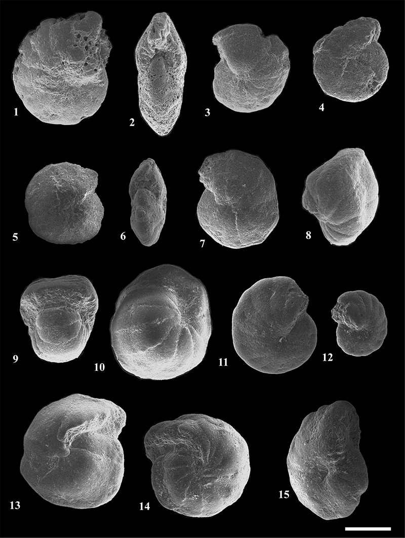
FIGURE 21. Scanning electron micrographs of Cyclamminidae from the Caltavuturo Formation cropping out at Portella Colla. 1-3, Reticulophragmium acutidorsatum (Hantken, 1868), PC3. 4-7, Reticulophragmium amplectens (Grzybowski, 1898), PC3. 8-10, Reticulophragmium rotundidorsatum (Hantken, 1875), PC060624. 11-12,Cyclammina cancellata Brady, 1879, PC22. 13-14, Reticulophragmium projectum Schröder-Adams and McNeil, 1994, PC060624. 15, Cyclammina placenta (Reuss, 1851), PC11. Scale bar equals 0.5 mm.


