FIGURE 1. Stratigraphic and paleobiogeographical distributions of the currently accepted members of Erymnochelyinae, including the European taxa described here, i.e., the representatives of the new genus Eocenochelus, belonging to the Erymnochelys group. The shell and skull symbols accompanying the name of each taxon indicate which of these elements are known. The star indicates continental Stereogenyina, and the pentagon indicates continental or littoral Stereogenyina.
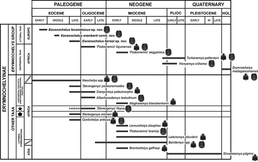
FIGURE 2. Geographic locations of the type localities of the European Eocene representatives of the podocnemidid Eocenochelus gen. nov.: Soulane (Jonquières, Aude, France), type locality of the lower Eocene (Ypresian) Eocenochelus lacombianus sp. nov.; Saint-Germain-en-Laye (Yvelines, Île-de-France, France), type locality of the middle Eocene (Lutetian) Eocenochelus eremberti comb. nov.; Can Beuloví (Sobremunt, Barcelona, Spain), type locality of the upper Eocene (Priabonian) Eocenochelus farresi sp. nov.
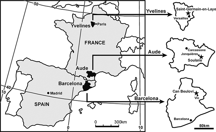
FIGURE 3. Skull of the holotype of Eocenochelus eremberti comb. nov., MNHN.F CGR 101, from the middle Eocene (Lutetian) of Saint-Germain-en-Laye (Yvelines, Île-de-France, France), in dorsal (1-2), ventral (3-4) and right lateral (5-6) views. Abbreviations: ane, apertura narium externa; ang, angular; bo, basioccipital; bs, basisphenoid; cm, condylus mandibularis; cpt, cavum pterygoidei; cv, cervical vertebra; den, dentary; ex, exoccipital; fct, foramen anterius chorda tympani; fo, fossa orbitalis; fpo, fenestra postotica; fpp, foramen palatinum posterius; fr, frontal; FR, frontal scute; fst, foramen stapedio-temporale; IP, interparietal scute; ju, jugal; MS, masseterian scute; mx, maxilla; MX, maxillary scute; op, opisthotic; pa, parietal; PA, parietal scute; pal, palatine; pf, prefrontal; pm, premaxilla; po, postorbital; pr, prootic; pt, pterygoid; ptp processus trochlearis pterygoidei; pw, pterygoid wing; q, quadrate; qj, quadratojugal; so, supraoccipital; SO, subocular scute.
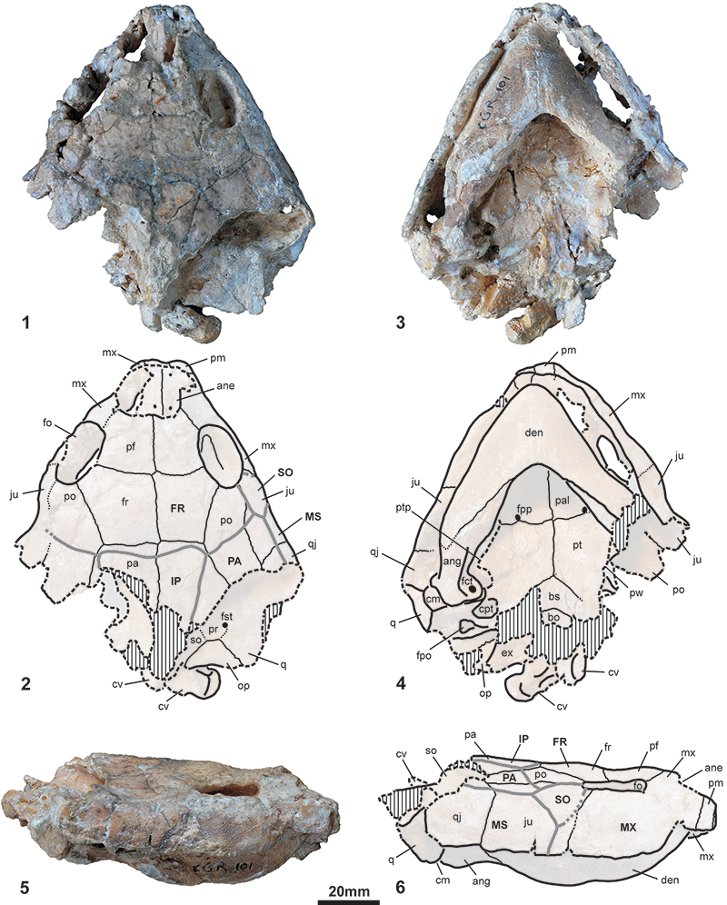
FIGURE 4. Some elements of the carapace of the holotype of Eocenochelus eremberti comb. nov., MNHN.F CGR 101, from the middle Eocene (Lutetian) of Saint-Germain-en-Laye (Yvelines, Île-de-France, France). 1-2, plates of the carapace, in dorsal and ventral views. 3-4, reconstruction of the dorsal view of the shell, showing the position of all these elements of the carapace. 4-6, details of the outer surface of some plates of the carapace: the second left peripheral (4), the distal region of the sixth right costal (5), and the proximal region of the fourth left costal (6). Abbreviations: c, costal; M, marginal scute; n, neural; p, peripheral; PL, pleural scute; py, pygal; V, vertebral scute.
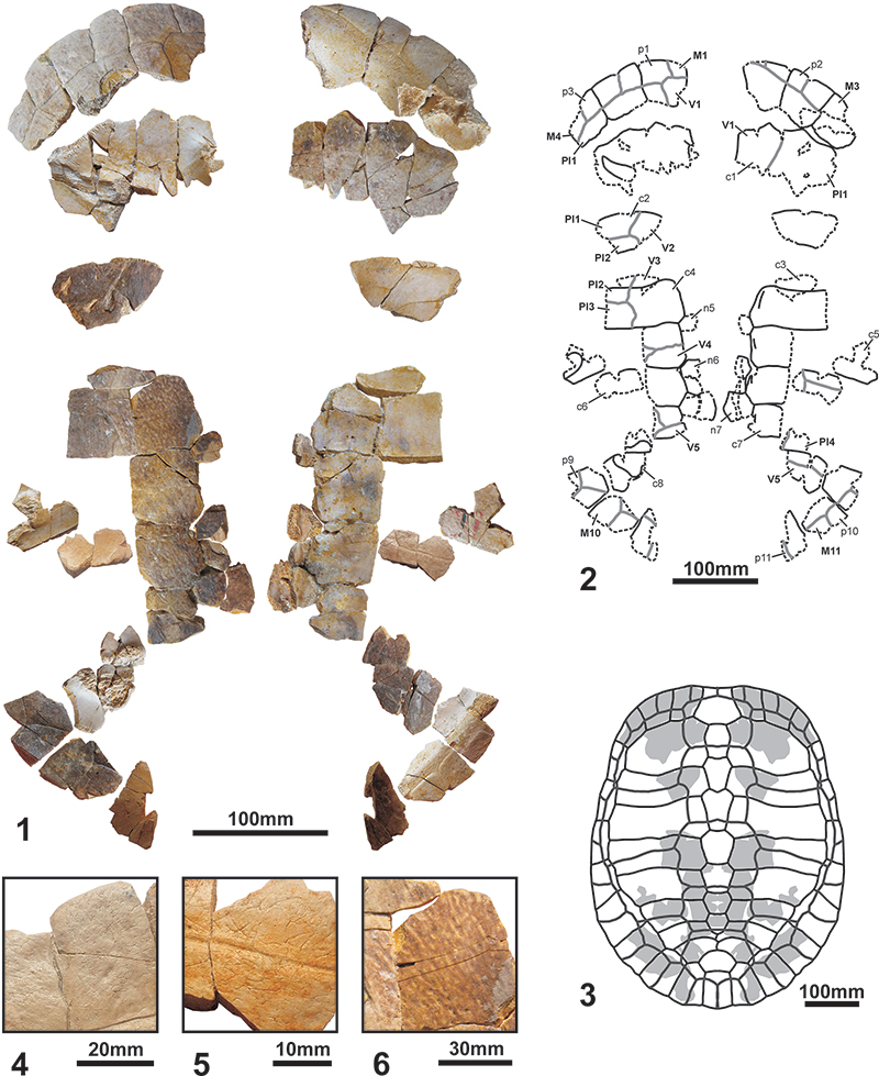
FIGURE 5. Plastron of the holotype of Eocenochelus eremberti comb. nov., MNHN.F CGR 101, from the middle Eocene (Lutetian) of Saint-Germain-en-Laye (Yvelines, Île-de-France, France), in ventral (1-2) and dorsal (3-4) views. Abbreviations: AB, abdominal scute; AN, anal scute; en, entoplastron; ep, epiplastron; FE, femoral scute; GU, gular scute; hp, hypoplastron; HU, humeral scute; hy, hyoplastron; IG, intergular scute; M, marginal scute; ms, mesoplastron; PC, pectoral scute; xi, xiphiplastron.
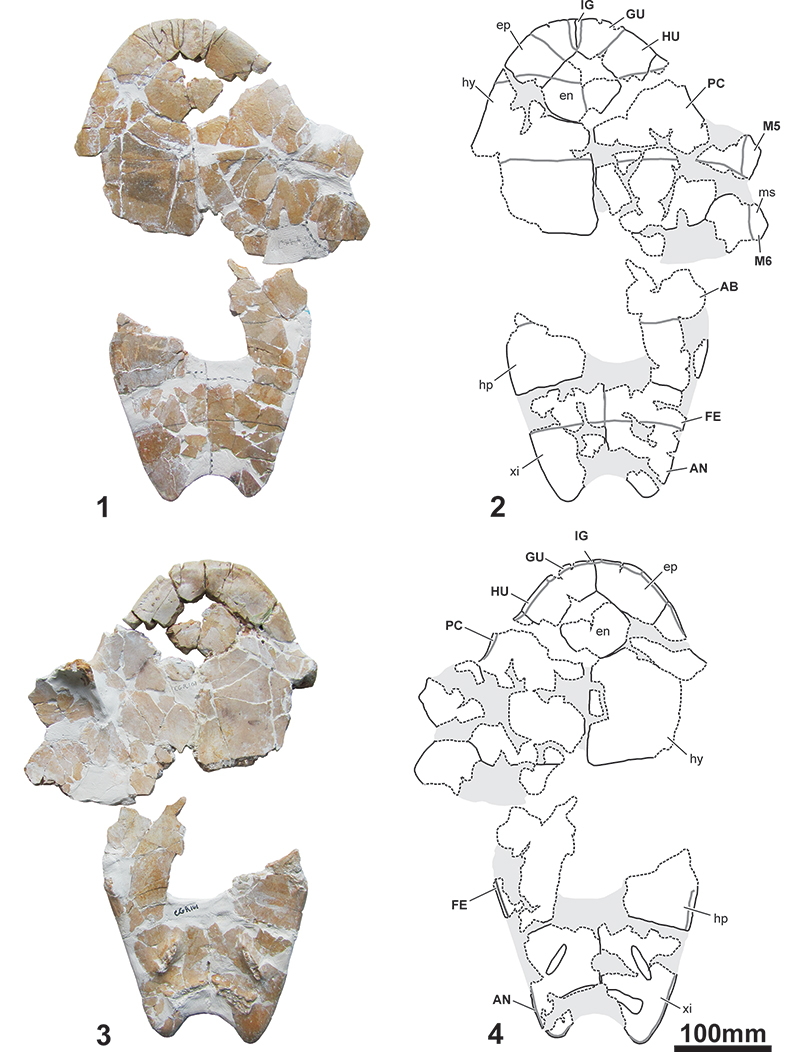
FIGURE 6. Fragmentary skeletal bones of the holotypes of Eocenochelus eremberti comb. nov., MNHN.F CGR 101, from the middle Eocene (Lutetian) of Saint-Germain-en-Laye (Yvelines, Île-de-France, France) (1-49), and Eocenochelus lacombianus sp. nov., MNHN.F EBA 534, from the lower Eocene (Ypresian) of Soulane (Jonquières, Aude, France) (50-57). 1-4, left hyoid horn, second branchial horn, in dorsal (1), medial (2), ventral (3) and lateral (4) views. 5-9, acromion of the right scapula with the partial glenoid articular region, in posterior (5), ventral (6), anterior (7) and dorsal (8) views, and articular part in ventrolateral view (9). 10-14, acromion of the left scapula with the partial glenoid articular region, in posterior (10), ventral (11), anterior (12) and dorsal (13) views, and proximal articular section (14). 15-19, left coracoid in dorsal (15), posteroventral (16), ventral (17) and anterior (18) views, and articular-glenoid facet part, in proximal view (19). 20-25, left ischium, in anterior (20), medial (21), posterior (22) and distal (23) views, and dorsal (24) and ventral (25) sections. 26-31, right ilium, in posterior (26), lateral (27), anterior (28) and medial (29) views, and dorsal (30) and ventral (31) sections. 32-37, left pubis, in anterior (32), medial (33), posterior (34) and lateral (35) views, proximal articular-acetabular region in dorsal view (36), and ventral section (37). 38-43, left humerus, in anterior (38), ventral (39), posterior (40) and dorsal (41) views, and proximal (42) and distal (43) sections. 44-49, right femur, in anterolateral (44), ventral (45), posteromedial (46) and dorsal (47) views, and proximal (48) and distal (49) sections. 50-53, right hemipelvis, in lateral (50), posterior (51), anterior (52) and medioventral (53) views. 54-57, left hemipelvis, in lateral (54), posterior (55), anterior (56) and medial (57) views.
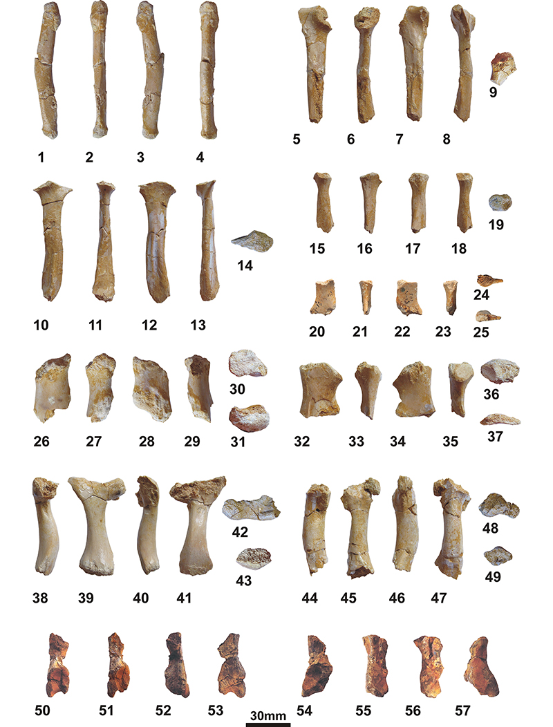
FIGURE 7. Shell of the holotype of Eocenochelus lacombianus sp. nov., MNHN.F EBA 534, from the lower Eocene (Ypresian) of Soulane (Jonquières, Aude, France). 1-2, carapace, in dorsal view. 3-4, carapace, in ventral view. 5, reconstruction of the dorsal view of the shell, showing the position of the preserved elements of the carapace. 6-7, plastron, in ventral view. 8-9, plastron, in dorsal view. Abbreviations: AB, abdominal scute; AN, anal scute; c, costal; en, entoplastron; ep, epiplastron; FE, femoral scute; GU, gular scute; hp, hypoplastron; HU, humeral scute; hy, hyoplastron; IG, intergular scute; M, marginal scute; ms, mesoplastron; n, neural; nu, nuchal; p, peripheral; PC, pectoral scute; PL, pleural scute; py, pygal; V, vertebral scute; xi, xiphiplastron.
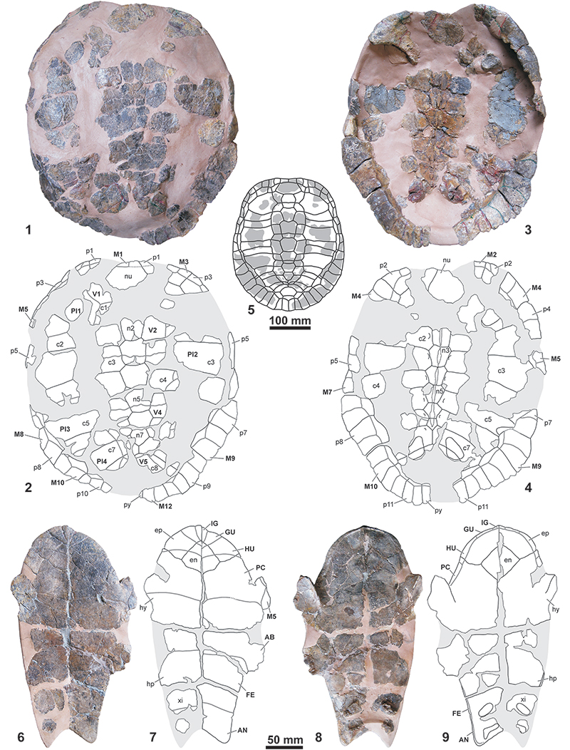
FIGURE 8. Shell of the holotype of Eocenochelus farresi sp. nov., MGSB 74.641-GLV.T.45, from the upper Eocene (Priabonian) of Can Beuloví (Sobremunt, Barcelona, Spain), in dorsal (1-2) and ventral (3-4) views. Abbreviations: AB, abdominal scute; AN, anal scute; c, costal; en, entoplastron; ep, epiplastron; FE, femoral scute; GU, gular scute; hp, hypoplastron; HU, humeral scute; hy, hyoplastron; IG, intergular scute; M, marginal scute; ms, mesoplastron; n, neural; p, peripheral; PC, pectoral scute; PL, pleural scute; py, pygal; V, vertebral scute; xi, xiphiplastron.
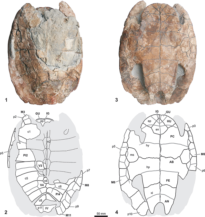
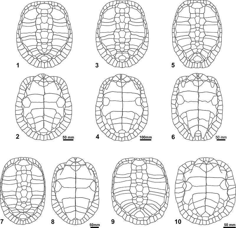
FIGURE 9. Reconstruction of the dorsal and ventral views of the shells of the three Podocnemididae species studied here, Eocenochelus lacombianus sp. nov. (1-2), Eocenochelus eremberti comb. nov. (3-4), and Eocenochelus farresi sp. nov. (5-6), compared to those of the extant Malagasy Erymnochelys madagascariensis (7-8), and the European Eocene Neochelys liriae (9-10).