FIGURE 1. Geographic location and Cenozoic stratigraphy of the Contamana area, Peru. 1, location map of the Contamana area in Peruvian Amazonia (Loreto Department). 2-3, synthetic stratigraphic units of the complete Contamana Cenozoic sequence along the Cachiyacu stream (modified after Antoine et al., 2016, figure 3), including fossil-bearing levels, among which Eocene rodent-yielding localities CTA-47, CTA-51, CTA-73, CTA-27, CTA-66, and CTA-29. Note also in the same section, the location of the other rodent-bearing localities, designated by an asterisk, in the Pozo, Chambira and Pebas Fm.; 2, NE Flank of the Maquía Anticline; 3, SW Flank of the Maquía Anticline. Modified from Antoine et al. (2012, 2016). Fm., Formation.
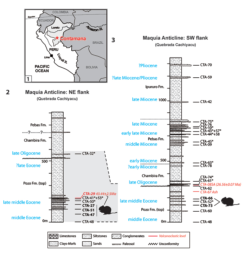
FIGURE 2. Dental nomenclature for upper teeth in occlusal view. 1, upper molar; 2, P4; 3, dP4. 1, paracone; 2, protocone; 3, metacone; 4, hypocone; 5, parastyle; 6, mesostyle; 7, anteroloph; 8, anterior arm of the protocone; 9, posterior arm of the protocone (= lingual protoloph); 10, posterior outgrowth of the protocone; 11, protoloph (= labial protoloph); 12, mure; 13, third transverse crest (= central transverse crest); 14, mesolophule; 15, mesoloph; 16, anterior arm of the hypocone; 17, metaloph; 18, posteroloph; 19, paraflexus; 20, hypoflexus/hypofossette; 21, confluence of the paraflexus with the hypoflexus; 22, mesoflexus/mesofossette; 23, metaflexus; 24, confluence of the metaflexus with the posteroflexus; 25, posteroflexus. Based on observations made on the new material, the dental terminology is modified after Wood and Wilson (1936), Fields (1957), Marivaux et al. (2004, 2017) and Antoine et al. (2012).
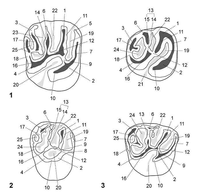
FIGURE 3. Dental nomenclature for lower teeth in occlusal view. 1, lower molar; 2, p4; 3, dp4. 1, protoconid; 2, metaconid; 3, mesoconid; 4, entoconid; 5, hypoconid; 6, mesostylid; 7, metalophulid I; 8, posterior arm of the metaconid; 9, posterior arm of the protoconid; 10, neomesolophid; 11, second transverse cristid; 12, mesolophid; 13, rest of the mesolophid?; 14, ectolophid; 15, mesial ectolophid; 16, distal ectolophid; 17, hypolophid; 18, anterior arm of the entoconid; 19, posterior arm of the entoconid; 20, anterior arm of the hypoconid; 21, posterior arm of the hypoconid; 22, anterior outgrowth of the hypoconid; 23, posterolophid; 24, anteroflexid/anterofossettid; 25, mesoflexid; 26, mesial mesoflexid; 27, distal mesoflexid; 28, confluence of the anteroflexid with the mesoflexid; 29, hypoflexid; 30, metaflexid; 31, confluence of the hypoflexid with the metaflexid. Based on observations made on the new material, the dental terminology is modified after Wood and Wilson (1936), Fields (1957), Marivaux et al. (2004, 2017) and Antoine et al. (2012).
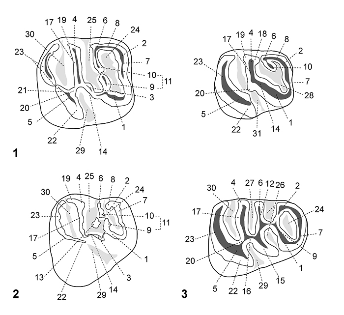
FIGURE 4. Scanning electron microscope images (in occlusal view) of fossil caviomorph teeth from CTA-27. Cachiyacuy contamanensis (1-6), Eobranisamys javierpradoi sp. nov. (7-8), Cachiyacuy kummeli (9-13), Canaanimys maquiensis (13-16) and cf. Eoespina sp. (17). 1, right m3 (MUSM 2713); 2, right m2 (MUSM 1914); 3, right m2 (MUSM 1915); 4, right p4 (MUSM 2678); 5, fragmentary right dp4 (MUSM 2670); 6, left M3 (MUSM 2758); 7, right dP4 (MUSM 2797); 8, fragmentary left M3 (MUSM 2801); 9, left dp4 (MUSM 2762); 10, left p4 (MUSM 2766); 11, right m3 (MUSM 2780, reversed); 12, left M1 (MUSM 2785); 13, left M2 (MUSM 2786); 14, fragmentary right M3 (MUSM 2794); 15, right M3 (MUSM 2793); 16, left M3 (MUSM 2792, reversed); 17, fragmentary left M2 (MUSM 2802). Top scale for 1-13, bottom scale for 14-17.
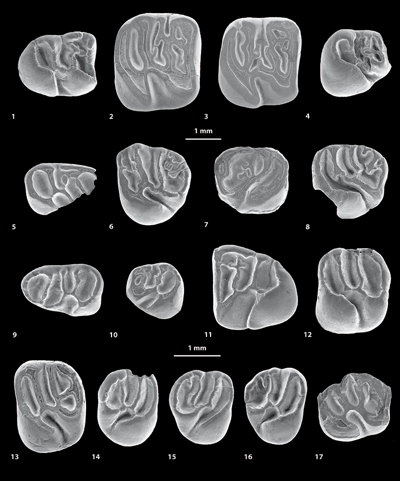
FIGURE 5. Scanning electron microscope images (in occlusal view) of fossil caviomorph teeth from CTA-47 (1-4), CTA-51 (5-12), CTA-73 (13-14) and CTA-66 (15-16). ?Canaanimys sp. (1-2), ?Cachiyacuy kummeli (3), Caviomorpha indet. 1 (4), Cachiyacuy cf. contamanensis 1 (5-7), Caviomorpha indet 2 (8-10), Cachiyacuy cf. kummeli (11), Eoespina sp. (12), Caviomorpha indet 3 (13), Caviomorpha indet 4 (14) and Eobranisamys sp. (15-16). 1, fragmentary right dp4 (MUSM 2645); 2, fragmentary left lower molar (MUSM 2646); 3, fragmentary left dP4 (MUSM 2648); 4, fragmentary right lower molar (MUSM 2647); 5, fragmentary right dp4 (MUSM 2651, reversed); 6, left m1 (MUSM 2652); 7, left m3 (MUSM 2653); 8, fragmentary right lower molar (MUSM 2656); 9, fragmentary left dP4 (MUSM 2657); 10, fragmentary left upper molar (MUSM 2658); 11, fragmentary left M1 (MUSM 2654); 12, right M2 (MUSM 2655); 13, fragmentary right lower molar (MUSM 2659); 14, fragmentary left upper molar (MUSM 2660); 15, fragmentary left upper molar? (MUSM 2841); 16, fragmentary left upper molar (MUSM 2842). Top scale for 1-12, bottom scale for 13-16.
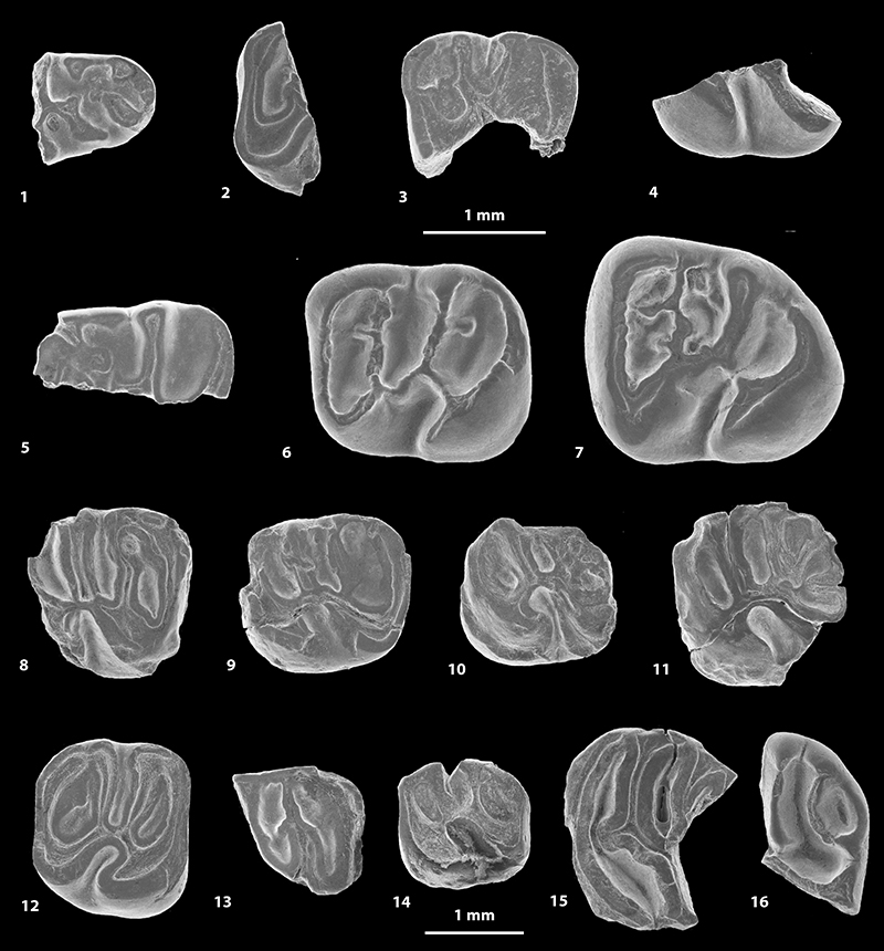
FIGURE 6. Scanning electron microscope images of fossil caviomorph teeth from CTA-29. Pozomys ucayaliensis gen. et sp. nov. (1-6), Cachiyacuy cf. contamanensis 2 (7-12), Caviomorpha indet. 5 (13), Caviomorpha indet. 6 (14) and Cavioidea or Chinchilloidea indet. (15-17). 1, right m1-2, occlusal view (MUSM 2822); 2, left p4, occlusal view (MUSM 2821, reversed); 3, fragmentary right M3, occlusal view (MUSM 2819); 4, right M2, occlusal view (MUSM 2833); 5, right m1-2, labial view (MUSM 2822); 6, right m1-2, lingual view (MUSM 2822); 7, left dp4, occlusal view (MUSM 2825); 8, right m1, occlusal view (MUSM 2827, reversed); 9, right dP4, occlusal view (MUSM 2828, reversed); 10, left M1, occlusal view (MUSM 2831); 11, right M2, occlusal view (MUSM 2563, reversed); 12, fragmentary left M2, occlusal view (MUSM 2832); 13, right dp4, occlusal view (MUSM 2838); 14, fragmentary right upper molar, occlusal view (MUSM 2839); 15, right m2, occlusal view (MUSM 2835); 16, right dp4, occlusal view (MUSM 2834); 17, fragmentary right upper molar, occlusal view (MUSM 2836, reversed).
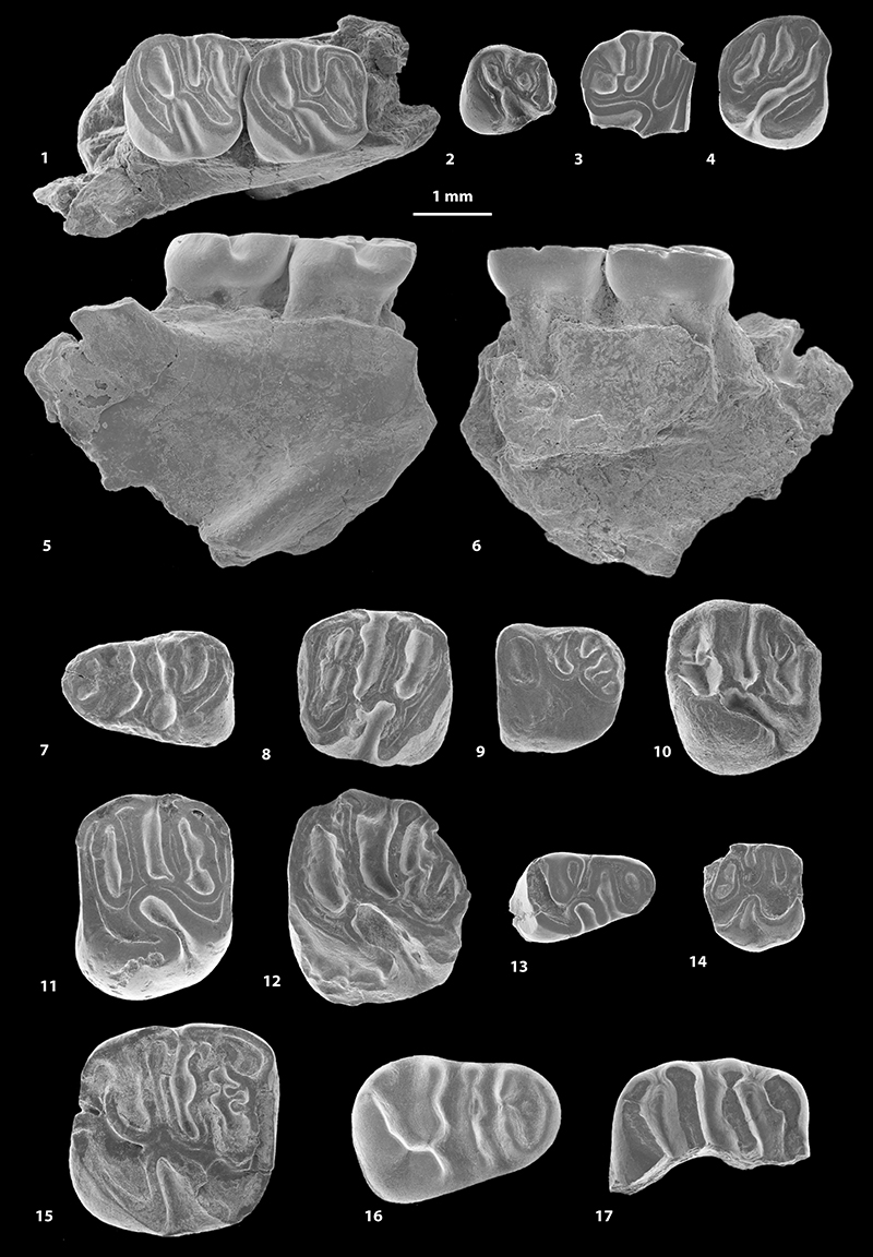
FIGURE 7. Morphological variation of the p4 (in occlusal view) of Cachiyacuy contamanensis from CTA-27. 1, MUSM 2674; 2, MUSM 2676; 3, MUSM 2677; 4, MUSM 1879; 5, MUSM 2678. Original computerized schemas (1-5) by Myriam Boivin.
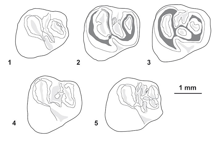
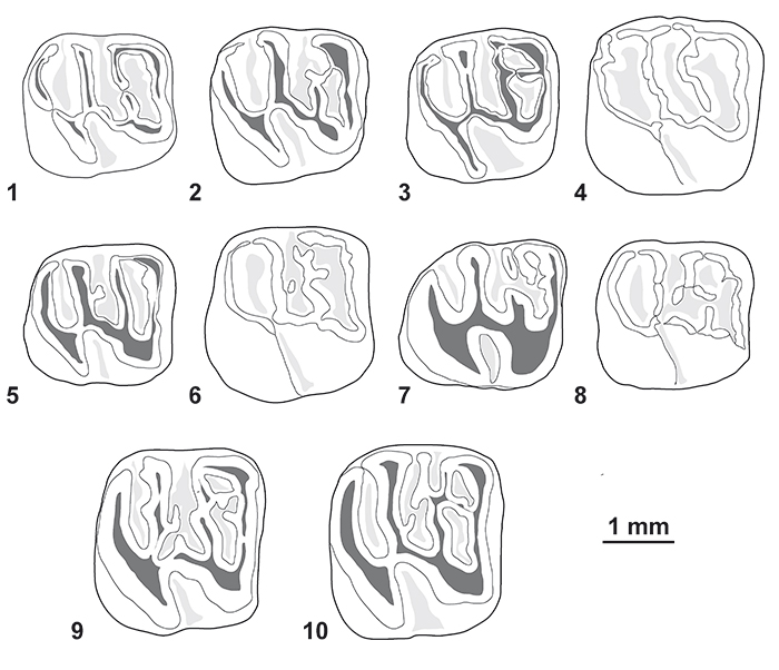
FIGURE 8. Morphological variation of the lower molars (in occlusal view) of Cachiyacuy contamanensis from CTA-27. 1, MUSM 1878; 2, MUSM 2704; 3, MUSM 2684; 4, MUSM 2708; 5, MUSM 2692; 6, MUSM 2701; 7, MUSM 2714; 8, MUSM 2689; 9, MUSM 1915; 10, MUSM 1914. Original computerized schemas (1-10) by Myriam Boivin.