FIGURE 1. General context of Lower Carboniferous sedimentation in north-western Europe showing the distribution of emergent areas and Waulsortian buildups at the end of the Tournaisian (modified from Ziegler, 1990; Devuyst and Dehantschutter, 2007).
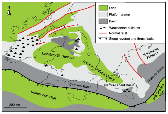
FIGURE 2. Schematic geological map of southern Belgium with location of the fossiliferous localities (modified from de Béthune, 1954). Abbreviations: D, Dréhance; F, Furfooz; G, Germany; HSMOTS, Haine-Sambre-Meuse Overturned Thrust Sheets; L, Grand Duchy of Luxemburg; N, The Netherlands; S, Sosoye.
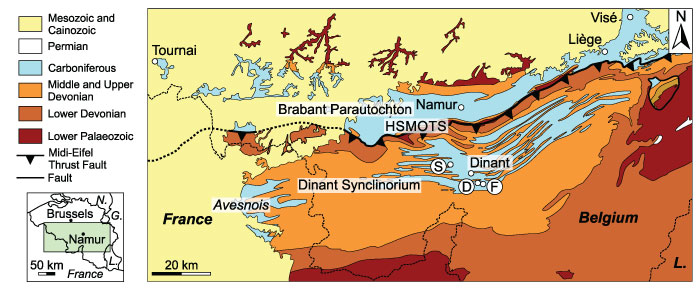
FIGURE 3. Late Tournaisian sedimentation areas in the Namur-Dinant Basin (not palinspastically restored; modified from Hance et al., 2001; Poty et al., 2006; Poty, 2016) with with location of the fossiliferous localities. Abbreviations: ASA, Avesnois sedimentation area; CSA, Condroz sedimentation area; D, Dréhance; DSA, Dinant sedimentation area; F, Furfooz; G, Germany; HSA, Hainaut sedimentation area; L, Grand Duchy of Luxemburg; N, The Netherlands; NSA, Namur sedimentation area; S, Sosoye; VSA, Visé-Maastricht sedimentation area.
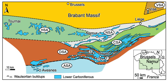
FIGURE 4. Distribution of the athyridides, orthotetides and strophomenides described and/or discussed herein within the essentially carbonate Tournaisian and Visean succession of southern Belgium (see text). Stratigraphy, lithostratigraphy (incompletely represented here for the Hainaut and Visé-Maastricht sedimentation areas; formations and members are in roman and italic letters, respectively), and biostratigraphy are adapted from Poty et al. (2002, 2006, 2014). The lenticular Vignobles Member is not represented here as it is rarely developed (it is comprised between the Vaulx Member of the Tournai Formation and the Antoing Formation). Note that the “Grande Brèche” is an informal stratigraphic unit resulting from the dissolution of evaporitic levels in the Grands Malades Formation; other brecciated horizons occur in the Lives Formation.
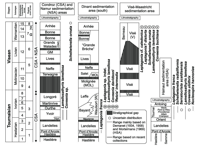
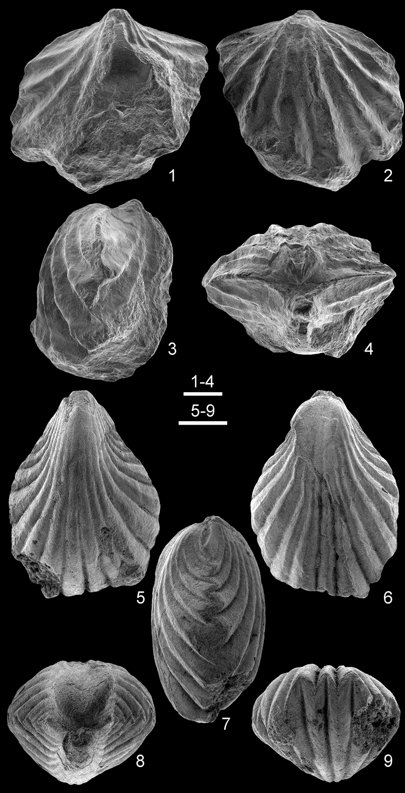
FIGURE 5. Leptagonia franca sp. nov. from the Tournai area, Tournai Formation (Tournaisian). 1-6, RBINS a5891 (holotype), articulated specimen, almost complete, in ventral (perpendicular to the surface of the alae), dorsal, lateral, posterior, anterior, and ventral (perpendicular to the disc) views. 7-12, RBINS a5892, articulated specimen, almost complete, in ventral (perpendicular to the surface of the alae), dorsal, lateral, posterior, anterior, and ventral (perpendicular to the disc) views. 13-18, RBINS a13099, articulated specimen (with an auloporid tabulate fixed to its right ventral flank), almost complete, in ventral (perpendicular to the surface of the alae), dorsal, lateral, posterior, anterior, and ventral (perpendicular to the disc) views. Scale bar equals 10 mm.
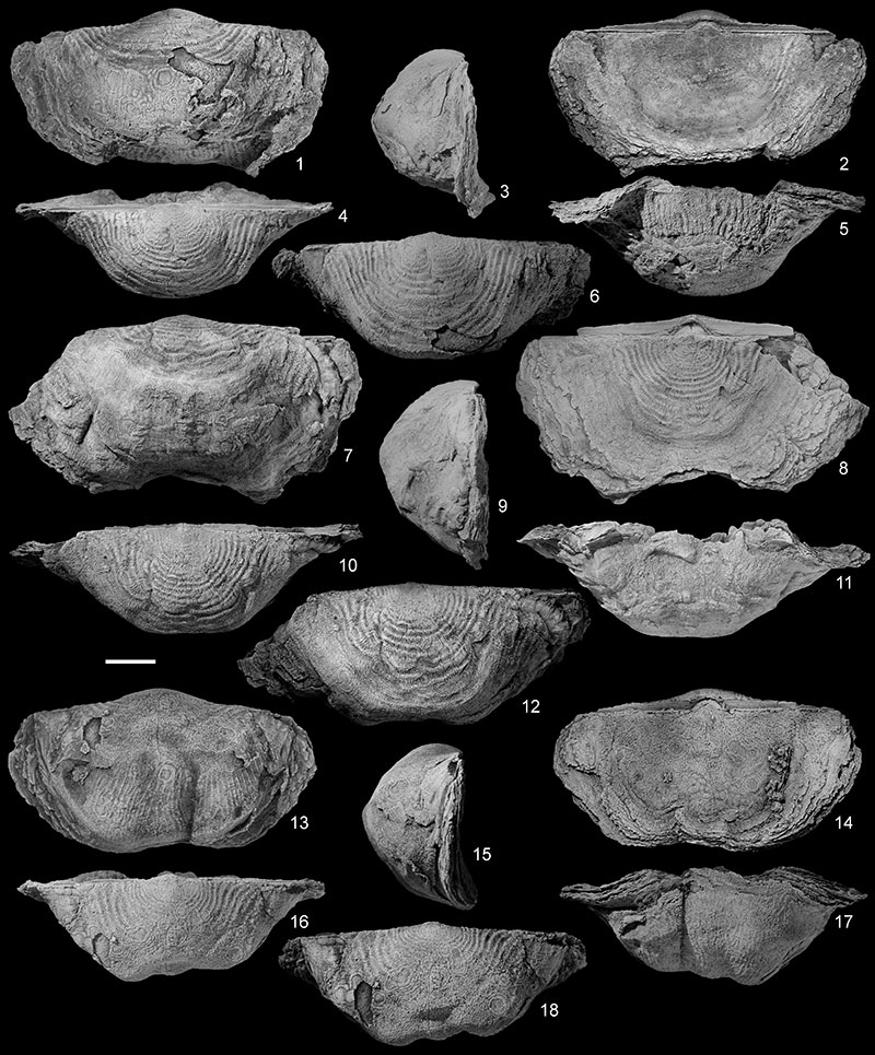
FIGURE 6. Leptagonia franca sp. nov. from the Tournai area, Tournai Formation (Tournaisian). 1-6, RBINS a5829, complete articulated specimen (with Petrocrania? ryckholtiana (de Koninck, 1843) attached to ventral and dorsal valves) in ventral (orientation as in Figure 5.1), ventral (perpendicular to the visceral disc), dorsal, lateral, posterior, and anterior views. 7-12, RBINS a5895, complete articulated specimen (with Petrocrania? ryckholtiana and microconchid attached to ventral valve), in ventral (orientation as in Figure 5.1), lateral, ventral (perpendicular to the visceral disc), dorsal, posterior and anterior views. 13-17, RBINS a13100, articulated specimen, almost complete, in ventral (orientation as in Figure 5.1), dorsal, lateral, posterior, and anterior views. 18-22, RBINS a13101, articulated specimen, almost complete, in ventral (orientation as in Figure 5.1), dorsal, lateral, posterior and anterior views. 23-27, RBINS a13102, articulated specimen, almost complete, in ventral (orientation as in Figure 5.1), dorsal, lateral, posterior, and anterior views. Scale bar equals 10 mm.
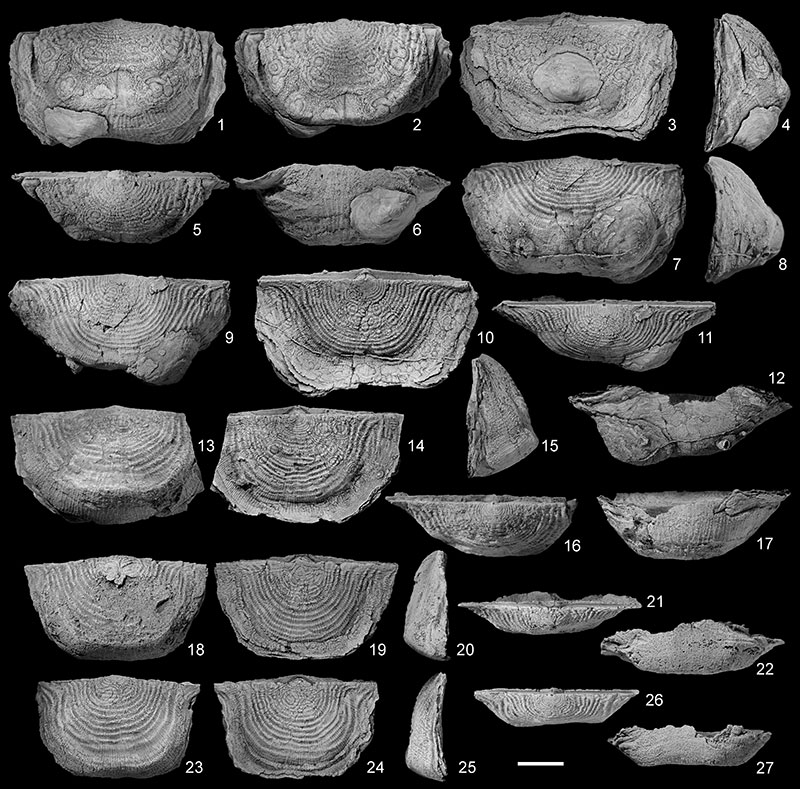
FIGURE 7. Leptagonia franca sp. nov. from the Tournai area, Tournai Formation (Tournaisian). 1-3, Detail of the small pseudodeltidium and apical foramen of three articulated specimens with the ventral valve on top: RBINS a5896 (1), RBINS a13103 (2), and RBINS a5895 (3). 4-5, RBINS a5902, incomplete ventral valve showing the posterior morphology. 6-7, RBINS a a13104, incomplete ventral valve interior showing the posterior morphology and posterior view notably displaying the prominent teeth. 8, RBINS a5903, incomplete ventral valve interior showing the posterior morphology. 9, RBINS a5899, almost complete dorsal valve interior in ventral view. 10-13, RBINS a5897, almost complete dorsal valve interior in posterior and ventral views, detail of the lateral vascular canals, and close-up of the cardinal process and the muscular field. Scale bars equal 2.5 mm (1-3), 5 mm (4-8, 12-13), 10 mm (9-11).
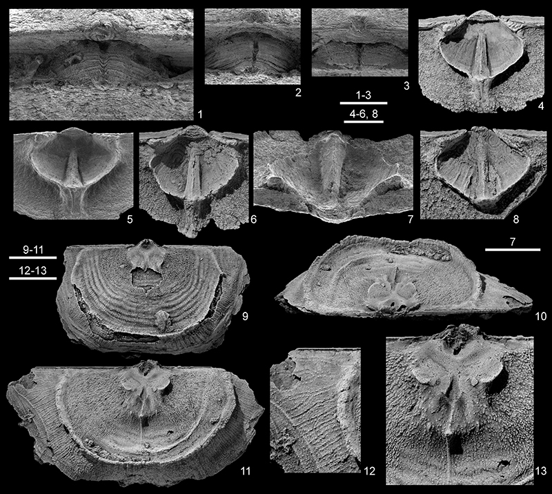
FIGURE 8. Scatter diagrams of Leptagonia franca sp. nov. N: number of specimens measured. 1, Relation between shell width and length. 2, Relation between shell width and shell length/shell width ratio. 3, Relation beween shell width and thickness. 4, Relation between shell width and shell thickness/shell witdth ratio. 5, Relation between unrolled ventral valve length and ratio between unrolled ventral trail length (Vtl) and unrolled ventral disc length (Vdl). 6, Relation between unrolled dorsal valve length and ratio between unrolled dorsal trail length (Dtl) and unrolled dorsal disc length (Ddl).
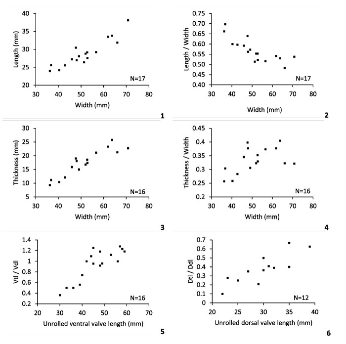
FIGURE 9. 1-4, Leptagonia sp. indet. from Sosoye, Waulsort Formation (Tournaisian, Ivorian), RBINS a11747, almost complete ventral valve in ventral, ventral (perpendicular to the disc), lateral and anterior views. 5-26, Leptagonia cf. caledonica Brand, 1972 from Visé, Visé Formation (Visean, Warnantian). 5-9, RBINS a5915, incomplete articulated specimen in ventral (perpendicular to the disc), dorsal, lateral, posterior, and anterior views. 10-14, RBINS a5914, incomplete articulated specimen in ventral (perpendicular to the disc), dorsal, lateral, posterior, and anterior views. 15-20, ULg.PA. 2016.12.25/1, articulated specimen, almost complete, in ventral, ventral (perpendicular to the disc), dorsal, lateral, posterior and anterior views. 21-26, ULg.PA.2016.12.25/2, articulated specimen, almost complete, in ventral, lateral, ventral (perpendicular to the disc), dorsal, posterior, and anterior views. Scale bar equals 10 mm.

FIGURE 10. Leptagonia cf. caledonica Brand, 1972 from Visé, Visé Formation (Visean, Warnantian). 1-5, 19-20, ULg.PA.2016.12.25/3, internal mould in ventral, dorsal, lateral, posterior and anterior views, and detail (SEM) of the muscular platform (latex cast). 6-10, 21-22, ULg.PA.2016.12.25/4, internal mould in ventral, dorsal, lateral, posterior, and anterior views, and detail (SEM) of the muscular platform (latex cast). 11-15, 23-24, ULg.PA.2016.12.25/5, internal mould in ventral, dorsal, lateral, posterior, and anterior views, and detail (SEM) of the muscular platform (latex cast). 16-18, ULg.PA.2016.12.25/6, distorted internal mould in dorsal view and close-up (SEM) of the muscular platform (latex cast). Scale bars equal 5 mm.
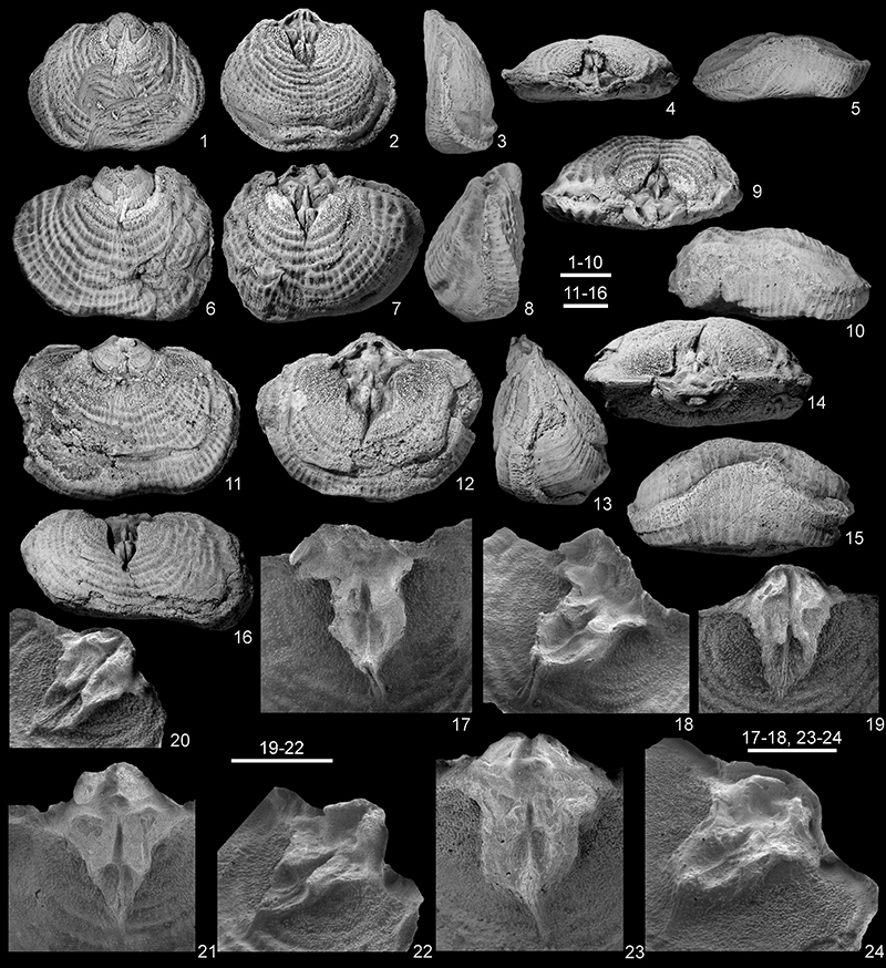
FIGURE 11. Leptagonia cf. caledonica Brand, 1972 from Visé, Visé Formation (Visean, Warnantian). 1-4, ULg.PA.2016.12.25/7, distorted, articulated internal mould in ventral view and detail of the latex mould of the muscular platform in three different views. Scale bar equals 5 mm (1), 2.5 mm (2-4).
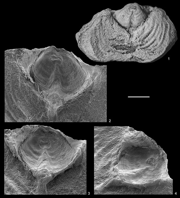
FIGURE 12. Schellwienella radialiformis Demanet, 1934 from the Tournai area, Tournai Formation (Tournaisian). 1-5, RBINS a5924, articulated specimen, almost complete, in ventral, dorsal, lateral, posterior, and anterior views. 6-10, RBINS a5928, articulated specimen, almost complete, in ventral, dorsal, lateral, posterior, and anterior views. 11-15, RBINS a5929, articulated specimen, almost complete, in ventral, dorsal, lateral, posterior and anterior views. 16-20, RBINS a5930 (lectotype), articulated specimen, almost complete, in ventral, dorsal, lateral, posterior, and anterior views. 21-25, RBINS a5926, almost complete articulated specimen (with a microconchid attached to the ventral valve, close to the anterior margin) in ventral, dorsal, lateral, posterior, and anterior views. Scale bars equal 10 mm.
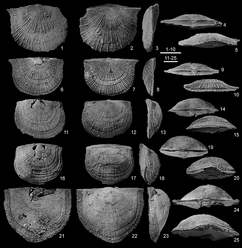
FIGURE 13. Schellwienella radialiformis Demanet, 1934 from the Tournai area, Tournai Formation (Tournaisian). 1-5, RBINS a5927, articulated specimen, almost complete, in ventral, dorsal, lateral, posterior and anterior views. 6-10, RBINS a13105, articulated specimen, almost complete, in ventral, dorsal, lateral, posterior, and anterior views. 11-12, RBINS a5924, detail (SEM) of the pseudodeltidium and of the koskinoid perforations penetrating in the umbonal part of the ventral valve. 13, RBINS a5928, detail of the microornament on dorsal valve (SEM). 14-17, RBINS a5925, incomplete ventral valve in external and internal views and detail (SEM) of the posterior internal and external morphology. 18-24, RBINS a13106, almost complete dorsal valve in dorsal, lateral, posterior, and internal views and detail (SEM) of the posterior morphology (cardinal process). Scale bars equal 10 mm (1-10, 14, 18-22), 1 mm (11), 0.25 mm (12), 0.5 mm (13), 2 mm (17, 23), 2.5 mm (24), 5 mm (15).
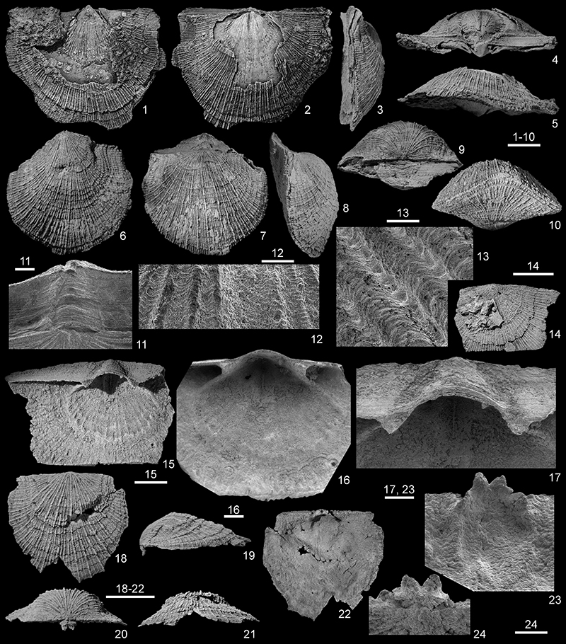
FIGURE 14. Schellwienella radialiformis Demanet, 1934 from the Tournai area, Tournai Formation (Tournaisian). 1-4, RBINS a13107, almost complete dorsal valve in dorsal, lateral and posterior views and detail of the chilidium and cardinal process lobes. 5-10, RBINS a5921, articulated internal mould with shelly remains in ventral, dorsal, lateral, posterior and anterior views and close-up of the ventral interarea (5). Scale bars equal 10 mm (1-2, 4-6), 1 mm (4, 5).
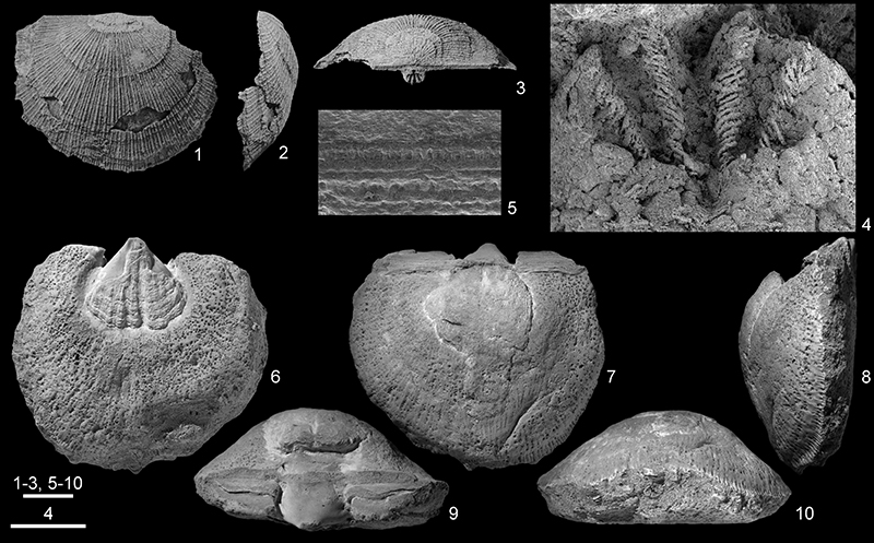
FIGURE 15. Schellwienella ornata Demanet, 1934 from Visé, Visé Formation (Visean, Warnantian). 1-8, RBINS a5922 (lectotype), articulated internal mould in ventral, enlarged ventral (showing the extent of the traces left by the dissolved dental plates), dorsal, lateral, posterior, and anterior views, and internal views of a latex cast (SEM) showing the internal morphology of the posterior part of the specimen (7-8). 9-14, RBINS a5923 (paralectotype), articulated specimen (partly sectioned, probably by Demanet) in ventral, dorsal, lateral, posterior, and anterior views, and close-up (SEM) of the parvicostellate ornamentation in the central part of the dorsal valve. Scale bars equal 10 mm (1, 3-6, 9-13), 5 mm (2), 2.5 mm (7-8), 1.25 mm (14).
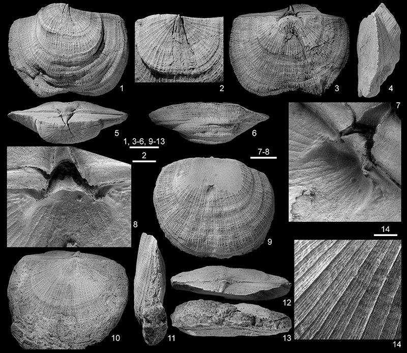
FIGURE 16. Schellwienella cylindrica (M’Coy, 1844), from the Visean (Brigantian) of Castle Espie (Comber, Co. Down, Ireland). 1-3, NMING:F6267 (lectotype), almost complete dorsal valve in dorsal, lateral, and posterior views. Scale bar equals 10 mm.
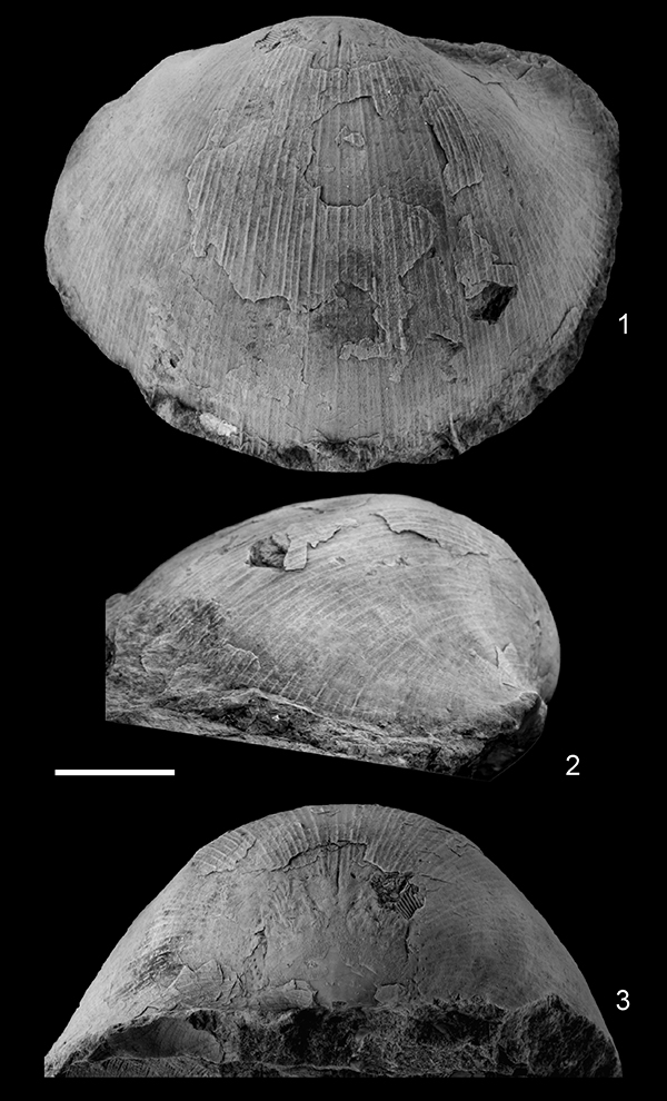
FIGURE 17. Serratocrista scaldisensis sp. nov. from the Tournai area, Tournai Formation (Tournaisian) (all SEM). 1-6, RBINS a13108, articulated specimen, almost complete, in ventral, dorsal, lateral, posterior, and anterior views, and close-up of the umbo showing the absence of dental plates. 7-11, RBINS a13109, articulated specimen in ventral, dorsal, lateral, posterior, and anterior views. 12-16, RBINS 13110 (holotype), articulated specimen in ventral, dorsal, lateral, posterior, and anterior views. 17-21, RBINS a13111, articulated specimen in ventral, dorsal, lateral, posterior, and anterior views. 22-25, RBINS a13112, almost complete ventral valve in ventral, lateral, posterior, and anterior views. Scale bars equal 1 mm (6), 5 mm (1-5, 7-25).
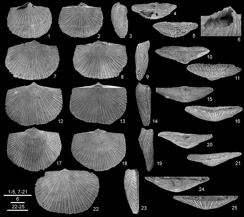
FIGURE 18. Serratocrista scaldisensis sp. nov. from the Tournai area, Tournai Formation (Tournaisian). 1, 6, RBINS a13110 (holotype), close-up of the strongly convex pseudodeltidium, and detail of the microornament (central part of the dorsal valve). 2-3, 19-21, RBINS a13111, detail of the strongly convex pseudodeltidium and of the cardinal process, and close-up of the koskinoid perforations developed in the ventral umbonal area and on the interarea. 4-5, RBINS a13113, incomplete articulated specimen in dorsal view and close-up of the posteriorly grooved lobes of the cardinal process. 7-13, RBINS a13114, dorsal valve in external, internal and posterior (10) views, detail of the chilidium (9), the cardinal process (11), and oblique views of the internal posterior region (12-13). 14-18, RBINS a13115, almost complete dorsal valve in external, internal and posterior views and oblique views of the internal posterior region. Scale bars equal 1 mm (1-3, 5-6, 9, 11, and 19-20), 2.5 mm (4), 5 mm (7-8, 10, 14-16, 12-13, and 17-18), 0.1 mm (21).
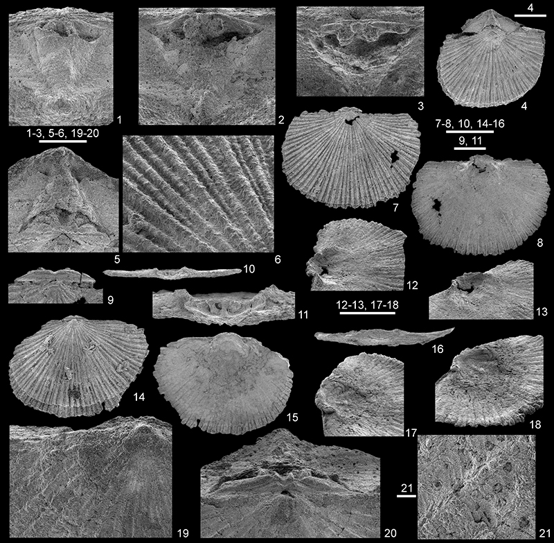
FIGURE 19. Serratocrista scaldisensis sp. nov. from the Tournai area, Tournai Formation (Tournaisian). 1-4, RBINS a13116, articulated specimen, incomplete, in dorsal view (1) with detail of the extropunctae arranged radially along the axis of the costellae (2, 3), and of the basal part of the pseudodeltidium and of the teeth (4). Scale bars equal 2 mm (1), 0.5 mm (2), 0.025 mm (3), 1 mm (4).
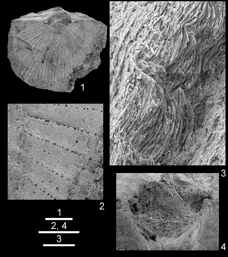
FIGURE 20. Lamellosathyris lamellosa (Léveillé, 1835) from the Tournai area, Tournai Formation (Tournaisian). 1-5, RBINS a13119, juvenile articulated specimen in ventral, dorsal, lateral, posterior and anterior views. 6-10, RBINS a13120, articulated specimen in ventral, dorsal, lateral, posterior, and anterior views. 11-15, RBINS a5468, articulated specimen in ventral, dorsal, lateral, posterior, and anterior views. 16-20, RBINS a13121, articulated specimen in ventral, dorsal, lateral, posterior, and anterior views. 21-25, RBINS a13122, articulated specimen (with two microconchids attached to the right dorsal flank) in ventral, dorsal, lateral, posterior and anterior views. 26-30, RBINS a13123, articulated specimen in ventral, dorsal, lateral, posterior, and anterior views. Scale bars equal 10 mm.
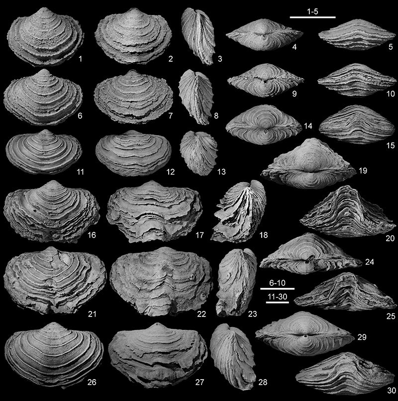
FIGURE 21. Lamellosathyris lamellosa (Léveillé, 1835) from the Tournai area, Tournai Formation (Tournaisian). 1-2, RBINS a13124, partial posterior view of an articulated specimen showing the foramen and detail of the lamellose ornamentation in the anterior part of the dorsal valve. 3-8, RBINS a13125, complete ventral valve (juvenile) in external and internal views and detail of the teeth and the dental plates. 9-11, RBINS a13126, complete ventral valve in external and internal views and detail of teeth supported by almost vertical dental plates. 12, RBINS a13127, incomplete internal mould of ventral valve. 13-18, ULg.PA.2016.12.25/8, complete dorsal valve (with two microconchids attached to its internal surface (arrows), close to the anterior margin) in external and internal view and detail of the cardinalia. 19-23, RBINS a5470, incomplete dorsal valve in internal view and detail of the cardinalia. Scale bars equal 5 mm (1-2, 3-4, 10, 11), 2 mm (5-8, 20-23), 2.5 mm (15-18), 10 mm (9, 12-14).
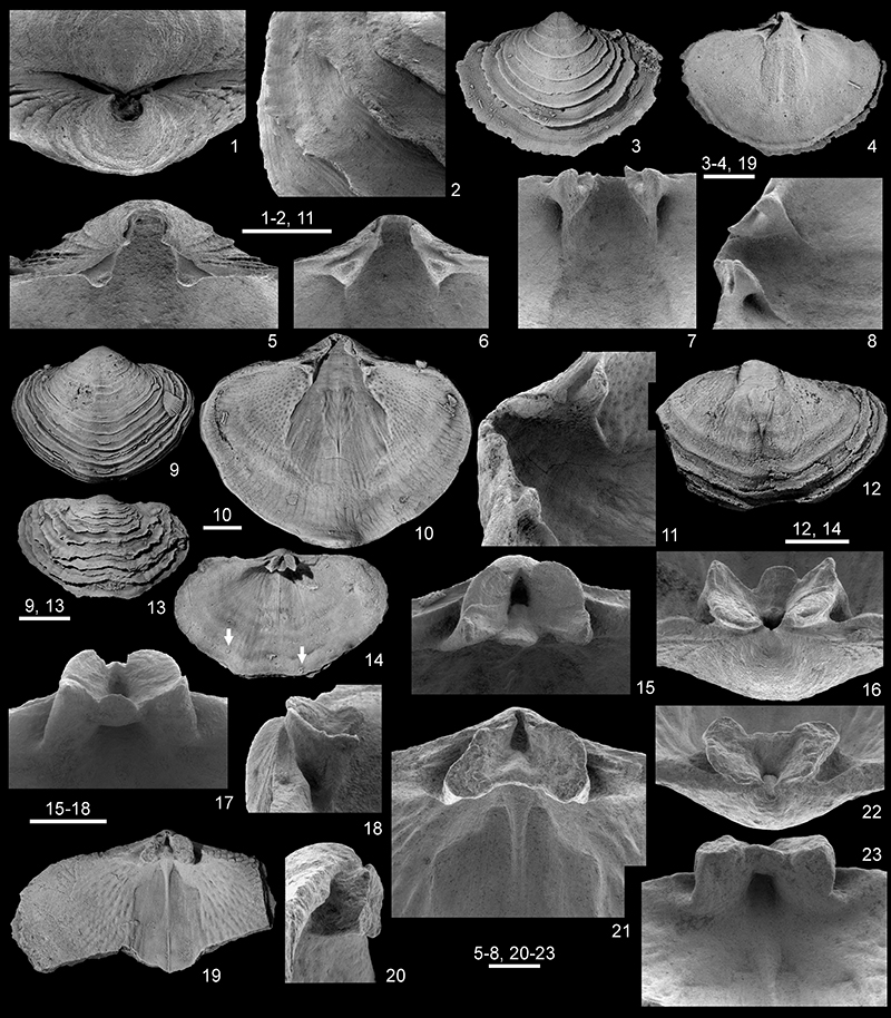
FIGURE 22. Lamellosathyris lamellosa (Léveillé, 1835) from the Tournai area, Tournai Formation (Tournaisian). 1-4, RBINS a13128, almost complete dorsal valve in external and internal views, with close-up of the cardinal plate in ventral and oblique lateral views. 5-6, RBINS a13129, incomplete ventral valve with partly preserved spiralia and detail of the jugal saddle and the primary lamella. 7-10, RBINS a13130, articulated specimen with top of the ventral valve removed and details of the crura, spiralia and fimbria. 11-13, ULg.PA.2016.12.25/9, articulated specimen with ventral valve partly removed in general view and details of the top of the jugal stem and the same in oblique view. Scale bars equal 10 mm (1, 7, 11), 5 mm (2, 5), 1 mm (3-4, 10), 2 mm (6, 9), 2.5 mm (8, 12-13).
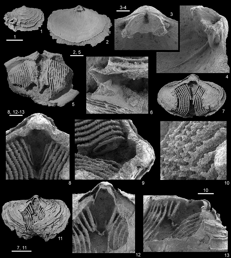
FIGURE 23.1-7, Lamellosathyris lamellosa (Lévéillé, 1835) from Visé, Visé Formation (Visean, Warnantian). 1-5, RBINS a5471, distorted specimen in ventral, dorsal, lateral, posterior and anterior views. 6-7, RBINS a13131, incomplete ventral valve in ventral view and anterior views. 8-17, Athyris vittata de Koninck, 1887 (here placed in synonymy with Lamellosathyris lamellosa (Léveillé, 1835)) from Furfooz, Waulsort Formation (Tournaisian, Ivorian). 8-12, RBINS a5472 (lectotype) distorted specimen in ventral, lateral, dorsal, posterior, and anterior views. 13-17, RBINS a13132 (paralectotype), distorted and incomplete specimen in ventral, dorsal, posterodorsal, posterior, and lateral views. Scale bars equal 10 mm.
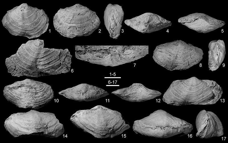
FIGURE 24. Scatter diagrams of Lamellosathyris lamellosa (Léveillé, 1835). N: number of specimens measured, r2: coefficient of linear regression. 1, Relation between shell width and length. 2, Relation between shell width and shell width/shell length ratio. 3, Relation beween shell width and thickness. 4, Relation between shell width and shell thickness/shell width ratio.
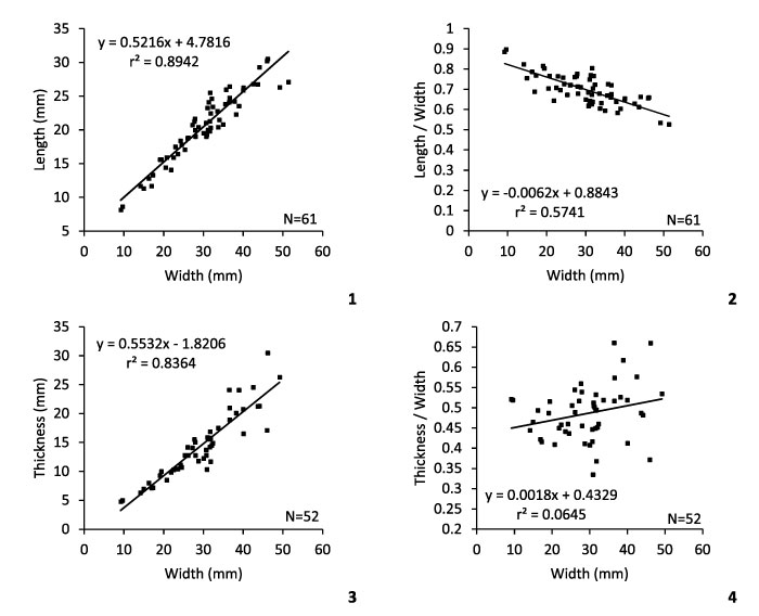
FIGURE 25. Nucleospira hannoniae nom. nov., from the Tournai area, Tournai Formation (Tournaisian). 1-5, RBINS a13134, articulated specimen, almost complete, in ventral, dorsal, lateral, posterior, and anterior views. 6-10, RBINS a5466 (paralectotype), articulated specimen, almost complete, in ventral, dorsal, lateral, posterior, and anterior views. 11-15, RBINS a5465 (lectotype), articulated specimen in ventral, dorsal, lateral, posterior, and anterior views. 16-21, RBINS a13135, articulated specimen in ventral, dorsal, lateral, posterior, and anterior views, and close-up of the interarea, Lemay quarry, Vaulx Member. 22-27, RBINS a13136, articulated specimen in ventral, dorsal, lateral, posterior, and anterior views, and close-up of the solid spines (right flank of the dorsal valve). Scale bars equal 2 mm (1-20, 22-26), 1 mm (21), 0.2 mm (21, 27).
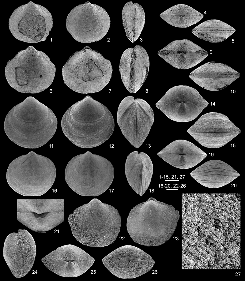
FIGURE 26. Nucleospira hannoniae nom. nov., from the Tournai area, Tournai Formation (Tournaisian). 1-2, RBINS a13137, distorted and incomplete specimen in dorsal view showing two whorls of the spiralium and detail of one of the cyrtomatodont teeth. 3-4, RBINS a13138, distorted articulated specimen in dorsal view displaying a circular drill hole and detail of the latter. 5, RBINS a13139, distorted articulated specimen with a circular drill hole in ventral view. Scale bars equal 2 mm (1, 3, 5), 0.5 mm (2, 4).
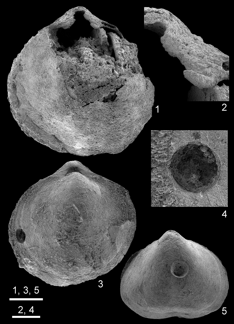
FIGURE 27. Scatter diagrams of Nucleospira hannoniae nom. nov. N: number of specimens measured. 1, Relation between shell width and length. 2, Relation between width and shell length/shell width ratio. 3, Relation beween shell width and thickness. 4, Relation between shell width and shell thickness/shell width ratio.
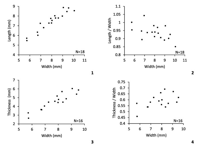
FIGURE 28. Coveenia ulothrix (de Koninck, 1843) from the Tournai area, Tournai Formation (Tournaisian). 1-5, 16-18, ULg.PA.2016.12.25/10, articulated specimen in ventral, dorsal, lateral, posterior and anterior views, and detail (SEM) of the foramen and the symphytium, and of circular drill hole developed close to the dorsal anterior margin. 6-10, RBINS a13140, articulated specimen in ventral, dorsal, lateral, posterior, and anterior views, Dutoit quarry, Pont-à-Rieu. 11-15, RBINS a13141, articulated specimen, almost complete, in ventral, dorsal, lateral, posterior and anterior views, Dutoit quarry, Pont-à-Rieu. Scale bar equals 10 mm (1-15), 1 mm (16-18).
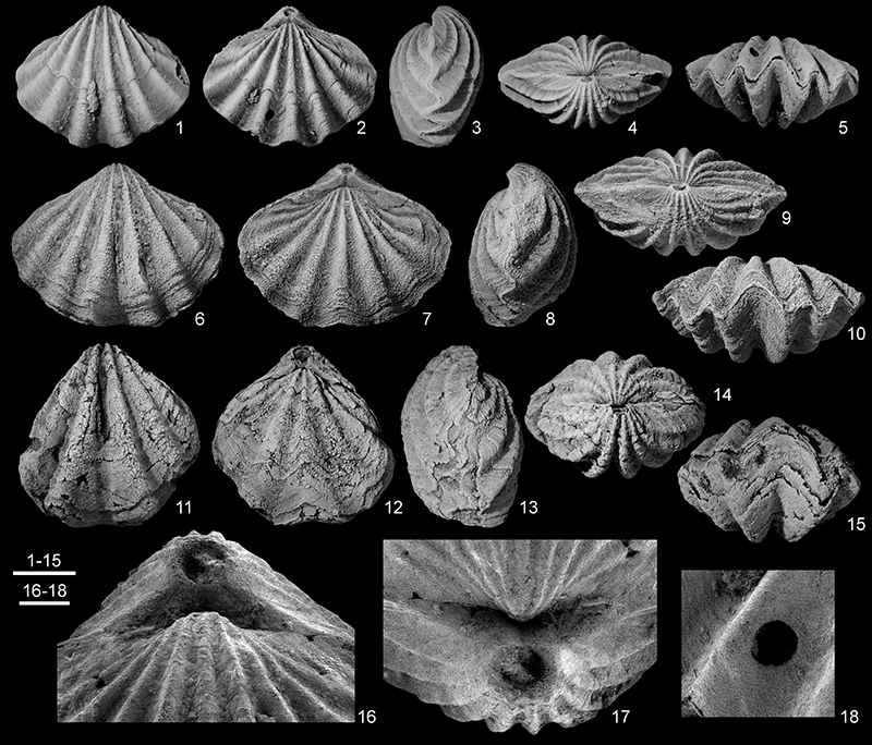
FIGURE 29. 1-4, Coveenia davidsoni (de Koninck, 1887), RBINS a5508 (holotype), articulated specimen in ventral, dorsal, lateral and posterior views, from Dréhance, Waulsort Formation (Tournaisian, Ivorian). 5-9, ‘Retzia’ intermedia de Koninck, 1887 (here considered as a rhynchonellide species), RBINS a5510 (lectotype), articulated specimen in ventral, dorsal, lateral, posterior and anterior views, from Furfooz, Waulsort Formation (Tournaisian, Ivorian). Scale bars equal 2 mm.