FIGURE 1. Elements histologically sampled for this analysis. 1, Digital reconstruction of Apatosaurus louisae (by K. Stevens) with sampled elements highlighted in red. Approximate location of sampling in dorsal ribs (2; from Gilmore, 1936), neural spines (3; from Hatcher, 1901), and femora (4; from Gilmore, 1936).
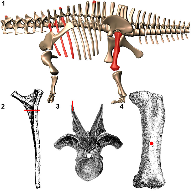
FIGURE 2. Schematic of coring bit method used in this analysis. A two part Bosch ™ core bit (1) with a coin placed on the bone surface prior to coring (2), the core bit separated post coring, with a bolt inserted to push on the coin (3) to extract the bone core (4).
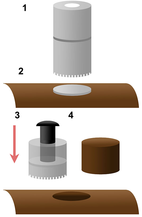
FIGURE 3. Ontogenetic development of neural spine bifurcation in Morrison Formation sauropods. Apatosaurinae - represented by the immature individuals CM 555 (Apatosaurus excelsus or Brontosaurus excelsus) and CM 3390 (Apatosaurus sp.) compared to the mature 3018. Diplodocus - represented by the immature MOR 592 (Diplodocus sp.) compared to the mature CM 84 (Diplodocus carnegii). Camarasaurus - represented by the immature CM 11338 (Camarasaurus lentus) compared to the mature AMNH 5761 (Camarasaurus supremus). Barosaurus - represented by the immature AMNH 7535 compared to the mature AMNH 6341. Images not to scale. Scale bars equal 10 cm.
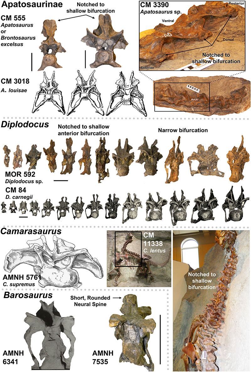
FIGURE 4. Presence of the Postparietal Aperture within Diplodocidea (indicated by red arrows). 1-4, Diplodocid specimens with the Postparietal Aperture: 1, Suuwassea (ANS 21122); 2, Galeamopus (SMA 0011); 3, Apatosaurus sp. (BYU 17096) (Balanoff, et al., 2010); 4, Kaatedocus (SMA 0004). Note 1 and 3 are dorsal views while 2 and 4 are posterior views. 5-6, Diplodocid skulls illustrating the presence of the Postparietal Aperture in immature individuals (5-image courtesy of the Science Museum of Minnesota), and its absence in mature individuals (6). For 5, from left to right: Kaatedocus (SMA 0004), Diplodocus sp. (SMM P. 84.15.3), Apatosaurus sp. (MOR 700), Galeamopus (SMA 0011; this skull is partially damaged, so the morphology of the foramen may be distorted), Suuwassea (ANS 21122), and Diplodocus sp. (MOR 592). For 6, note that the Diplodocus and Apatosaurus skulls are stylized renderings based on multiple specimens (from Whitlock [2011b]; Diplodocus sp. to scale of USNM 2672, and Apatosaurus louisae to CM 11162). Skulls in 5 and 6 to scale.
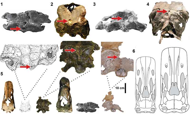
FIGURE 5. Macroscopic pneumatic architecture in diplodocid cervical vertebrae. 1, diplodocid indeterminate MOR 714 7-22-3-53; 2, Diplodocus sp. MOR 790 8-10-96-204; 3, D. carnegii CM 84 (from Hatcher, 1901). Increasing pneumatic complexity from 1 to 3. pfs = Pneumatic Fossa, acl = Accessory Lamina, pfm = Pneumatic Foramen (Wedel, 2003). Not to scale. Scale bar equals 10 cm.
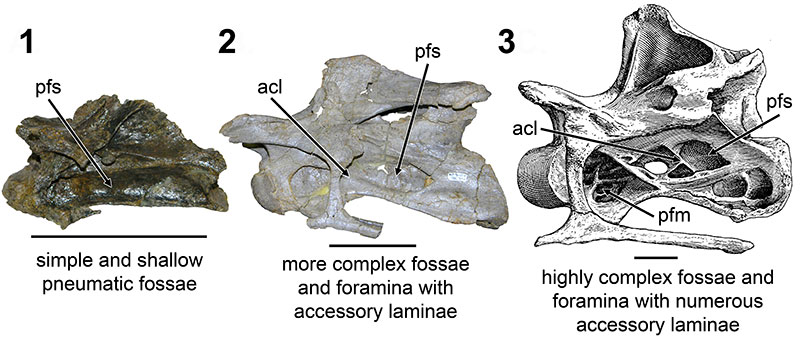
FIGURE 6. CT scans of diplodocid anterior cervical vertebrae. 1, diplodocid indeterminate MOR 714 7-22-3-53; 2, Diplodocus sp. MOR 790 8-10-96-204. Colored planes in 1 and 2 correspond to CT scan sections for each respective vertebrae. pfs = Pneumatic Fossa, cmr = Camera (Wedel, 2003). Red = frontal plane through the centrum, Blue = transverse plane through the anterior portion of the pfs, Green = transverse plane through the posterior portion of the pfs. Ant = anterior; Pos = posterior.
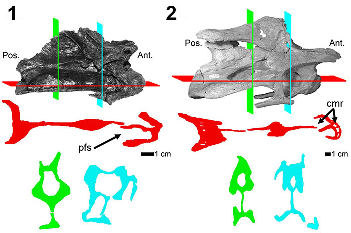
FIGURE 7. CT scans of diplodocid anterior dorsal vertebrae. 1, Diplodocus sp. MOR 790 8-21-95-238; 2, Diplodocus sp. MOR 592 8-22-90-15. Colored planes in 1 and 2 correspond to CT scan sections for each respective vertebrae. pfs = Pneumatic Fossa, cmr = Camera, cml = Camella (Wedel, 2003). Red = frontal plane through the centrum, Blue = transverse plane through the pfs.
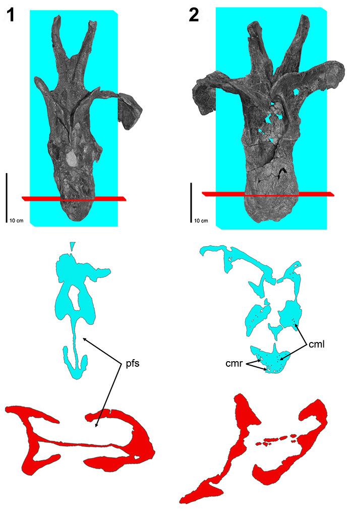
FIGURE 8. CT scans of diplodocid posterior dorsal vertebrae. 1, Diplodocus sp. MOR 790 7-8-95-17; 2, Diplodocus sp. MOR 592 8-22-90-77; 3, Apatosaurus sp. MOR 957 6-29-92#1. Colored planes in 1, 2, and 3 correspond to CT scan sections for each respective vertebrae. pfs = Pneumatic Fossa, cmr = Camera, cml = Camella (Wedel, 2003). Red = frontal plane through the centrum, Blue = transverse plane through the pfs.
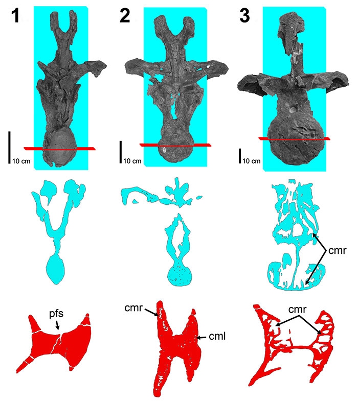
FIGURE 9. Coronal histologic sections of diplodocid posterior cervical and anterior dorsal vertebrae neural spines. 1, Posterior cervical Diplodocus sp. MOR 790 (un-numbered 1); 2, Anterior dorsal Diplodocus sp. MOR 790 8-21-95-238; 3, Posterior cervical Diplodocus sp. MOR 592 8-24-90-91; 4, Anterior dorsal Diplodocus sp. MOR 592 8-22-90-15. R.L. (right lateral), L.L (left lateral), M. (medial), L (lateral). Not to scale. Scale bar equals 1 cm.
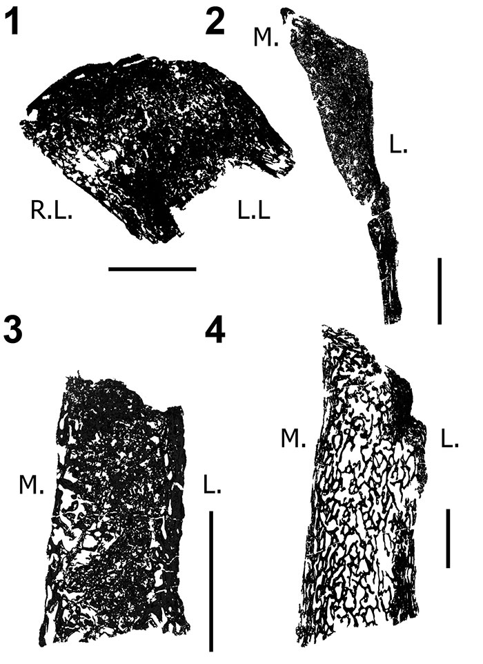
FIGURE 10. Transverse histologic sections of the H-MOS Stage 2 Diplodocus sp. 1, MOR 790 7-24-96-95 (10x) and 2, MOR 790 7-27-8-96 dorsal ribs (4x). 1, MOR 790 7-24-96-95 records a minimum of two annuli (blue lines). Red insert box highlights bone microstructure differences between the anterior intercostal ridge and lateral margins with longitudinal vascular canals. Green insert box highlights a sample of the segments that comprise one of the growth markers (an annulus, highlighted in blue). 2, MOR 790 7-27-8-96 records a minimum of six LAGs (blue lines; note some of these LAGs do extend past the demarcated blue lines). Red insert box highlights the microstructure and shows the vascularity patterns while the white arrows denote two of the LAGs present.
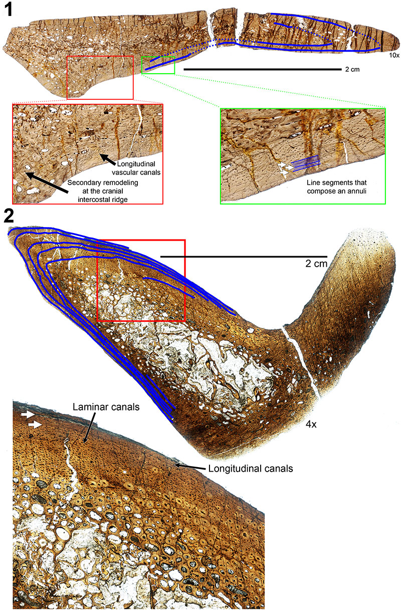
FIGURE 11. Transverse histologic section of the H-MOS Stage 3 Diplodocus sp. MOR 592 dorsal rib at 4x. MOR 592 records a minimum of eight LAGs (blue lines). Red insert box highlights the organization of longitudinal vascular canals in radial rows (alternating rows highlighted in blue). Green insert box highlights the vascularity patterns with white arrows denoting four of the LAGs present.
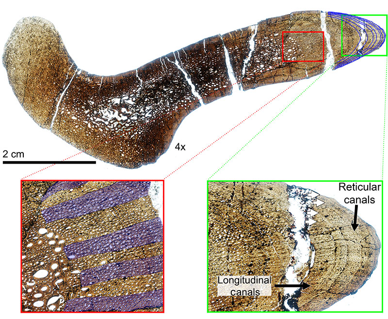
FIGURE 12. Transverse histologic section of the H-MOS Stage 4 Diplodocus CM 94 (Diplodocus carnegii paratype) dorsal rib at 4x. CM 94 records a minimum of 24 LAGs (blue lines). Red insert box highlights the episodic zones of large (LDC) and small (SDC) diameter longitudinal vascular canals with white arrows denoting some of the LAGs present. Green insert box highlights the outermost cortex which records the presence of an EFS. White arrows denote LAGS present, with the EFS bracketed.
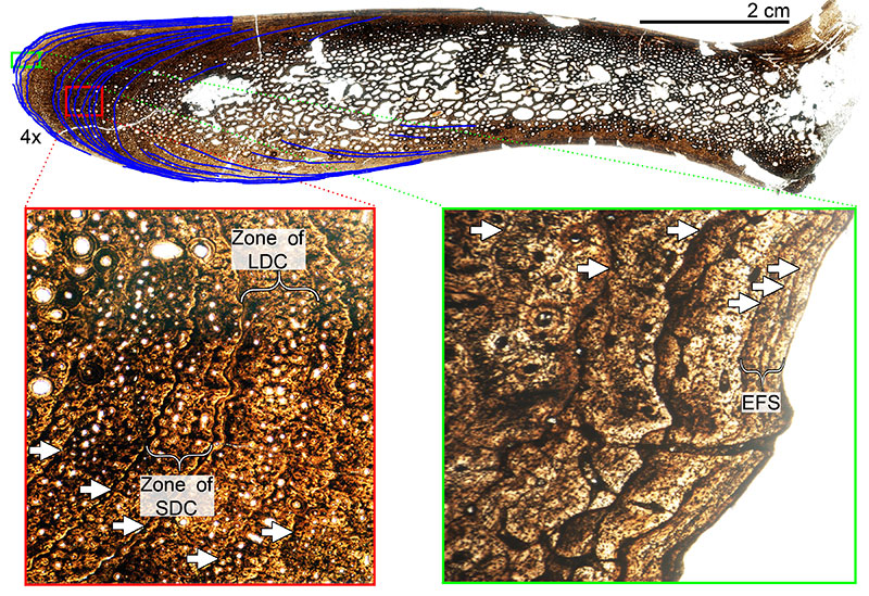
FIGURE 13. Transverse histologic section of the H-MOS Stage 2 Diplodocus sp. MOR 790 7-23-95-122 femur at 4x. MOR 790 7-23-95-122 is HOS 7 out of 13. No LAGs are present.
FIGURE 14. Transverse histologic section of the H-MOS Stage 3 Diplodocus sp. MOR 592-35 femur at 4x. White arrow marks one observable LAG. MOR 592 is HOS 9 out of 13.
FIGURE 15. Selection of the diplodocid femoral dataset used throughout this ontogenetic analysis. Top row diplodocines, from left to right: CM 33991 (Diplodocus longus), CM 21788 (D. longus), CM 30762 (D. longus), MOR 790 7-5-97-7 (Diplodocus sp.), MOR 790 7-23-95-122 (Diplodocus sp.), MOR 592-35 (Diplodocus sp.), CM 21752 (Diplodocus sp.), SMA 0013 (Diplodocus sp.), CM 84 (D. carnegii); Bottom row apatosaurines, from left to right: AMNH 613 (Apatosaurus sp.), OMNH 1279 (Apatosaurus sp.), AMNH 606 (Apatosaurus sp.), MWC 5439 (Apatosaurus sp.), CM 21784 (A. louisae), CM 33997 (A. louisae), MOR 700 7-24-91-31 (Apatosaurus sp.), AMNH 353 (Apatosaurus sp.), SMA 0014 (Apatosaurus sp.), CM 85 (Apatosaurus sp.), MOR 857 7-16-92-30 (Apatosaurus sp.), MWC “Moffit Co. Apato.” (Apatosaurus sp.). Scale bar equals 1 m. Note the colored line on CM 84 and MWC “Moffit Co. Apato.” illustrate how femoral positions and trends were examined. Colored lines to the far right of each row indicate the general allometric changes of each femur scaled to the same length (Black line = femoral length). Diplodocus row: Yellow lines = MOR 790 7-5-97-7, Blue lines = MOR 592-35, Red lines = CM 84. Apatosaurus row: Green lines = AMNH 613, Yellow lines = CM 33997, Blue lines = MOR 700 7-24-91-31, Red lines = MWC “Moffit Co. Apato.”
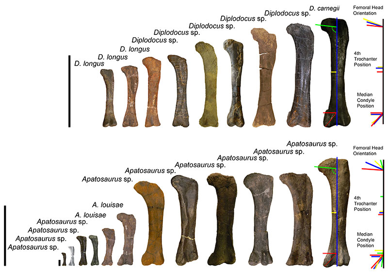
FIGURE 16. Calculated body masses for the Morrison diplodocids Apatosaurus (1) and Diplodocus (2) using the formula of Mazzetta et al. (2004) (log Body Mass = 2.955 x log Femur Circumference − 4.166); 3, Dorsal rib LAG count vs. femur circumference for Diplodocus; 4, Femur length vs. femur circumference for Diplodocus; 5, Dorsal rib LAG count vs. body mass for Diplodocus with posterior cervical vertebrae marking each size range to illustrate the correlation between mass and spine morphology. Data for all diplodocid specimens can be found in Appendix 3.
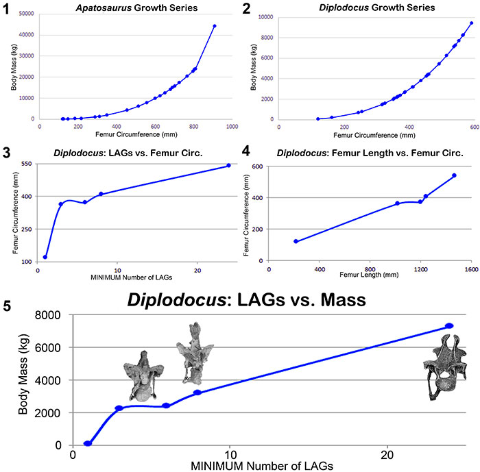
FIGURE 17. Histologic and morphologic commonalities of Diplodocus sp. (MOR 592) and Suuwassea emilieae (ANS 21122) which demonstrate immature maturational states for both animals (H-MOS Stage 3). Histologic section of Suuwassea tibia from Hedrick et al. (2014). The Suuwassea tibia histologic section is from Acta Palaeontologica Polonica, articles are distributed under the terms of the Creative Commons Attribution License (CC BY), which permits unrestricted use, distribution, and reproduction in any medium, provided the original author and source are credited. For licence details please see http://creativecommons.org/licenses/by/4.0/.
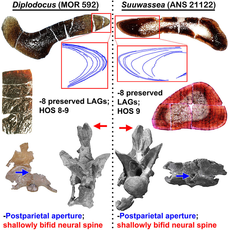
FIGURE 18. The degrees and variation of neurocentral fusion in diplodocid cervical vertebrae indicate that fusion is not uniformly indicative of maturity. 1, The posteriorly unfused H-MOS Stage 2 Diplodocus sp. MOR 790 8-10-96-204; 2, The completely fused H-MOS Stage 1 diplodocid indeterminate MOR 714 7-22-3-53; 3, The completely unfused H-MOS Stage 3 Apatosaurus excelsus (or Brontosaurus excelsus) CM 555; 4, The anteriorly unfused H-MOS Stage 3 Diplodocus sp. MOR 592. Red inset boxes highlight the visible sutures (in blue). Not to scale. Scale bar equals 10 cm.
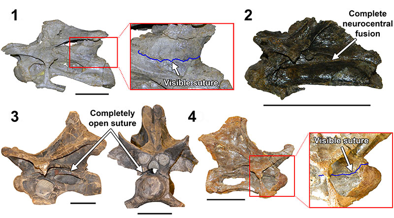
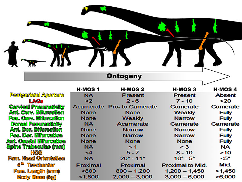
FIGURE 19. An ontogenetic trajectory in consideration of morphologic and histologic attributes for the Morrison diplodocid Diplodocus. Note diplodocids, and possibly all other sauropods, did not skeletally develop along an isometric trajectory. Human scale bar is Dante Alighieri from Domenico di Michelino’s “La commedia illumina Firenze”, depicting Dante as 1.63 m tall. Modified silhouette of Diplodocus carnegii CM 84 by S. Hartman and available via PhyloPic under the Creative Commons Attribution-ShareAlike 3.0 Unported License.