FIGURE 1. Geology map of western Newfoundland and southern Labrador showing main geological terranes and outlining the location of the study areas discussed in this article and illustrated in more detailed maps in Figure 2, Figure 4, Figure 7.
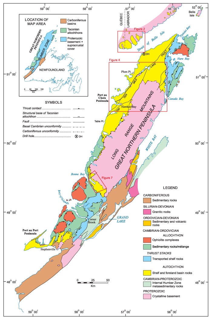
FIGURE 2. Geology map of the south coast of Labrador showing localities in the Forteau Formation, Labrador Group that were studied and yielded a rich brachiopod fauna (map based on Bostock et al., 1983 and Gower, 2010). LLQ - L’Anse-au-Loup quarry, DCQ - Diablo Cove quarry
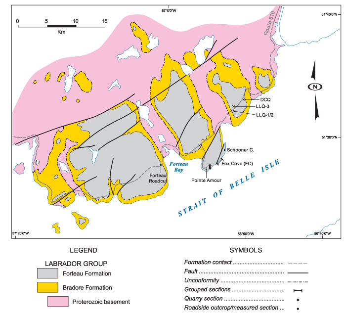
FIGURE 3. Sections measured in the Forteau Formation of south Labrador coast. FC1 (open shelf facies of James and Kobluk, 1978) and FC2a (Patch reef facies of James and Kobluk, 1978; bioherm of Debrenne and James, 1981) are adjacent to each other on the foreshore east of Fox Cove; FC2 is divided into a lower (FC2a) and upper part (FC2b) separated by a vertical fault that cuts out most of the Middle shale; FC3 is measured to the southwest of FC2 and is a composite of three short sections that are correlated over more than 50 m on the base of the Upper limestone (upper biostrome of Debrenne and James, 1981). Legend is applicable to all stratigraphic figures (Figure 5-Figure 6, Figure 8-Figure 9).
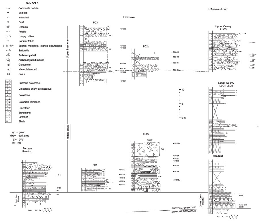
FIGURE 4. Geology map of central part of the Great Northern Peninsula showing faunally productive localities studied at Mount St. Margaret, along Route 432 north of the Long Range Proterozoic massif, and along the Big East River resource road at the south end of Highlands of St. John. MSM - Mount St. Margaret, ECP - East Castor Pond road, R432-5 etc - Route 432; BER-5 etc - Big East River resource road.
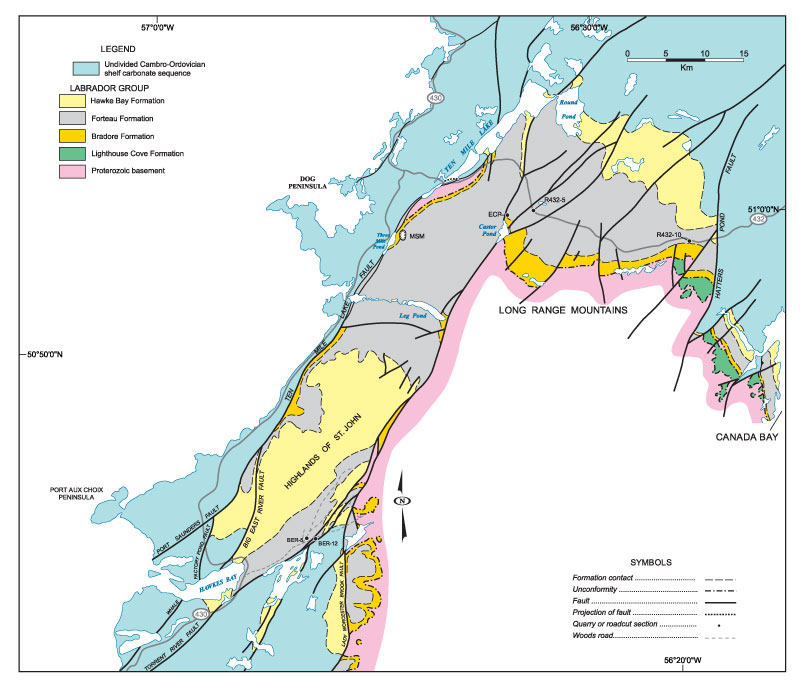
FIGURE 5. Stratigraphic section through the incomplete Forteau Formation exposed at Mount St. Margaret quarry (MSM). The archaeocyathid interval at top of section is partly based on scattered roadside outcrops above the quarry. The succession is host to brachiopod assemblage 1 and 2.

FIGURE 6. Measured sections displaying samples collected from rocks of the upper heterolithic interval of the Upper limestone, Forteau Formation on Route 432 east of Ten Mile Lake (R432-5) and Big East River resource road northeast of Hawkes Bay (BER-5) (see Figure 4). Both sections host brachiopod assemblage 2. The succession at R432-5 comprises skeletal carbonate and shale overlain by calcareous siltstone and white quartz arenite. The incomplete succession at BER-5 is lithologically similar to the lower carbonate interval of R432-5 and may tentatively suggest they are distant correlatives of the same parasequence. The brachiopod fauna hosted by lithofacies of small-scale transgression are inherited from the restricted fauna associated with the long-term regressive phase of the Forteau shelf sedimentation.
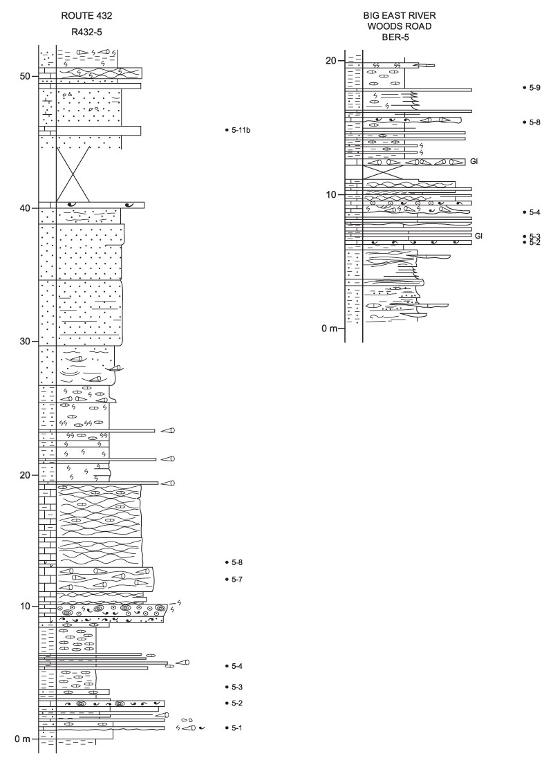
FIGURE 7. Geology map of the Gros Morne area showing location of sections in the Forteau Formation (based on Knight, 2013, figure 3). Samples GM07 -1 etc. were collected randomly in 2007 and are linked to localities GM11-1 etc. that were mapped, sectioned, and sampled in 2011 (see Figure 8-Figure 9).
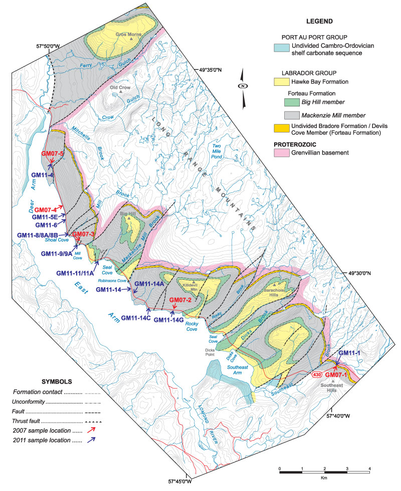
FIGURE 8. Stratigraphic sections through the Forteau Formation in Gros Morne National Park (based on Knight, 2013, figure 4) showing the correlation of limestone intervals and samples that yielded assemblage 3 brachiopod fauna. DAL - Deer Arm Limestone. DCL – Devils Cove limestone.
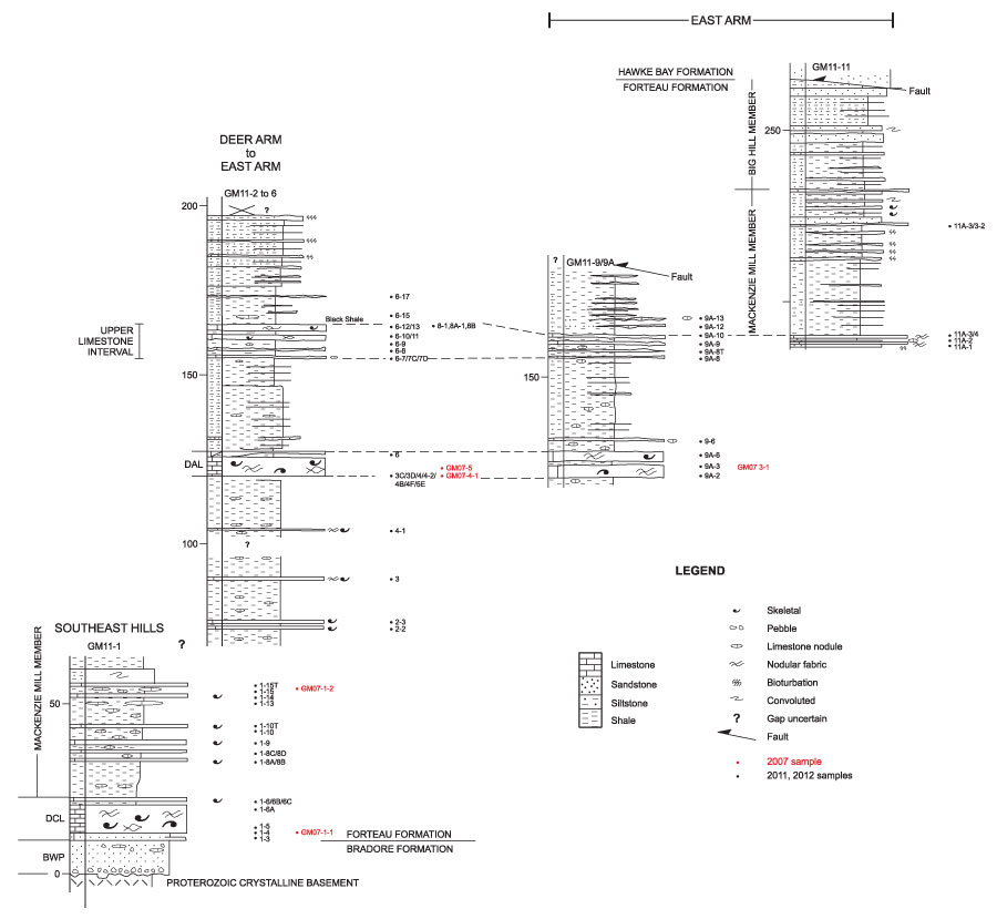
FIGURE 9. Sections for localities GM11-14, 14A and 14G at the eastern end of East Arm (see Figure 7) showing location of samples that yielded assemblage 3 brachiopod fauna. The folded and faulted succession there is difficult to correlate with any certainty to the stratigraphy of Figure 8. Nonetheless, two limestone intervals in GM11-14 are tentatively correlated with the Deer Arm (DAL) and Upper limestone interval of Figure 8. The offset of sections (GM11-14 and 14A) reflects the stratigraphic uncertainty of the structurally displaced succession although GM11-14G may correlate with the upper GM11-14A section.
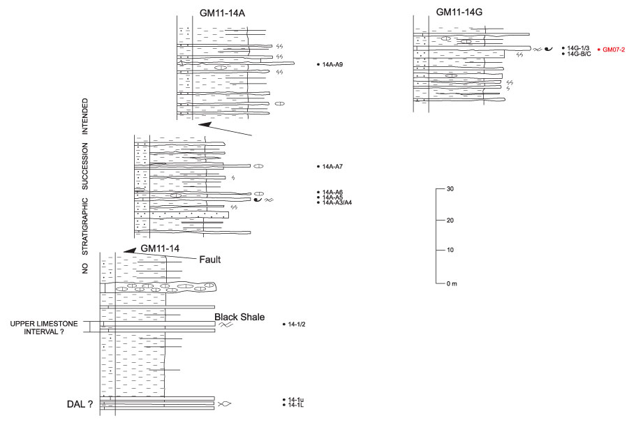
FIGURE 10. Distribution of brachiopod taxa recovered from sections measured in the Forteau Formation of the south Labrador coast. Shaded area indicates known stratigraphic range of individual taxa. Correlation of adjacent sections is approximate only. For more exact correlation of sections and samples see Figure 3.
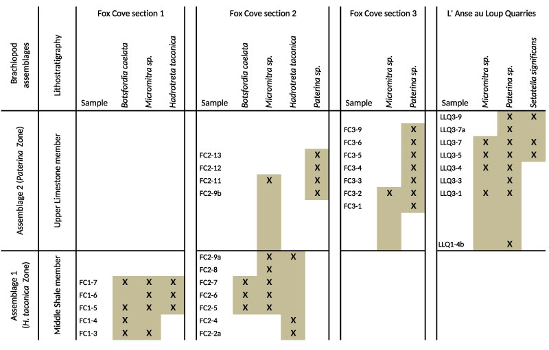
FIGURE 11. Distribution of brachiopod taxa recovered from sections measured in the Forteau Formation of the Great Northern Peninsula of western Newfoundland. Shaded area indicates known stratigraphic range of individual taxa. Correlation of adjacent sections is approximate only. For more exact correlation of sections and samples see Figure 6-Figure 7.
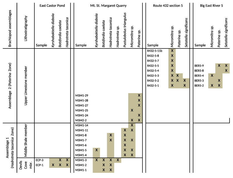
FIGURE 12. Distribution of brachiopod taxa recovered from sections measured in the Forteau Formation of the Gros Morne National Park of western Newfoundland. Shaded area indicates known stratigraphic range of individual taxa. Correlation of adjacent sections is approximate only. For more exact correlation of sections and samples see Figure 9-Figure 10.
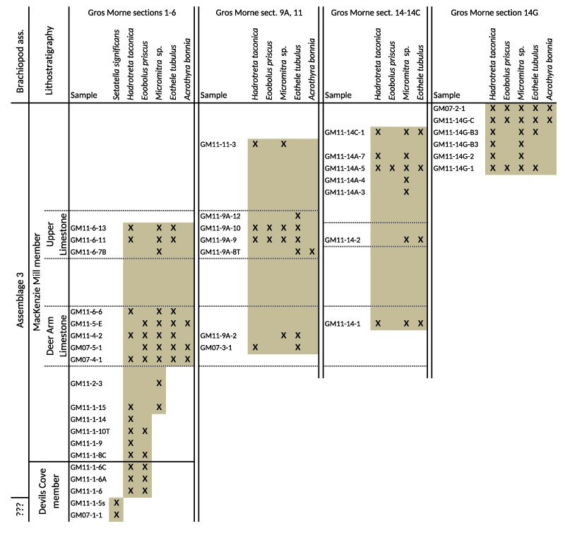
FIGURE 13. Environmental preferences of studied brachiopod taxa distributed along a simplified cross section of the eastern Laurentian shelf during the early Cambrian with the relative position of the three general study areas indicated. A. Representing the environment during the deepening phase of the transgressive systems tract (Middle shale of Labrador and the Great Northern Peninsula and Devils Cove member and lower part of the Mackenzie Mill member in Gros Morne area). B. Representing the environment during the regressive high stand system tract with the development of a prograding shelf (Upper limestone of Labrador and the Great Northern Peninsula and the upper part of the Mackenzie Mill member in Gros Morne area).
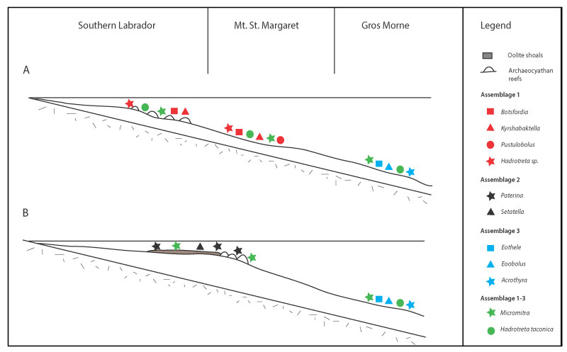
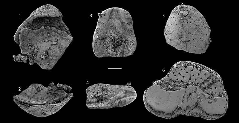
FIGURE 14. Kyrshabaktella diabola n. sp. from the Forteau Formation of southern Labrador and western Newfoundland. 1-2, Ventral valve NFM F-2516 from Devils Cove member, East Castor Pond, sample ECP-01; 1, external view; 2, detail of larval shell and resorbed pedicle groove. 3-4, Ventral valve NFM F-2517 (Holotype) from Devils Cove member, Mount St. Margaret, sample MSM-03; 3, internal view; 4, oblique posterior view showing pedicle groove. 5, Ventral valve exterior NFM F-2518 from Devils Cove member, Mount St. Margaret, sample ICS1520. 6-8, Dorsal valve NFM F-2519 from Lower Shale member, Mount St. Margaret, sample MSM-5; 6, internal view; 7, oblique internal view; 8, detail of internal surface showing vascula lateralia and median tongue with muscle scars. 9-10, Dorsal valve NFM F-2520 from the Lower shale, Fox Cove, sample ICS1575; 9, internal view; 10, detail of dorsal interior showing dorsal pseudointerarea and median tongue. All scalebars equal 500 µm except in figure 2 where the bar equals 125 µm
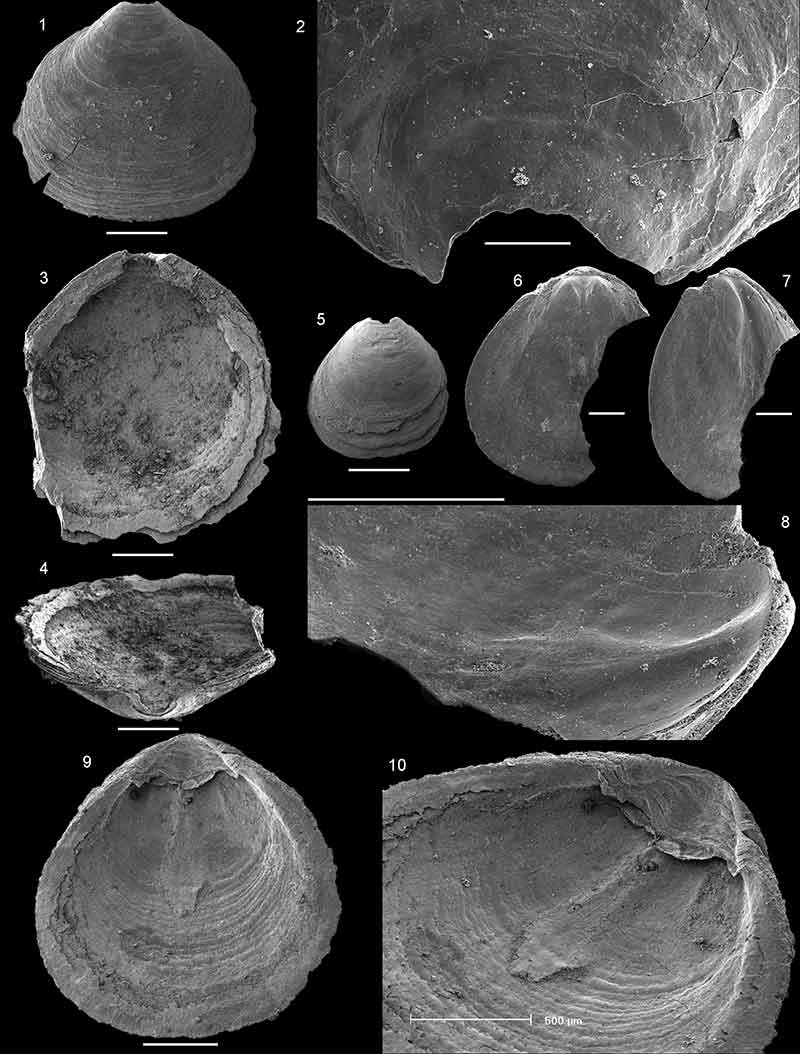
FIGURE 15. Eoobolus priscus (Poulsen, 1932) from the Forteau Formation of western Newfoundland. 1, Ventral valve exterior NFM 662 from Deer Arm Limestone, sample JSP 1982-01. 2-3, Ventral valve NFM F-2570 from Deer Arm Limestone, sample GM07-4-1; 2, external view; 3, detail of pustulose adult surface ornament. 4, Ventral valve interior NFM F-2571 from Deer Arm Limestone, sample GM07-4-1 showing arrangement of muscle scars. 5-6, Ventral valve NFM F-2574 from Deer Arm Limestone, sample GM07-5-1; 5, internal view; 6, oblique interal view showing imprints of pedicle nerves. 7, Ventral valve interior NFM F-2572 from Deer Arm Limestone, sample GM07-4-1. 8, Dorsal valve NFM F-2522 from Deer Arm Limestone, sample ICS 1421. 9, Ventral valve interior NFM F-2566 from Mackenzie Mills member, sample GM11-9A-10. 10-12, Dorsal valve NFM F-2567 from Mackenzie Mills member, sample GM11-9A-10; 10, internal view; 11, oblique internal view; 12, detail of columnar shell structure from naturally broken edge. Scalebars equal 250 µm in figures 1-2 and 4-11, 50 µm in figure 3 and 10 µm in figure 12.
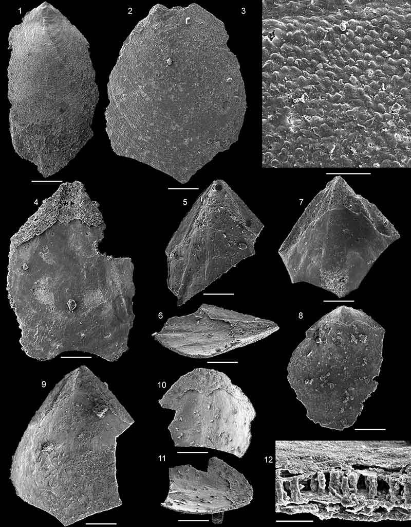
FIGURE 16. Pustulobolus triangulus n. gen et. n. sp. from the Forteau Formation, Lower shale, Mount St. Margaret of western Newfoundland. 1-2, Ventral valve NFM F-2523, sample MSM-11; 1, oblique lateral view; 2, external view. 3-4, Ventral valve NFM F-2524, sample MSM-11; 3, detail of larval shell; 4, detail of pitted larval shell ornament. 5, Ventral valve exterior NFM F-2525, sample MSM-11. 6, Ventral valve exterior NFM F-2526, sample MSM-5. 7-8, Ventral valve NFM F-2527, sample MSM-5; 7, exterior view; 8, detail of pustulose adult surface ornament. 9-10, Dorsal Valve NFM F-2528, sample MSM-7; 9, exterior view; 10, detail of larval shell in oblique view. 11, Conjoined shell NFM F-2529, sample MSM-11, showing outline of dorsal valve and triangular ventral pseudointerarea. Scalebars equal 250 µm in figures 1-2, 5-7, 9 and 11; 50 µm in figures 3 and 10; 20 µm in figure 8 and 5 µm in figure 4.
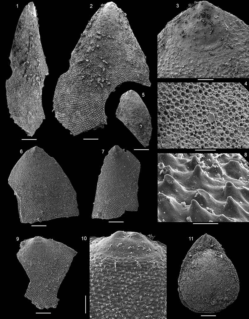
FIGURE 17. Pustulobolus triangulus n. gen et. n. sp. from the Forteau Formation, Lower shale, Mount St. Margaret of western Newfoundland, all from sample MSM-11. 1, 4, Ventral valve interior NFM F-2530, 1, interior view; 4, oblique view of interior. 2-3, Ventral valve NFM F-2531 (holotype); 2, interior view; 3, detail of ventral pseudointerarea with pustulose ornament lateral to flexure lines. 5, Ventral valve NFM F-2532, oblique view of interior showing cavities under pseudointerarea. 6, Ventral valve interior NFM F-2533. 7, Ventral valve interior NFM F-2534. 8-10, Dorsal valve NFM F-2535; 8, interior view; 9, detail of posterior margin; 10, detail of larval shell. Scalebars equal 250 µm in figures 1-2 and 4-8; 100 µm in figures 3 and 9; 20 µm in figure 10.
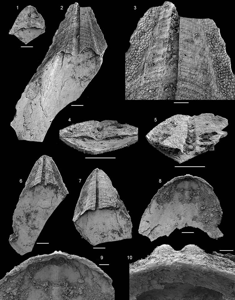
FIGURE 18. Botsfordia caelata (Hall, 1847) from the Forteau Formation of southern Labrador and western Newfoundland. 1, 8, Ventral valve NFM F-2536 from sample ICS1565; 1, exterior view; 8, detail of larval shell with pitted ornament. 2-4, Ventral valve NFM F-2537 from Devils Cove member, East Castor Pond, sample ECP-03; 2, external view; 3, oblique posterolateral view; 4, detail of pustulose adult shell ornament. 5-6, Ventral valve NFM F-2538 from Devils Cove member, East Castor Pond, sample ECP-03; 5, interior view; 6, oblique interior view. 7, Ventral valve exterior NFM F-2539 from Devils Cove member, East Castor Pond, sample ECP-03. 9-10, Ventral valve NFM F-2540 from Devils Cove member, East Castor Pond, sample ECP-03; 9, interior view; 10, detail interior surface in oblique view showing imprints of pedicle nerves. 11-13, Dorsal valve NFM F-2541 from Devils Cove member, East Castor Pond, sample ECP-03; 11, interior view; 12, oblique interior view; 13, detail of internal surface with dorsal pseudointerarea and posterolateral muscle scars. Scalebars equal 500 µm in figures 1-3, 5-7, 9 and 11-12; 100 µm in figures 4, 10 and 13; 10 µm in figure 8.
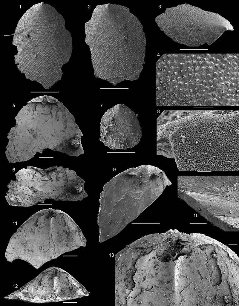
FIGURE 19. Eothele tubulus Ushatinskaya in Voronova et al., 1987 from the Forteau Formation of western Newfoundland. 1-3, Ventral valve NFM F-2575 from Deer Arm Limestone, sample GM07-5-1; 1, external view; 2, detail of larval shell showing pitted larval and pustulose adult shell ornament; 3, detail of posterior margin in oblique view showing oval opening of pedicle tube. 4, Ventral valve NFM F-2573 from Deer Arm Limestone, sample GM07-4-1. 5-6, Ventral valve NFM F-2568 from Deer Arm Limestone, sample GM07-3-1; 5, internal view; 6, oblique view showing median tongue and imprints of vascula lateralia. 7-8, Dorsal valve NFM F-2576 from Deer Arm Limestone, sample GM07-5-1; 7, external view; 8, oblique posterior view. 9-10, Dorsal valve NFM F-2521 from Deer Arm Limestone, sample GM07-5-1; 9, oblique lateral view; 10, oblique posterior view. 11-12, Dorsal valve NFM F-2569 from Mackenzie Mill member, sample GM07-2-1; 11, internal view showing dorsal pseudointerarea; 12, oblique lateral view. All scalebars equal 500 µm except in figures 2-3 that equals 100 µm.
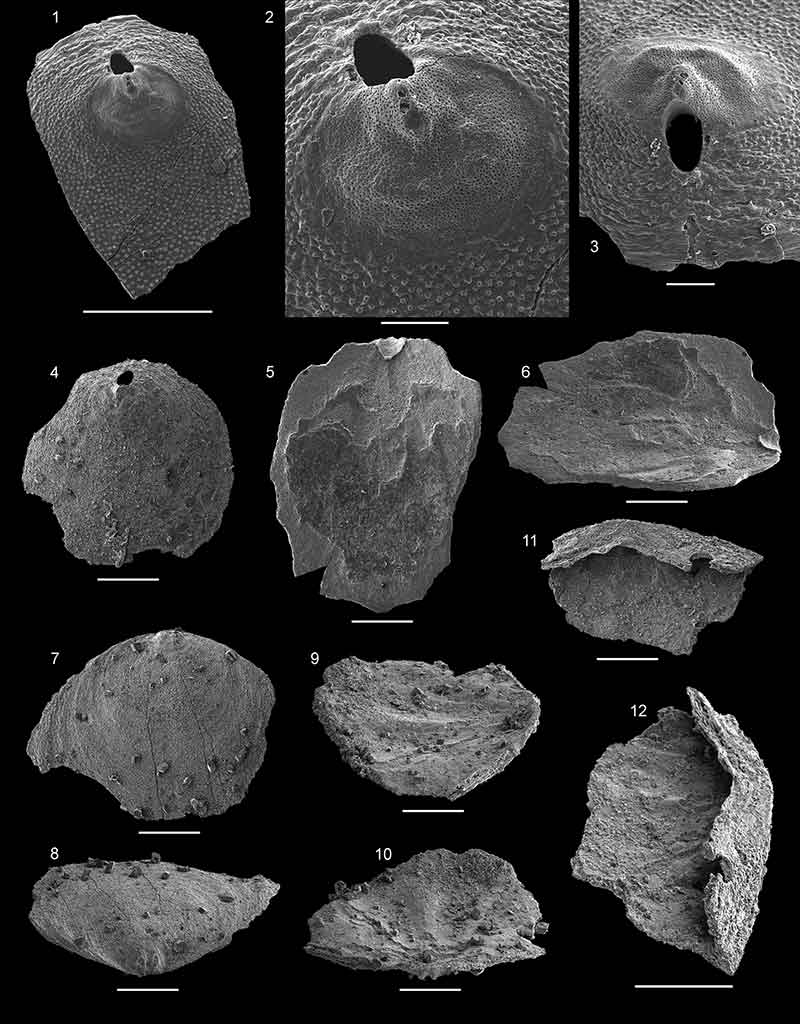
FIGURE 20. Acrothyra bonnia n. sp. from the Forteau Formation of western Newfoundland, Deer Arm Limestone, sample GM07-5-1. 1-3, Ventral valve NFM F-2559; 1, external view; 2, oblique lateral view; 3, detail of larval shell and pedicle foramen. 4, Ventral vave NFM F-2560, oblique lateral view. 5-6, Dorsal valve NFM F-2561; 5, external view; 6, oblique posterior view. 7-9, Ventral valve NFM F-2562 (Holotype), 7, internal view; 8, oblique lateral view of posterior margin and apical process; 9, detail of oblique anterior view of shell interior showing internal opening of pedicle foramen and muscle scars lateral to apical process. 10-11, Ventral valve NFM F-2563, 10, internal view; 11, oblique anterior view. 12-13, Ventral valve NFM F-2564, 12, internal view; 13, detail of ventral pseudointerarea in oblique posterior view showing triangular intertrough. 14, Dorsal view NFM F-2565, oblique lateral view showing low median ridge. All scalebars equal 250 µm except in figures 3, 9 and 13 that equals 100 µm.
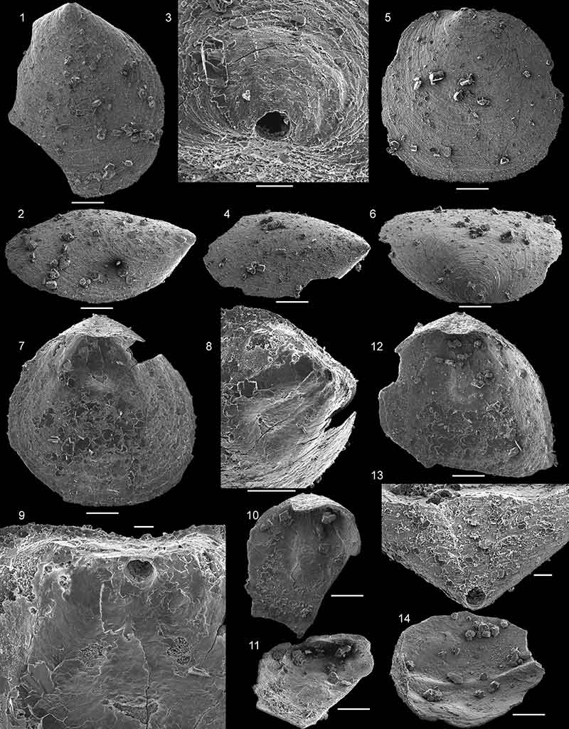
FIGURE 21. Hadrotreta taconica (Walcott, 1887) from the Forteau Formation of southern Labrador and western Newfoundland. 1-4, Ventral valve NFM F-2542 from sample ICS1577; 1, external view; 2, oblique lateral view; 3, oblique posterolateral view; 4, detail of apex showing larval shell. 5-6, Ventral valve NFM F-2543 from sample ICS1577; 5, external view; 6, detail of adult shell ornament. 7, Ventral valve NFM F-2544 from sample ICS1565 in external view. 8, Ventral valve NFM F-2545 from sample ICS1565, detail of posterior margin in oblique lateral view showing position of pedicle foramen at junction between larval and adult shell. 9, Conjoined shell NFM F-2546 ICS1575 in lateral view. 10-12, Ventral valve NFM F-2547 from sample ICS1545; 10, internal view; 11, oblique anterior view; 12, detail of apical process, pedicle foramen and apical pits. 13, Ventral valve NFM F-2548 from sample ICS1553; oblique lateral internal view. 14, Dorsal valve NFM F-2549 from sample ICS1548; external view. 15-17, Dorsal valve NFM F-2550 from sample ICS1553; 15, internal view; 16, detail of posterior margin showing dorsal pseudointerarea; 17, oblique lateral view. 18-19, Dorsal valve NFM F-2551 from sample ICS1575; 18, internal view; 19, oblique lateral view. Scalebars equal 200 µm in figures 1-3, 5, 7 and 9-19, 100 µm in figures 4 and 6 and 50 µm in figure 8.
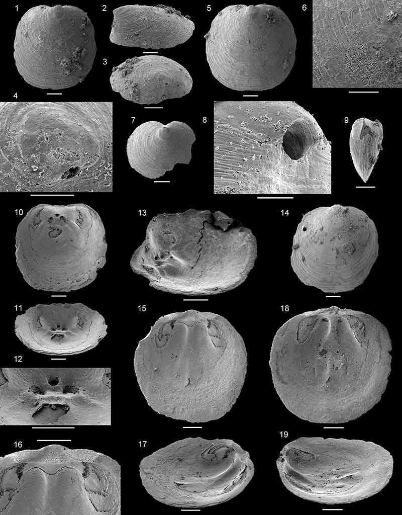
FIGURE 22. Hadrotreta sp. from the Forteau Formation of southern Labrador and western Newfoundland. 1-2, Ventral valve NFM F-2552 from sample ICS1553; 1, oblique posterior view showing procline pseudointerarea; 2, external view. 3-4, Ventral valve NFM F-2553 from sample ICS1577; 3, external view; 4, detail of larval shell with drop-shaped pedicle foramen. 5-6, Ventral valve NFM F-2554 from Lower shale, Mount St. Margaret, sample MSM-3; 5, internal view showing vascula lateralia and apical process; 6, oblique anterior view showing apical pits and internal opening of pedicle foramen. Scalebars equal 500 µm in figure 3, 200 µm in figures 1-2, 5-6 and 100 µm in figure 4.
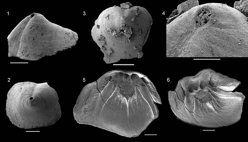
FIGURE 23. Paterinids from the Forteau Formation. 1-5, Paterina sp. 1-3, ventral valve NFM F-2555 from sample ICS1548; 1, posterior view; 2, external view; 3, lateral view. 4-5, Dorsal valve NFM F-2556 from sample LLQ3-5; 4, posterior view; 5, external view. 6-9, Micromitra sp. 6-8, Ventral valve NFM F-2557 from sample MSM-11; 6, posterior view; 7, external view; 8, lateral view. 9, Dorsal valve NFM F-2558 from sample MSM-7; oblique posterolateral view. All scalebars equal 500 µm.
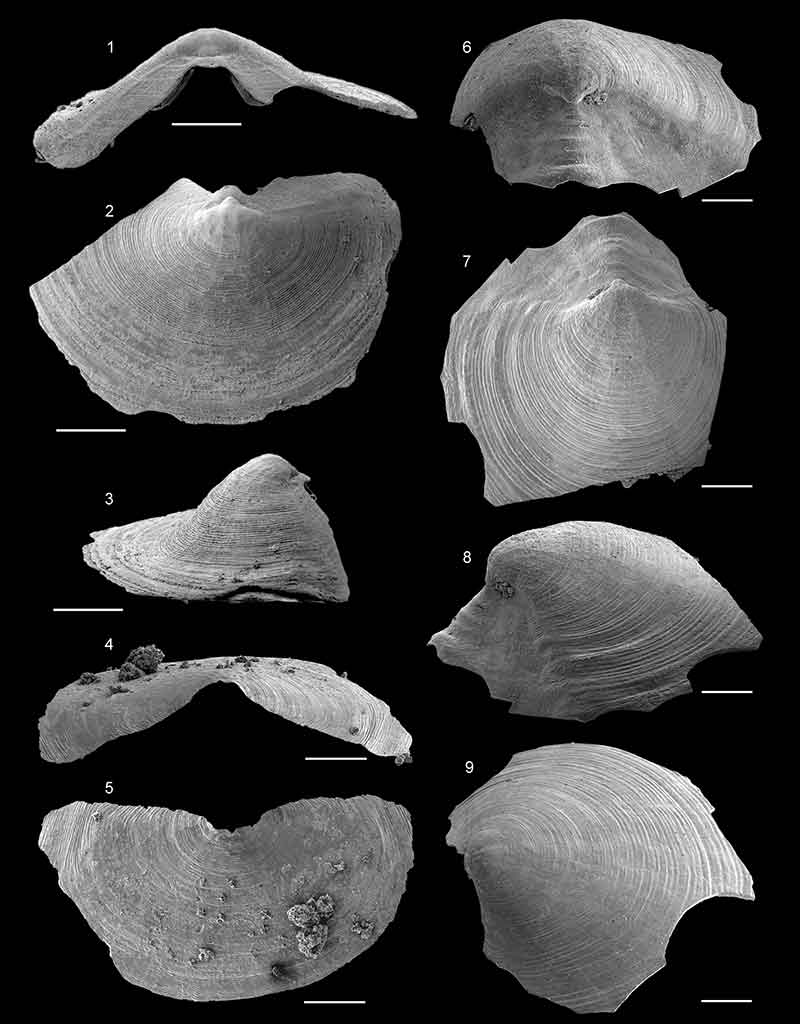
FIGURE 24. Setatella significans Skovsted, Streng, Knight and Holmer, 2010 from the Forteau Formation, The upper biostrome, L’anse au Loupe, southern Labrador. 1-2, Ventral valve NFM F-792 sample LLQ3-7; 1, internal view of posterior margin; 2, posterior view showing pseudointerarea. 3-4, Ventral valve NFM F-790, sample LLQ3-9; 3, internal view; 4, oblique lateral view. 5, Ventral valve NFM F-789, sample LLQ3-7, external view. 6, Shell fragment NFM F-793, sample LLQ3-9. All scalebar equals 250 µm.