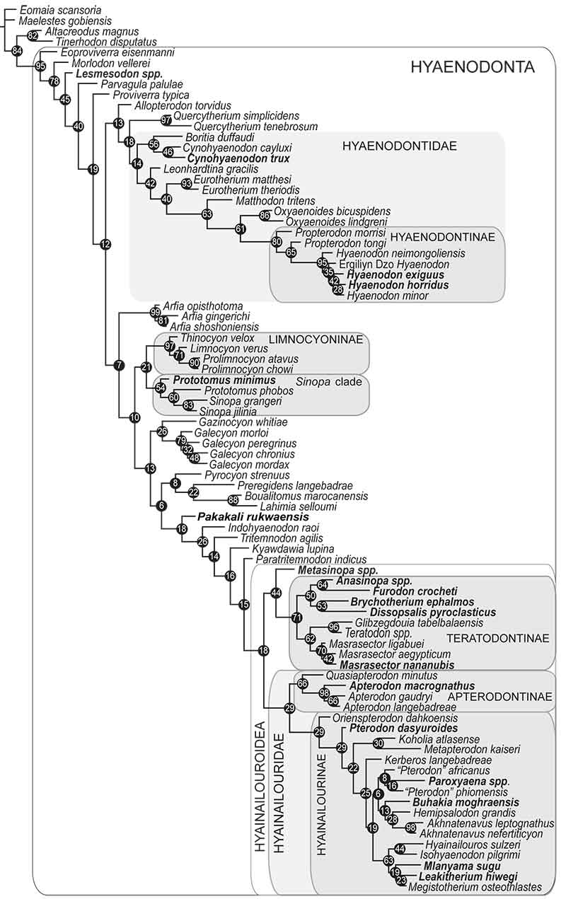
FIGURE 1. Apterodon macrognathus left maxilla fragment with dP4 and alveoli of dP3 (DPC 4126) from Quarry M (early Oligocene) in the Fayum Depression, Egypt in 1) occlusal view; 2) buccal view; and 3) lingual view. Scale bar is 10 mm.
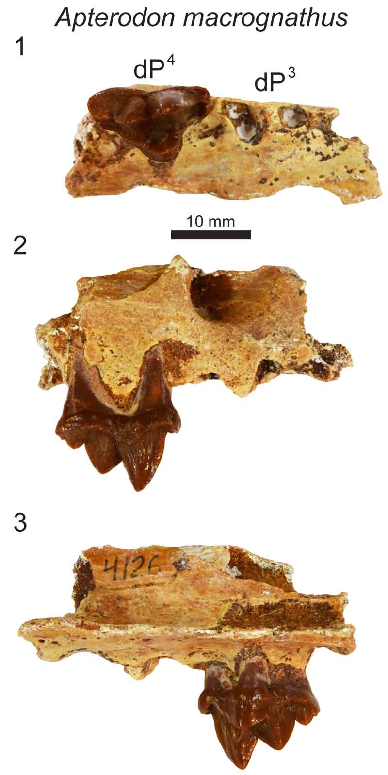
FIGURE 2. Apterodon macrognathus left dentary with dP3, dP4, M1 and M2 in crypt (DPC 8217) from Quarry M (early Oligocene) in the Fayum Depression, Egypt. Original specimen figured with digital model based on µ-CT scans reconstructed in Avizo. Mandibular corpus rendered transparent to reveal developing dentition. Grey, deciduous dentition. Yellow, permanent dentition. 1) Specimen occlusal view; 2) model occlusal view; 3) specimen lingual view; 4) model lingual view; 5) specimen buccal view; 6) model buccal view. Scale bar is 10 mm.
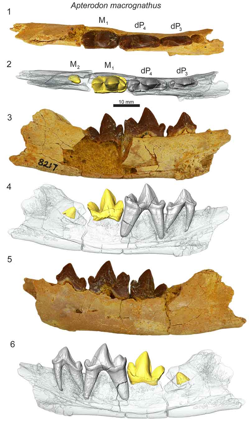
FIGURE 3. Leakitherium hiwegi maxilla fragments from Rusinga Island (early Miocene), Kenya. KNM-RU 2949 (CMF 4025 in Savage, 1965) is a left maxilla fragment dP3 and dP4 shown in 1) occlusal view; 2) buccal view; and 3) lingual view. KNM-RU 15182 is a right maxilla fragment with dP3 and dP4 and the germ of P4 shown in 4) occlusal view; 5) buccal view; 6) lingual view; 7) dorsal view; 8) and distal view. The well-developed germ of P4 is best viewed in dorsal and distal view. Specimens are to scale and scale bar is 10 mm.
FIGURE 4. Anasinopa leakeyi left maxilla cast with dP2, dP3 and dP4 (KNM-RU 2385) from Rusinga Island (early Miocene), Kenya in 1) occlusal view; 2) buccal view; and 3) lingual view. Scale bar is 10 mm.
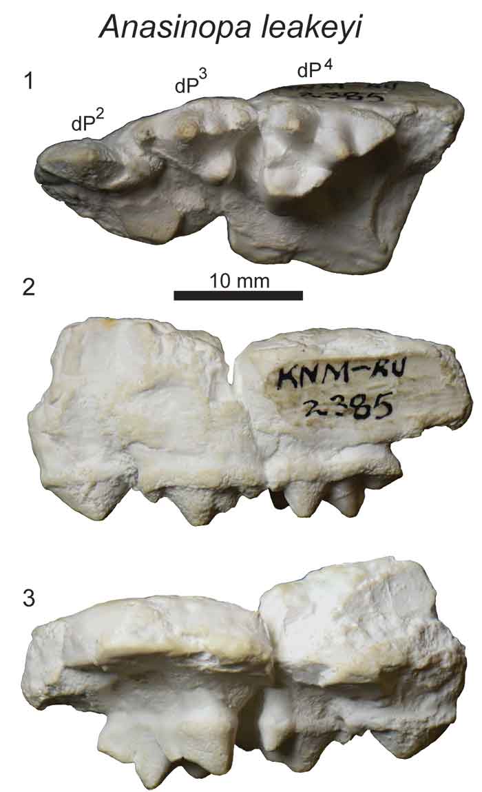
FIGURE 5. Anasinopa libyca left dentary with dP3 and dP4 from Gebel Zelten (middle Miocene), Libya (BMNH M82378) in 1) occlusal view; 2) lingual view; 3) buccal view. Scale bar is 10 mm.
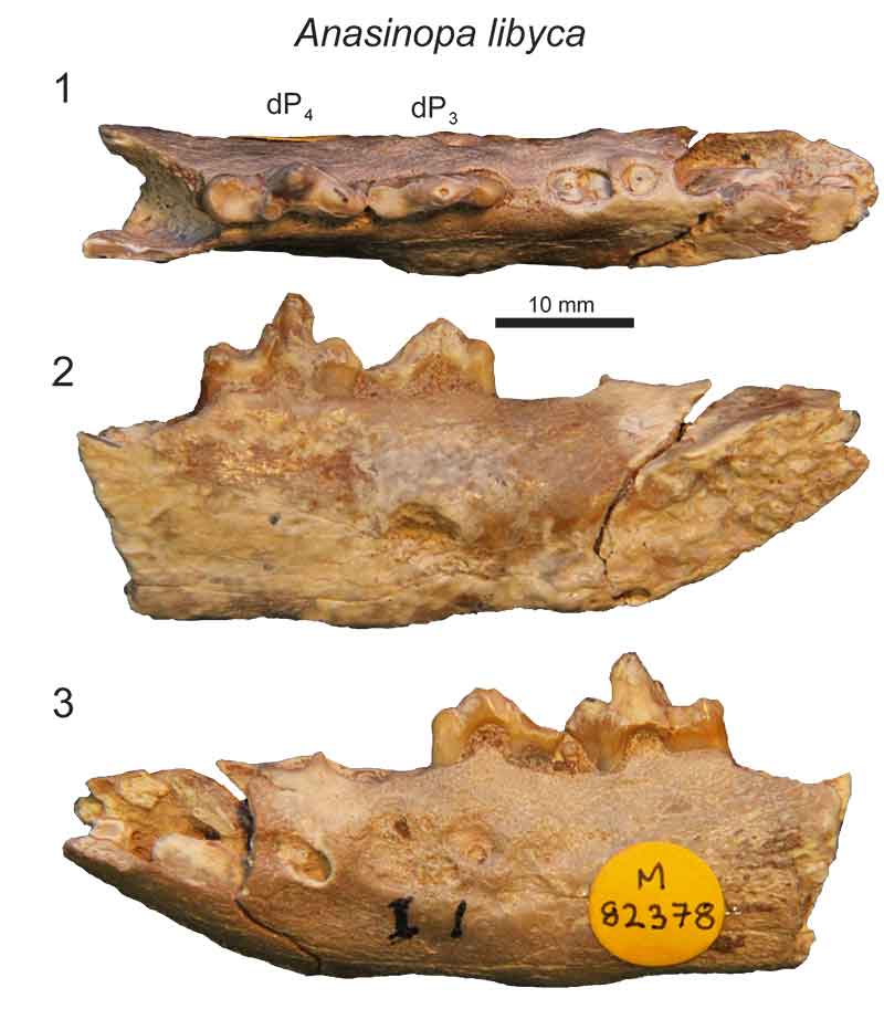
FIGURE 6. Dissopsalis pyroclasticus left maxilla with dP3 and dP4 (KNM-FT 3357) from Fort Ternan (early Miocene), Kenya in 1) occlusal view; 2) buccal view; and 3) lingual view. Scale bar is 10 mm.
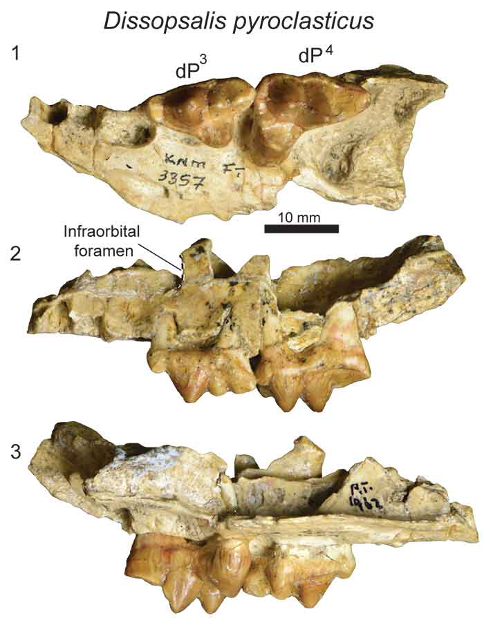
FIGURE 7. Masrasector nananubis left maxillae from Locality 41 (latest Eocene), Fayum Depression, Egypt. Original specimens figured with digital models based on µ-CT scans reconstructed in Avizo. Maxilla rendered transparent to reveal developing dentition. Grey, deciduous dentition. Yellow, permanent dentition. DPC 20882 is a left maxilla fragment that preserves dP3 and dP4 and no indication of developing permanent dentition. 1) specimen occlusal view; 2) model occlusal view; 3) specimen buccal view; 4) model buccal view; 5) specimen lingual view; 6) model lingual view. DPC 13837 is a left maxilla fragment that preserves erupted dP3, dP4, and M1 and P3 and P4 developing in crypts. 7) specimen occlusal view; 8) model occlusal view; 9) specimen buccal view; 10) model buccal view; 11) specimen lingual view; 12) model lingual view. Scale bar is 10 mm.
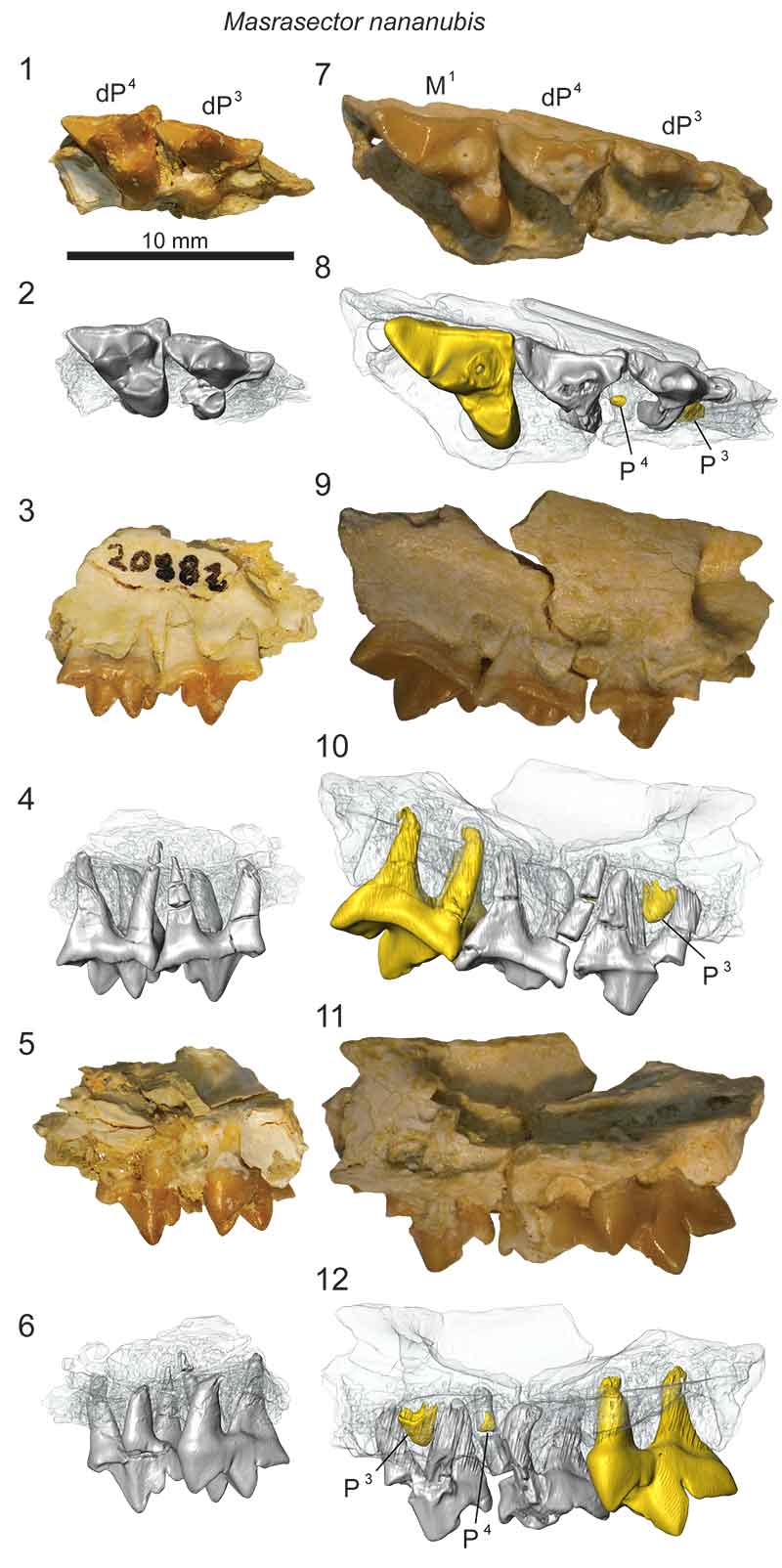
FIGURE 8. Masrasector nananubis right dentary with erupted dC, dP2, dP3, and dP4, M1 alveoli and P2, P3, and M2 in crypts (DPC 18276) from Locality 41 (latest Eocene), Fayum Depression, Egypt. Original specimen figured with digital model based on µ-CT scans reconstructed in Avizo. Dentary rendered transparent to reveal developing dentition. Grey, deciduous dentition. Yellow, permanent dentition. 1) specimen occlusal view; 2) model occlusal view; 3) specimen lingual view; 4) model lingual view; 5) specimen buccal view; 6) model buccal view. Scale bar is 10 mm.
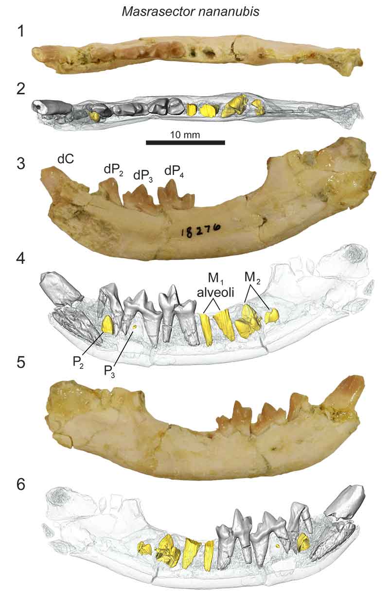
FIGURE 9. Masrasector nananubis left dentary with erupted dP3, dP4, M1 and M2 and P2, P3, and M3 in crypts (DPC 10358) from Locality 41 (latest Eocene), Fayum Depression, Egypt. Original specimen figured with digital model based on µ-CT scans reconstructed in Avizo. Dentary rendered transparent to reveal developing dentition. Grey, deciduous dentition. Yellow, permanent dentition. 1) specimen occlusal view; 2) model occlusal view; 3) specimen lingual view; 4) model lingual view; 5) specimen buccal view; 6) model buccal view. Scale bar is 10 mm.
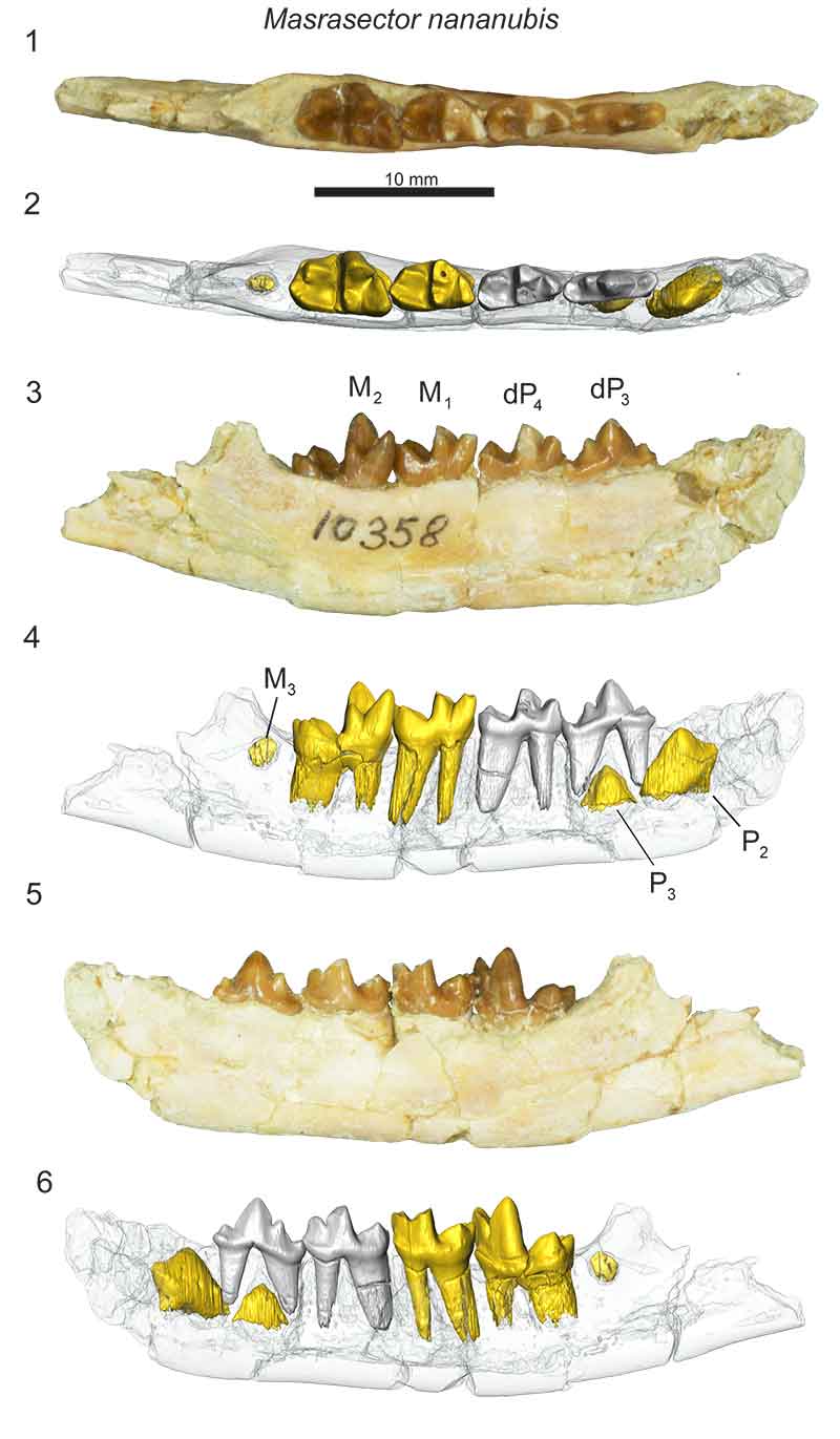
FIGURE 10. Metasinopa sp. left maxilla fragments with dP3, dP4, and M1 (DPC 10199) from Quarry V (early Oligocene), Fayum Depression, Egypt in 1) occlusal view; 2) buccal view; and 3) lingual view. Note that there is matrix around the roots of the specimens that has been cropped to make the dentition easier to view. Scale bar is 10 mm.
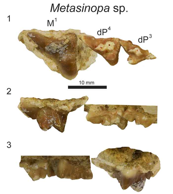
FIGURE 11. Metasinopa sp. right dentary with dC, dP4, M1, M2 and I, C, P2, P3, P4 M3 in crypts (DPC 4544) from Quarry V (early Oligocene), Fayum Depression, Egypt. Original specimen figured with digital model based on µ-CT scans reconstructed in Avizo. Dentary rendered transparent to reveal developing dentition. Grey, deciduous dentition. Yellow, permanent dentition. 1) specimen occlusal view; 2) model occlusal view; 3) specimen lingual view; 4) model lingual view; 5) specimen buccal view; and 6) model buccal view. Scale bar is 10 mm.
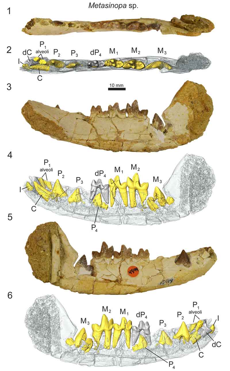
FIGURE 12. Results of the phylogenetic analysis of Hyaenodonta character-taxon matrix including deciduous dental characters. Results are visualized as an “allcompat” (majority rule plus compatible groups) consensus tree. Posterior probabilities (PP) are placed over each node. Bold taxa are scored for deciduous dental characters. Major, named clades recovered or discussed in this analysis and recovered in other analyses are illustrated.