FIGURE 1. Map locality, 1) state of Baja California Sur, Mexico; 2) Oligocene outcrops of San Juan Member, El Cien Formation; 3) El Saladito and mesa El Tosoro fossilferous localities, north to San Juan de la Costa village.

FIGURE 2. General stratigraphy and observations at mesa El Tesoro; 1) view of the strata over the mined phosphatic Humboldt bed; 2-3) position of the gray phosphatic sandstone over the Humboldt bed; 4) fossils Tlaxcallicetus cf. guaycurae in situ in the gray phosphatic sandstone and 5) ventral view, left ear region of Tlaxcallicetus sp., field observation at mesa El Tesoro.

FIGURE 3. Tlaxcallicetus guaycurae (MU EcSj5/06/31), cranium, dorsal view.

FIGURE 4. Tlaxcallicetus guaycurae (MU EcSj5/06/31), cranium, ventral view.

FIGURE 5. Tlaxcallicetus guaycurae (MU EcSj5/06/31), cranium, frontal view.

FIGURE 6. Tlaxcallicetus guaycurae (MU EcSj5/06/31), cranium, posterior view.

FIGURE 7. Tlaxcallicetus guaycurae (MU EcSj5/06/31), cranium, lateral view.

FIGURE 8. Tlaxcallicetus guaycurae (MU EcSj5/06/31), ear region (1) with the left periotic bone (2-4), ventral view. Pars cochlearis (3), posteromedial view.

FIGURE 9. Detailed features of Tlaxcallicetus guaycurae (MU EcSj5/06/31), (1) pitted edge in the glenoid fossa that suggest a fibrocartilaginous joint; (2) inflated and rounded squamosal prominence; (3) external occipital sulcus on the posterior middle part of the supraoccipital.

FIGURE 10. Ear region and left periotic comparison between Tlaxcallicetus species, ventral view. Tlaxcallicetus sp. (MU EcSj5/18/95) (1-2), Tlaxcallicetus guaycurae (MU EcSj5/06/31) (3-4), and specimen found at mesa El Tesoro, Tlaxcallicetus sp., field observation (5-6).

FIGURE 11. Tlaxcallicetus sp. (MU EcSj5/18/95), cranium, dorsal view.

FIGURE 12. Tlaxcallicetus sp. (MU EcSj5/18/95), cranium, ventral view.

FIGURE 13. Tlaxcallicetus sp. (MU EcSj5/18/95), cranium, frontal view.

FIGURE 14. Tlaxcallicetus sp. (MU EcSj5/18/95), cranium, posterior view.

FIGURE 15. Tlaxcallicetus sp. (MU EcSj5/18/95), cranium, lateral view.

FIGURE 16. Left periotic with anatomical terms, Tlaxcallicetus sp. (MU EcSj5/18/95), medial (1), lateral (2), ventral (3), dorsal (4), posterior (5), and anterior (6) views.

FIGURE 17. Left periotic, Tlaxcallicetus sp. (MU EcSj5/18/95), medial (1), lateral (2), ventral (3), dorsal (4), posterior (5), and anterior (6) views.

FIGURE 18. Left periotic, Tlaxcallicetus sp. (MU EcSj5/18/95), closer view of internal acoustic meatus, dorsal view.

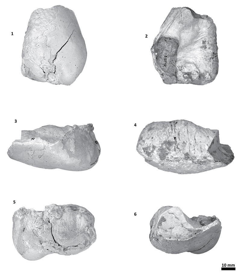
FIGURE 19. Left bulla with anatomical terms, Tlaxcallicetus sp. (MU EcSj5/18/95), ventral (1), dorsal (2), lateral (3), medial (4), posterior (5), and anterior (6) views.

FIGURE 20. Left bulla, Tlaxcallicetus sp. (MU EcSj5/18/95), ventral (1), dorsal (2), lateral (3), medial (4), posterior (5), and anterior (6) views.

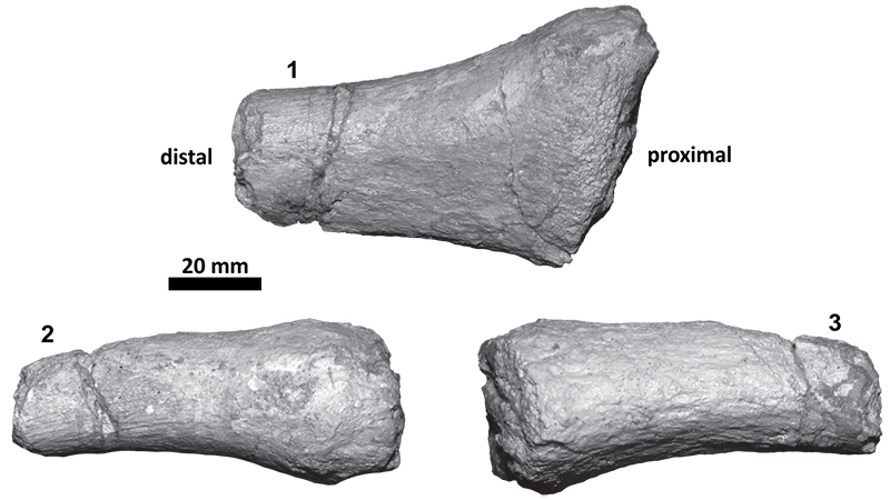
FIGURE 21. Left thyrohyoid, Tlaxcallicetus sp. (MU EcSj5/18/95), dorsal (1), anterior (2), and posterior (3) views.

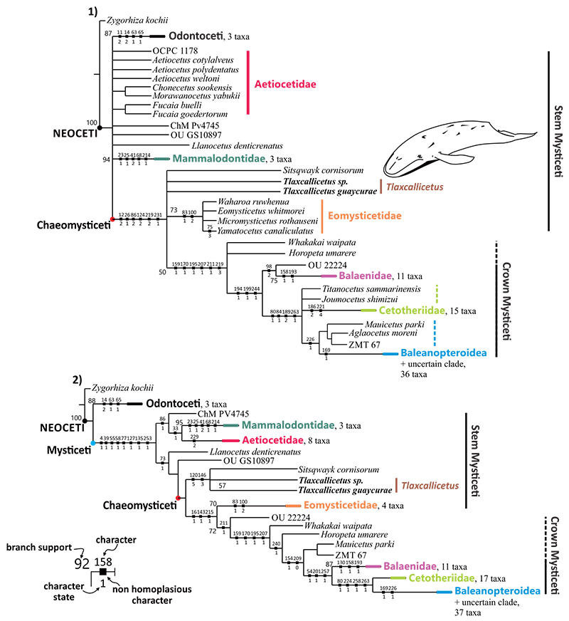
FIGURE 22. Philogenetic position of Tlaxcallicetus. 1) Under equal weights, consensus tree recovered from 260 parsimonious trees (1327 steps, consistency index CI= 0.246, retention index RI= 0.686, rescaled consistency index RCI= 0.168756, tree total length 1474). 2) Under implied weights: k = 6, consensus tree from 3 parsimonious trees (85.63773 steps, CI= 0.271, RI= 0.724, RCI= 0.196204, tree total length 1338. Black squares mark unequivocal characters that support the different groups (see legend). Support branch belongs only to those clades with 50% or more consistency. Dashed lines suggest a probable inclusion of taxa in clade.


