FIGURE 1. Geographical location of the sampling stations (red dots) in northeastern (1) and southeastern Sicily (2); and the external front of the Apenninic Chain (3) and overthrust of the Calabride Units (4). Some rights reserved: Imagery © 2015 TerraMe-trics, Map Data © 2015 Google. Note: The authors are the 'sole responsible' for the usage made of texts, illustrations (tables and drawings), photos and videos provided and used in their respective publications.
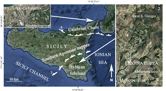
FIGURE 2. Stratigraphycal log (1) with the position and number of samples of the Villafranca Tirrena section (2).
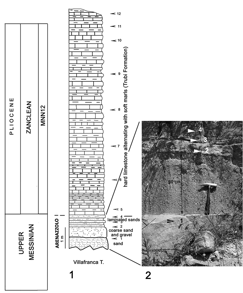
FIGURE 3. Stratigraphycal log (1) with the position and number of samples of the Abbeveratoio section (2) and particular of the Arenazzolo outcrop (3).
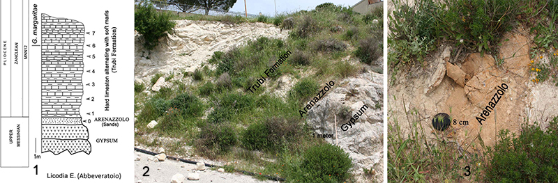
FIGURE 4. Stratigraphycal log (1) with the position and number of samples of the Cave San Giorgio section (2).
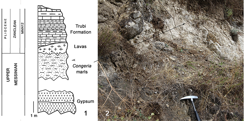
FIGURE 5. Stratigraphical log (1) with the position and number of samples of the Serpellizza section (2). The arrow indicates the boundary between the gypsum and the Trubi.
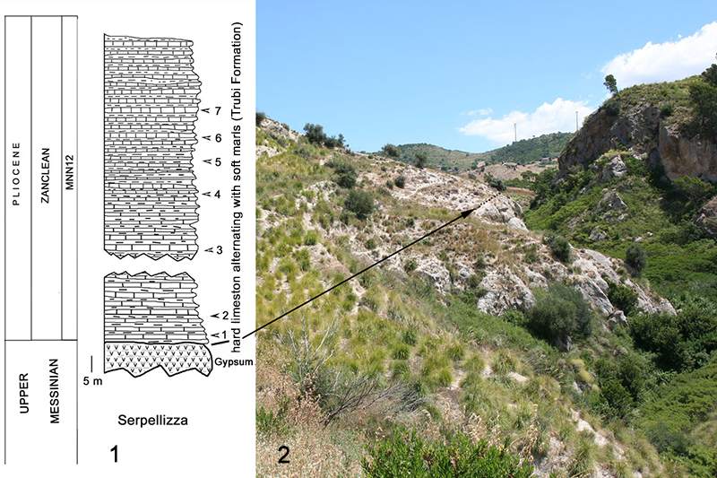
FIGURE 6. Scanning electron microscope (SEM) photographs of Late Messinian “Lago-Mare” ostracods. 1-3, Tyrrhenocythere pontica (Livental, 1929) from Cave San Giorgio, PMC. OFS 30, right valve in lateral view (1); PMC. OFS 31, right valve in lateral view (2); and PMC. OFS 32, left valve in external lateral view (3). 4-7, Tyrrhenocythere pulcherrima sp. nov. from Cave San Giorgio, PMC. O 20 H 19/5/2017 (holotype), right valve in lateral view (4); PMC. O 73 P 19/5/2017 (paratype), left valve in lateral view (5); PMC. O 74 P 19/5/2017 (paratype), left valve in internal view (6); and PMC. O 20 H 19/5/2017 (holotype), right valve, close-up of normal pores (7). 8, Cyprideis aff. anlavauxensis Carbonnel, 1979 from Cave San Giorgio, PMC. OFS 33, right valve in lateral view. 9, Cyprideis agrigentina Decima, 1964 from Cave San Giorgio, PMC. OFS 34, left valve in internal view. 10-12, Cyprideis ex C. torosa (Jones, 1850) group from Cave San Giorgio, PMC. OFS 35, right valve in internal view (10) and PMC. OFS 36, right valve in internal view (11); from Licodia Eubea, PMC. OFS 37, in dorsal view (12). 13-14, Loxoconcha eichwaldi Livental, 1929 from Cave San Giorgio, PMC. OFS 38, female, right valve in lateral view (13) and PMC. OFS 39, male, right valve in lateral view (14). 15-16, Loxoconcha muelleri (Méhes, 1908) from Cave San Giorgio, PMC. OFS 40, right valve in lateral view (15) and PMC. OFS 41, left valve in lateral view (16). 17, Loxoconcha n. sp. from Cave San Giorgio, PMC. OFS 42, right valve in lateral view. Scale bars equal 200 µm except 7 (50 µm) and 17 (100 µm).
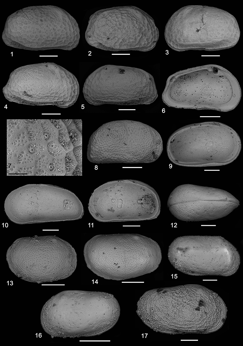
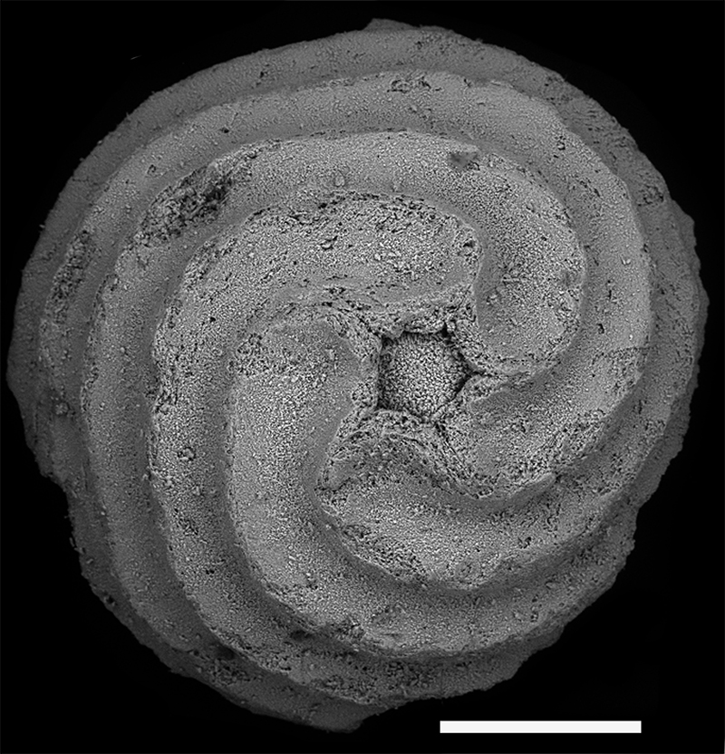
FIGURE 7. Scanning electron microscope (SEM) photographs of Chara hispida Linnaeus, 1753 from Cave San Giorgio, PMC. Gt.FS 48, basal view of gyrogonites. Scale bar equals 200 µm.