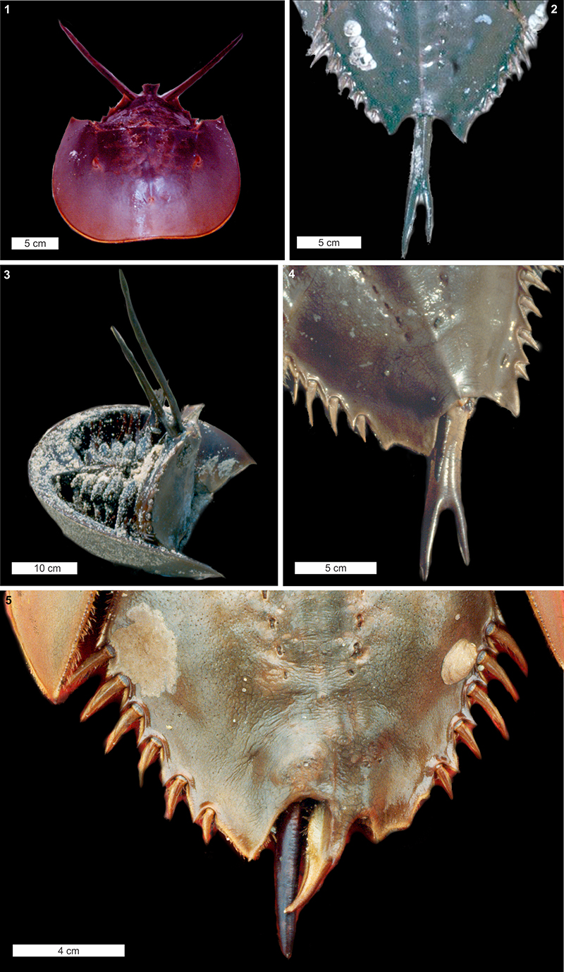
FIGURE 1. Examples of live Limulus polyphemus specimens with telson teratologies. (1, 3) Double telson–divided at the opisthosomal joint. (2) Forked telson asymmetrically bifurcated. (4) Forked telson and an over-developed axial spine. (5) Damaged telson spine and spine-like overdevelopment of the posterior opisthosomal margin. (1, 2, 4, 5) Images in dorsal view. (3) Image in ventral view. No specimens were accessioned into collections.

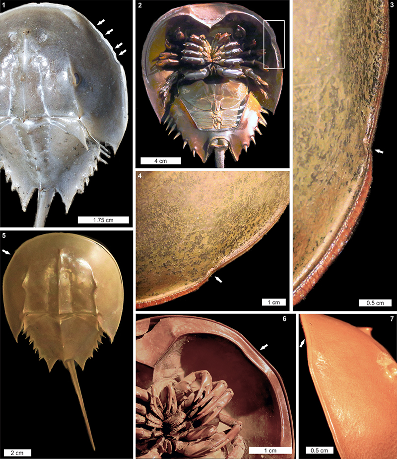
FIGURE 2. Examples of Limulus polyphemus with cephalothoracic injuries. (1) Four ‘U’ shaped embayments on right anterior cephalothorax (white arrows). (2–4) Small ‘U’ shaped embayment on left anterior cephalothorax. (2) Entire specimen. (3, 4) Close-ups of cephalothoracic buckling (white arrows). (5–7) Injury to left anterior cephalothorax (white arrows). (5) Entire specimen. (6, 7) Close-ups of slight cephalothoracic buckling (white arrows). (1, 4–5) images in dorsal view. (2, 5) Images in ventral view. (3, 7) Images in lateral view. No specimens were accessioned into collections.

FIGURE 3. A Tachypleus tridentatus carcass with a cephalothoracic and opisthosomal injury (OUMNH.ZC.25121). (1, 4) Entire specimen with boxes around the injuries. (2, 5) Close-ups of large, buckled ‘U’ shaped embayment on anterior right cephalothorax. (3, 5) Close-ups of ‘V’ shaped injury to posterior left opisthosoma. Note missing fourth moveable spine. (1–3) Images in dorsal view. (4–5) Images in ventral view. Images courtesy of Oxford Museum of Natural History.

FIGURE 4. Cephalothorax injuries to Mesolimulus walchi (Jurassic, Solnhofen Limestone, Bavaria, Germany). (1, 3) MCZ 106372. Large ‘W’ shaped embayment (white arrow) on left cephalothorax. Injury extends into the opisthosoma. Box in (1) enlarged in (3). (2, 4) MCZ 106382. Prominent ‘V’ shaped injury (white arrow) to anterior left cephalothorax. Image credit: (1-4) Museum of Comparative Zoology, Harvard University.

FIGURE 5. Cephalothorax injuries to Euproops danae (Francis Creek Shale, Will County, Illinois, USA, upper Carboniferous). (1, 2) FMNH.PE 21972. Parallel linear distortions to anterior left cephalothorax. (3, 4) MCZ 4674. ‘W’ shaped injury (white arrow) to right cephalothorax. (5) FMNH.P 29193. ‘V’ shaped injury (black arrow) on anterior right cephalothorax. (6): ROMIP5827. Complete, uninjured specimen for comparison. Image credit: (1) Mane Pritza, (3, 4) Museum of Comparative Zoology, Harvard University.

FIGURE 6. Cephalic injuries to Cambrian trilobites. (1, 2) Redlichia takooensis Lu, 1950 from the lower Cambrian (Series 2, Stage 4) Emu Bay Shale, Kangaroo Island, South Australia; SAM P48187. Cicatrised ‘W’ shaped injury to left cephalon. Injury was incurred during a recent moult. (3, 4) Olenellus thompsoni (Hall, 1859) from the Cambrian (Series 2, Stage 4) Kinzers Formation, Pennsylvania; USNM PAL 90809. Asymmetric, cicatrised ‘U’ shaped injury to left cephalon. Image credit: (1, 2) Originally figured in Bicknell and Paterson (2018); (3, 4) Frederick Cochard.


