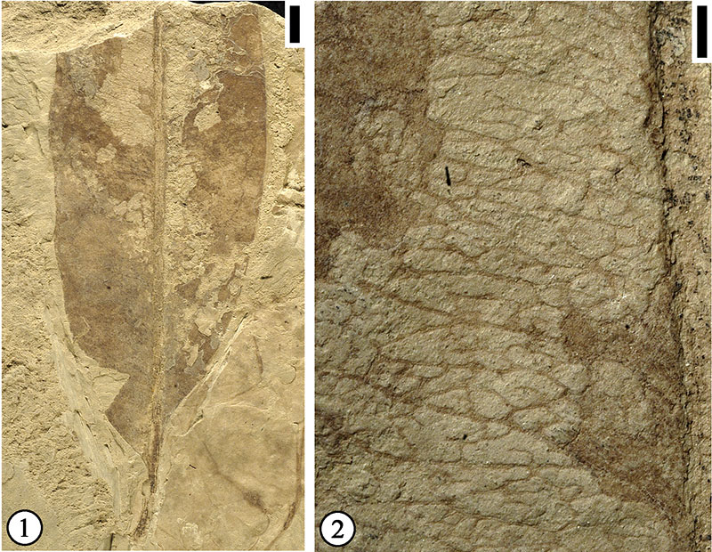
FIGURE 1. Regional outcrop of the Dakota Formation and the location of the Hoisington III locality and other plant megafossil localities discussed in the text. 1, Hoisington III, Kansas (UF 15706); 2, Braun Ranch, Kansas (UF15709); 3, Rose Creek, Nebraska (UF15713); and 4, Courtland I, Minnesota (UF18267). “*” indicates type area of the Dakota Formation. Map extends from southern Minnesota to north central Kansas along the eastern margin of the Western Interior Seaway in the United States. The inset map shows the location of the study area in USA. State boundaries are dashed. Outcrop map is based on figure 1 of Witzke and Ludvigson (1996).
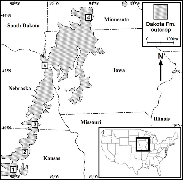
FIGURE 2. Chart shows the Albian, Cenomanian and Turonian stratigraphic units in Kansas, the Tri-state area (Iowa, Nebraska, and South Dakota; modified from figure 4 of Brenner et al., 2000), and Minnesota (modified from figure 8 of Setterholm, 1994). 1, Courtland I clay pit, Minnesota; 2, Rose Creek, Nebraska; 3, Braun Ranch, Kansas; and 4, Hoisington III, Kansas.
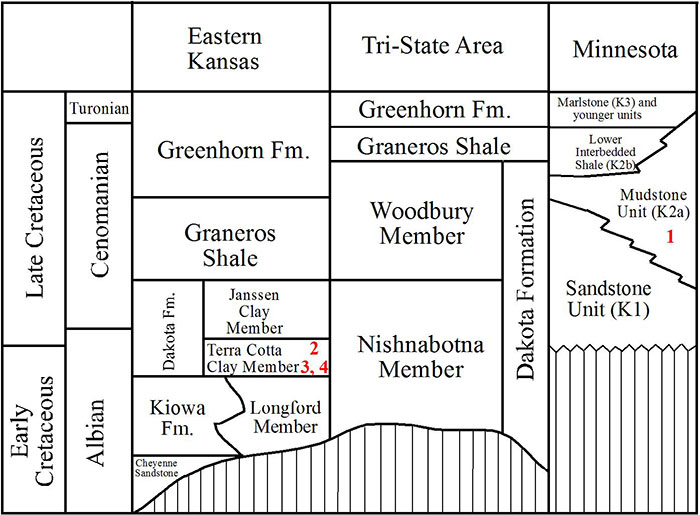
FIGURE 3. Cross section of the plant-bearing layer at the southwest end of the north clay pit at the Hoisington III locality, Kansas (Modified from figure 2 of Retallack and Dilcher, 2012).
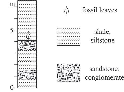
FIGURE 4. 1, Crassidenticulum decurrens (Lesquereux) Upchurch and Dilcher, 1990, UF15706-24648, fragment of leaf lamina. Note fine teeth on margin (indicated by arrow). Scale bar equals 2 mm. 2, Crassidenticulum trilobum Dilcher and Wang, 2006a, UF15706-24677, a trilobed leaf. Note thin and long petiole. Scale bar equals 1 cm. 3-4, cf. Crassidenticulum trilobum Dilcher and Wang, 2006a, UF15706-24684, a five lobed leaf (scale bar equals 1 cm) (3) and an enlargement of Figure 4.3 to show decurrent lamina tissue between two adjacent lobes (indicated by arrow) (scale bar equals 1 mm) (4).
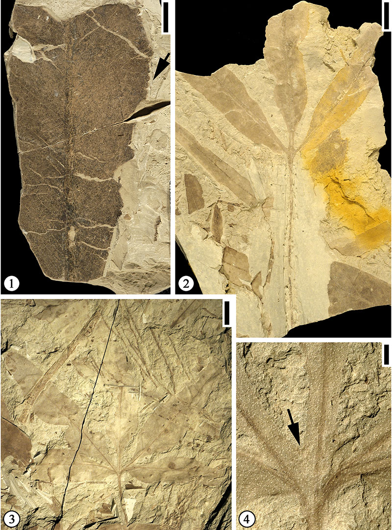
FIGURE 5. 1-2, Aquatifolia fluitans Wang and Dilcher 2006b, UF15706-8263′, leaf showing the spherical float on the petiole (1) and UF15706-24120, leaf showing cordate base and thin high order venation (2). Scale bars equal 5 mm. 3-4, Brasenites kansense Wang and Dilcher, 2006b, UF15706-14806, specimen (3) and line drawing (4) to show suborbiculate leaf shape, entire leaf margin, peltate central base, major primary veins. Scale bars equal 1 cm.
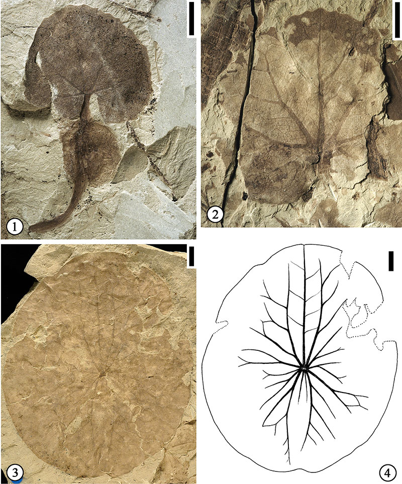
FIGURE 6. Longstrethia aspera (Lesquereux) comb. nov. 1, UF15706-24578, middle and lower portion of lamina. Scale bar equals 1 cm. 2, UF15706-24560, specimen showing secondary veins and toothed margin. Scale bar equals 5 mm. 3, Enlargement of Figure 6.1 (left middle portion of lamina; indicated by arrow) to show fine venation. Scale bar equals 2 mm.
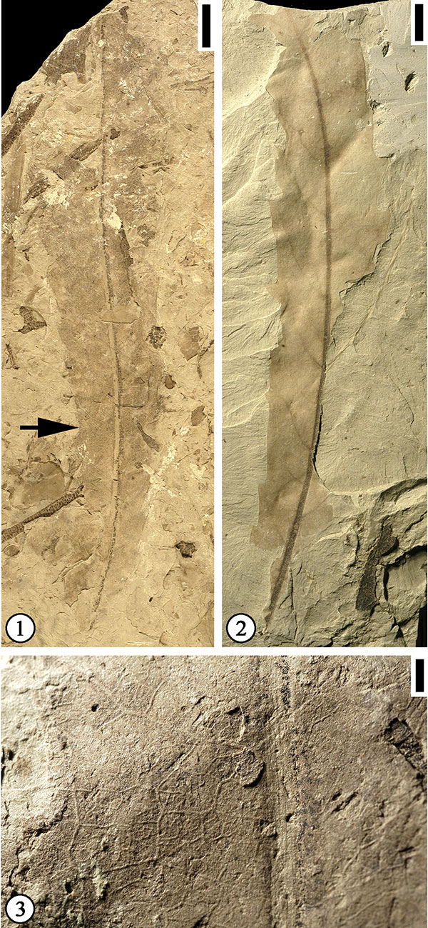
FIGURE 7. Pabiania variloba Upchurch and Dilcher, 1990. 1-3, UF15706-24423, specimen showing suprabasal actinodromous primary venation (scale bar equals 1 cm) (1), enlargement of a sinus area to show sinus bracing by secondary veins (scale bar equals 2 mm) (2); and enlargement of basal portion of leaf to show two pairs of basal secondary veins (scale bar equals 2 mm) (3). 4-5, UF15706-30154, specimen showing a long petiole and ocrea-like structure at the base (scale bar equals 5 mm) (4) and enlargement of the petiole to show the ocrea-like structure (scale bar equals 2 mm) (5). 6, UF15706-14832, specimen showing suprabasal actinodromous primary venation and acute apices of the lobes. Scale bar equals 1 cm.
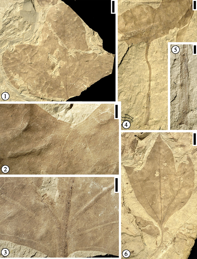
FIGURE 8. Pabiania variloba Upchurch and Dilcher, 1990. 1, UF15706-14823, leaf showing basal actinodromous primary venation and basal secondary veins. Scale bar equals 1 cm. 2, UF15706-24464, leaf showing rounded lobe apex. Scale bar equals 1 cm. 3, UF15706-24587, specimen showing a small lobe on the left and entire margin on the right of the leaf. Scale bar equals 5 mm. 4, Enlargement of Figure 8.2 to show straight primary vein extending to lobe apex and two series of loops in the excostal region. Scale bar equals 1 mm.
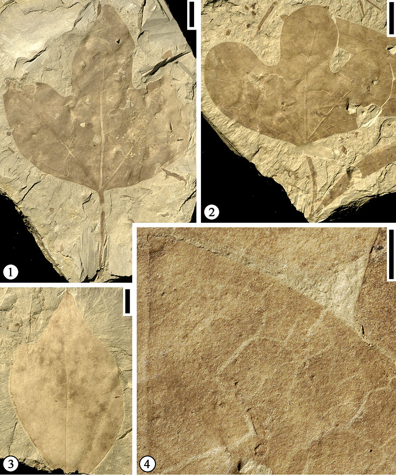
FIGURE 9. 1 and 3, Rogersia dakotensis Wang and Dilcher, 2009, UF15706-24798, general leaf shape, note thin secondary veins forming intramarginal veins (scale bar equals 5 mm) (1) and enlargement to show intramarginal veins formed by looping secondary veins (scale bar equals 1 mm) (3). 2 and 4, Rogersia parlatorii Dilcher and Wang, 2006, UF15706-7529, specimen showing leaf shape (scale bar equals 5 mm) (2) and enlargement to show secondary venation (scale bar equals 5 mm) (4).
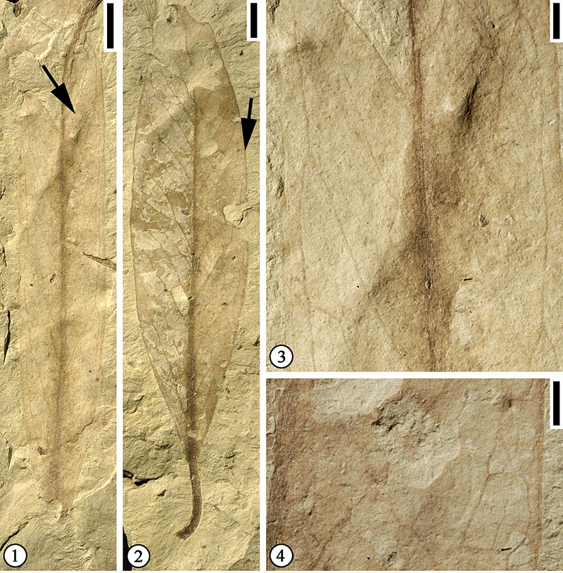
FIGURE 10. 1, Rogersia dakotensis Wang and Dilcher, 2009, UF15706-24620, general leaf shape. Note short petiole. Scale bar equals 5 mm. 2-3, Wolfiophyllum pfaffianum (Heer) Wang and Dilcher, 2009, UF15706-14815, basal and middle portion of leaf, note strong primary vein and entire margin (scale bar equals 1 cm) (2) and enlargement of Figure 10.2 (area indicated by arrow) to show eucamptodromous venation, note intersecondary veins (Scale bar equals 1 mm) (3).
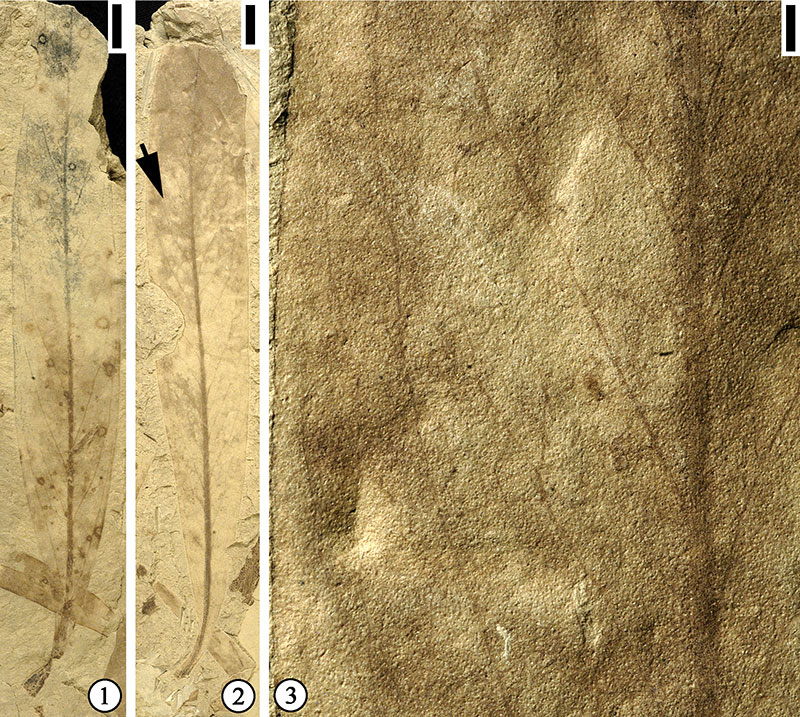
FIGURE 11. 1, Jarzenia kanbrasota Wang and Dilcher, 2009, UF15706-3171, showing elliptic leaf shape and secondary venation. Scale bar equals 1 cm. 2, Liriophyllum kansense Dilcher and Crane, 1984, UF15826-3188, showing deeply lobed leaf, long petiole, and secondary venation. Scale bar equals 1 cm. 3, Credneria cyclophylla (Heer) Wang and Dilcher, 2009, UF15706-14821, leaf showing craspedodromous venation. Note all secondary veins and their exmedial branches terminating on leaf margin, resulting in a wavy appearance of leaf margin. Scale bar equals 1 cm.
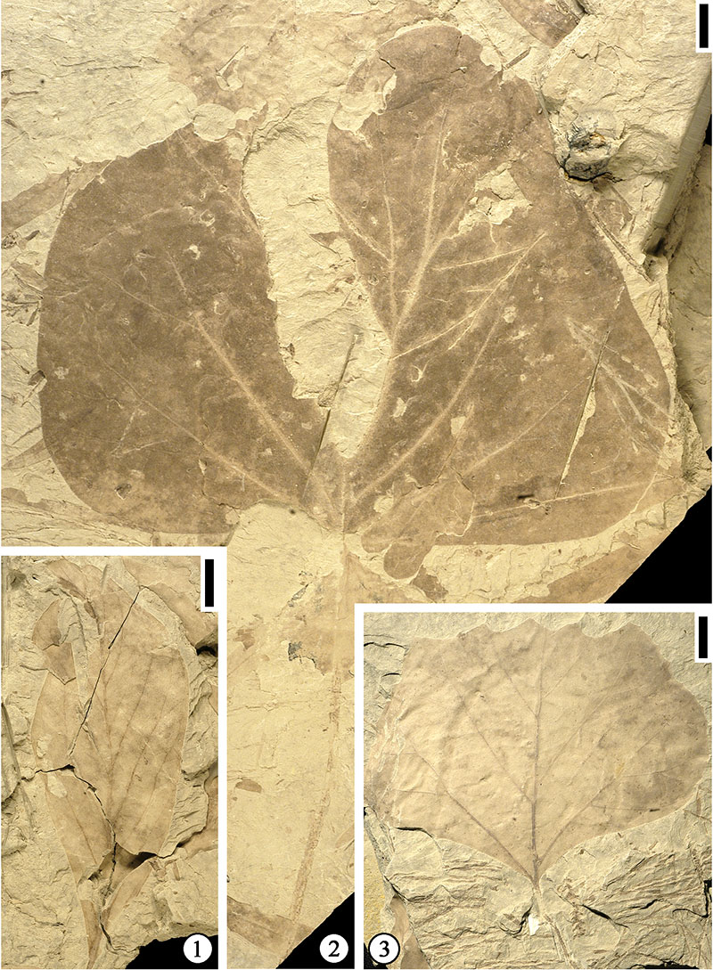
FIGURE 12. Paleonelumbo cf. macroloba Knowlton, 1930. 1, UF15826-24649, specimen showing primary venation (scale bar equals 5 mm) and 2, enlargement to show high order venation and toothed margin (scale bar equals 1 mm).
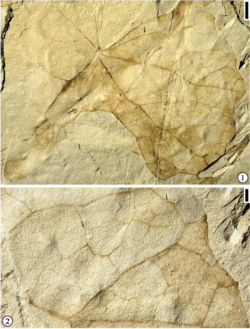
FIGURE 13. Sapindopsis powelliana (Lesquereux) comb. nov. 1, UF15706-14830. leaf with four leaflets. Note two ultimate leaflets appearing to be bilobed (indicated by arrow on the left), two oppositely arranged lateral leaflets, and stipule at the base. Scale bar equals 1 cm. 2, Enlargement of Figure 13.1 (indicated by arrow on the right) to show secondary and tertiary venation. Scale bar equals 2 mm. 3, Enlargement to of Figure 13.1 to show venation of the stipule. Scale bar equals 2 mm. 4, UF15706-14814, an elliptic leaflet with acuminate apex. Scale bar equals 1 cm. 5, Enlargement of Figure 13.4 to show secondary and tertiary venation. Scale bar equals 3 mm.
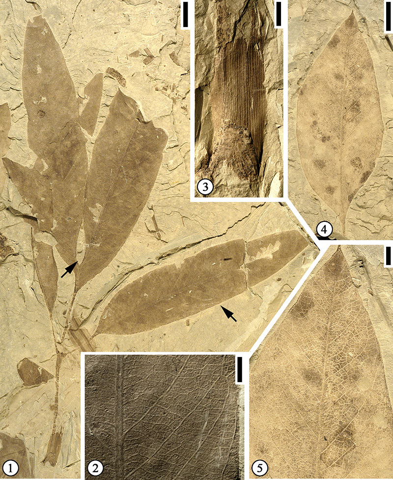
FIGURE 14. Sapindopsis powelliana (Lesquereux) comb. nov. 1, UF15706-24670, leaf with three leaflets. Note bilobed ultimate leaflet. Scale bar equals 1 cm. 2, UF15706-4812, leaf with narrow oblong leaflets. Scale bar equals 1 cm.
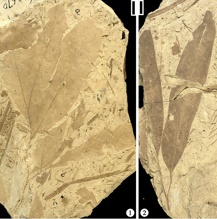
FIGURE 15. Sapindopsis powelliana (Lesquereux) comb. nov. UF15706-24719, leaf with two opposite lateral leaflets, one ultimate leaflet with long and thin petiolule, and a stipule at the base of petiole (indicated by arrow). Scale bar equals 1 cm.
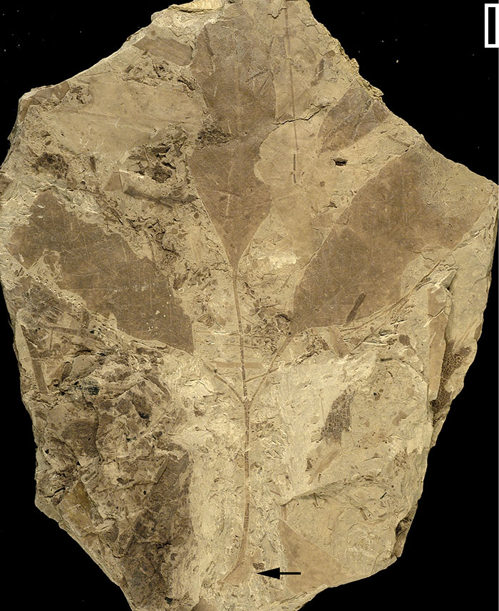
FIGURE 16. Sapindopsis powelliana (Lesquereux) comb. nov. 1, UF15706-24711, leaf with six leaflets (indicated by numbers). Note eucamptodromous secondary venation and strong secondary veins. Scale bar equals 1 cm. 2, UF15706-24675, leaf with two ultimate lobed leaflets. Note that the apex of each lobe of the two leaflets dissects one more time to form two smaller lobes (indicated by arrows). Scale bar equals 1 cm.
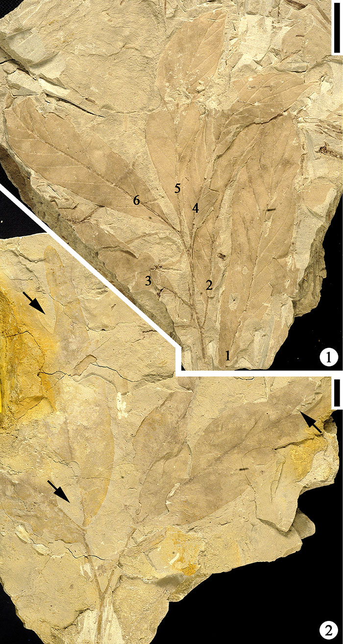
FIGURE 17. Sapindopsis retallackii sp. nov. 1, UF15706-3153, showing a leaflet with entire margin. Note the petiolule at the base (indicated by arrow). Scale bar equals 5 mm. 2, Enlargement of Figure 17.1 to show numerous thin secondary and intersecondary veins. Scale bar equals 1 mm.
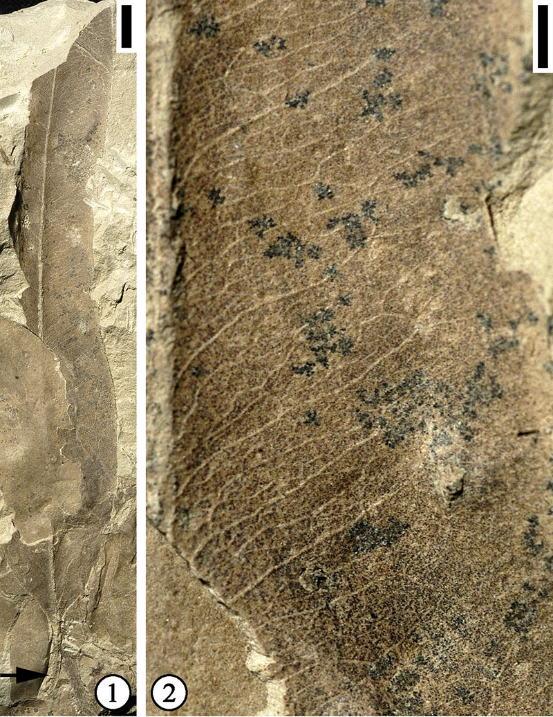
FIGURE 18. Anisodromum wolfei Upchurch and Dilcher, 1990. 1, UF15706-14818, general leaflet shape. Note long petiolule. Scale bar equals 1 cm. 2, Enlargement of Figure 18.1 to show strong secondary veins and percurrent tertiary veins oriented perpendicular to the primary vein. Scale bar equals 2 mm. 3, UF15706-24566, general leaflet shape. Scale bar equals 5 mm. 4, Enlargement of Figure 18.3 to show percurrent tertiary veins oriented almost perpendicular to the primary vein. Scale bar equals 2 mm.
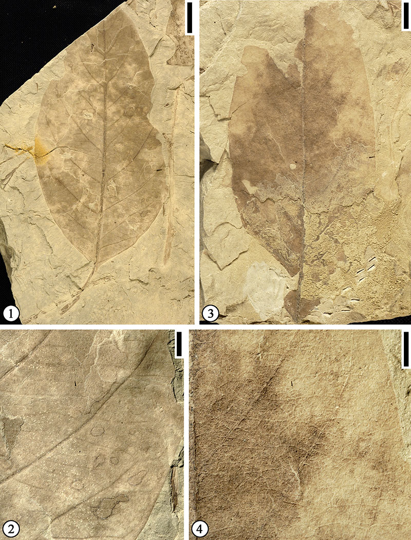
FIGURE 19. 1-2, Anisodromum upchurchii sp. nov., UF15706-24576, middle portion of lamina showing strong primary and secondary veins (scale bar equals 1 cm) (1) and enlargement of an area in Figure 19.1 (indicated by arrow) to show percurrent tertiary veins (scale bar equals 2 mm) (2). 3-4, Anisodromum schimperi (Lesquereux) comb. nov., UF15706-24635, leaf showing uneven spacing of secondary veins and reticulate tertiary veins (scale bar equals 5 mm) (3) and ; UF15706-24633, leaf showing secondary venation and reticulate tertiary veins (scale bar equals 5 mm) (4).
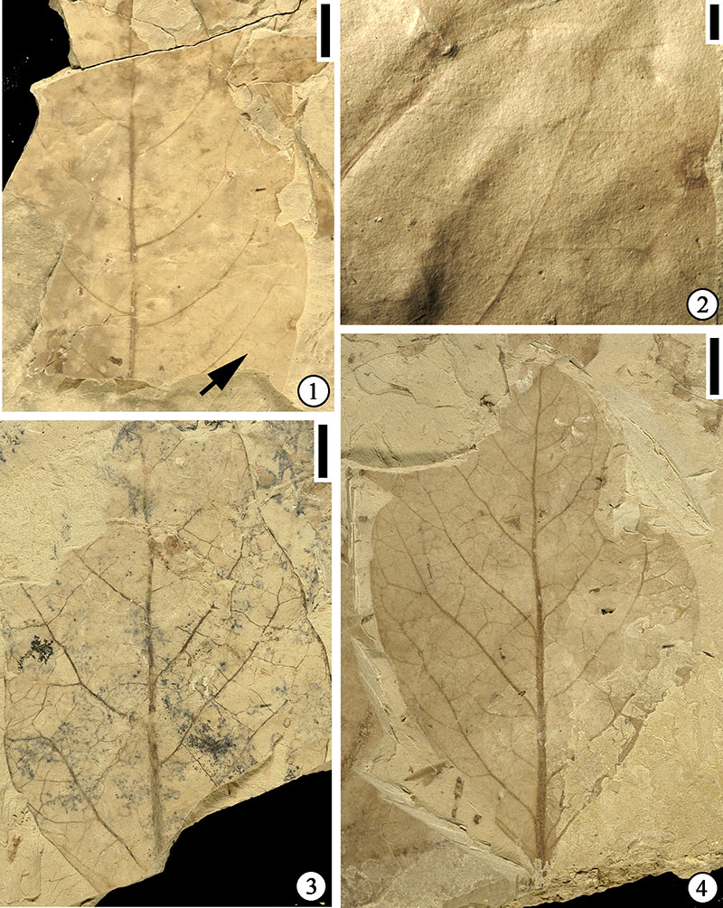
FIGURE 20. Citrophyllum aligera Lesquereux, 1892. 1, UF15706-24646, base of leaf showing lamina wing on petiole. Scale bar equals 5 mm. 2, UF15706-24645, base of leaf showing a long petiole with lamina wing and inflated petiolar base. Scale bar equals 5 mm. 3, UF15706-24332. leaf with a long petiole with thin lamina wing and inflated petiolar base. Scale bar equals 1 cm. 4, Enlargement of an area in Figure 20.3 (indicated by arrow) to show looping marginal veins. Scale bar equals 1 mm.
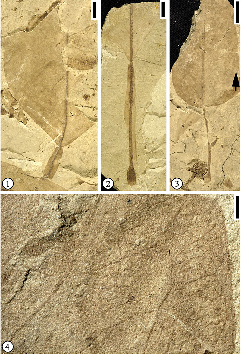
FIGURE 21. Wingia expansolobum (Upchurch and Dilcher) comb. nov. 1, UF15706-30158a, specimen showing a complete leaf. Note the long petiole with a swollen base and the extension of lamina tissue on the petiole. Scale bar equals 1 cm. 2, Enlargement of the petiole in Figure 21.1 to show the swollen base. Scale bar equals 2 mm. 3, UF15706-30158b, enlargement of a leaf on the back of the same specimen to show high order venation and glandular teeth. Scale bar equals 3 mm. 4, UF15706-24788, specimen showing a deeply lobed leaf. Note apically curved outer lateral lobes. Scale bar equals 1 cm.
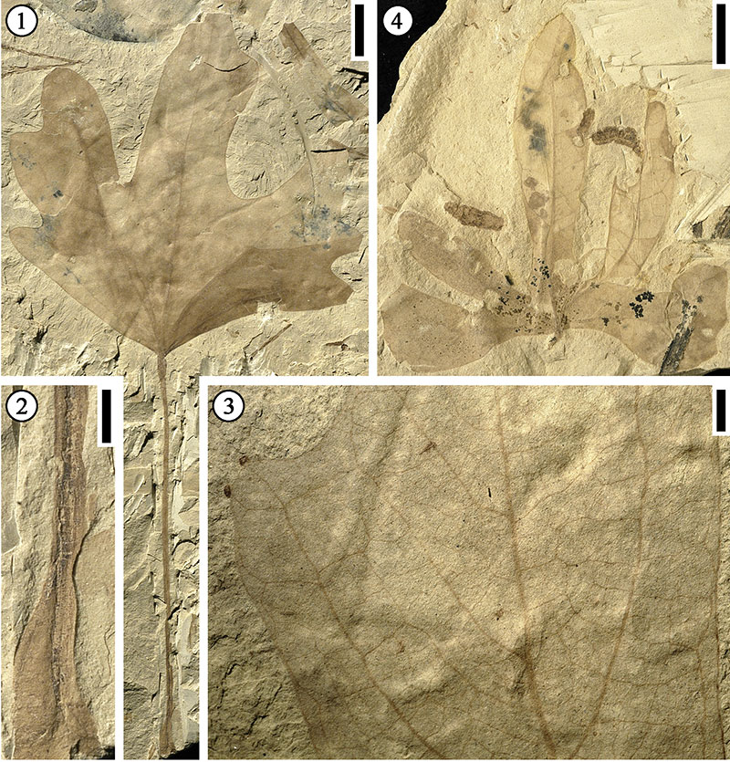
FIGURE 22. Wingia expansolobum (Upchurch and Dilcher) comb. nov. 1, UF15706-14825, specimen showing a large leaf with irregular shape of lateral lobes. Note the structurally reinforced margin on sinus, pluvinus lamina extension on the thin and long petiole. Scale bar equals 1 cm. 2, Enlargement of Figure 22.1 to show the pluvinus lamina extension on the petiole. Scale bar equals 2 mm. 3, UF15706-24461, enlargement of Figure 22.4 (area indicated by arrow) to show looping of secondary veins near the lamina margin. Scale bar equals 1 mm. 4, UF15706-24461, specimen showing a small leaf. Scale bar equals 5 mm.
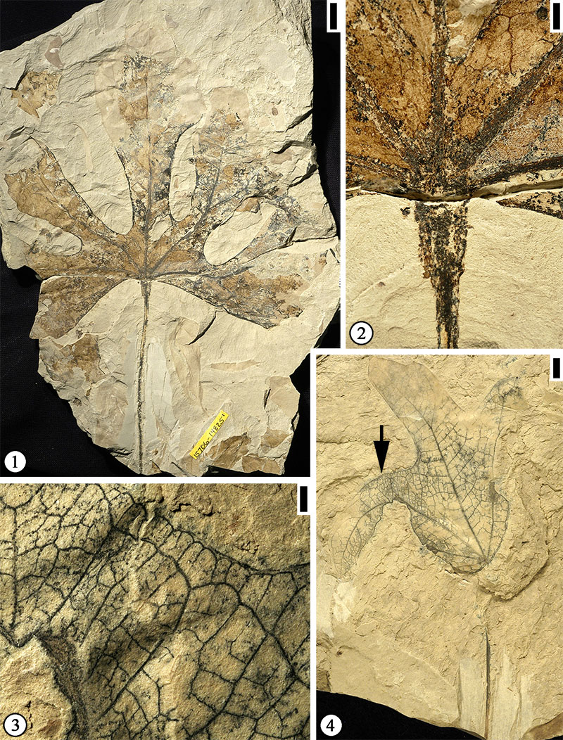
FIGURE 23. Wingia expansolobum (Upchurch and Dilcher) comb. nov. 1, UF15706-14828, specimen showing a multi-lobed leaf. Scale bar equals 1 cm. 2, Enlargement of Figure 23.1 (area indicated by the upper arrow) to show secondary and tertiary venation. Scale bar equals 1 mm. 3, Enlargement of Figure 23.1 (area indicated by the lower arrow) to show venation near the lamina margin. Scale bar equals 2 mm.
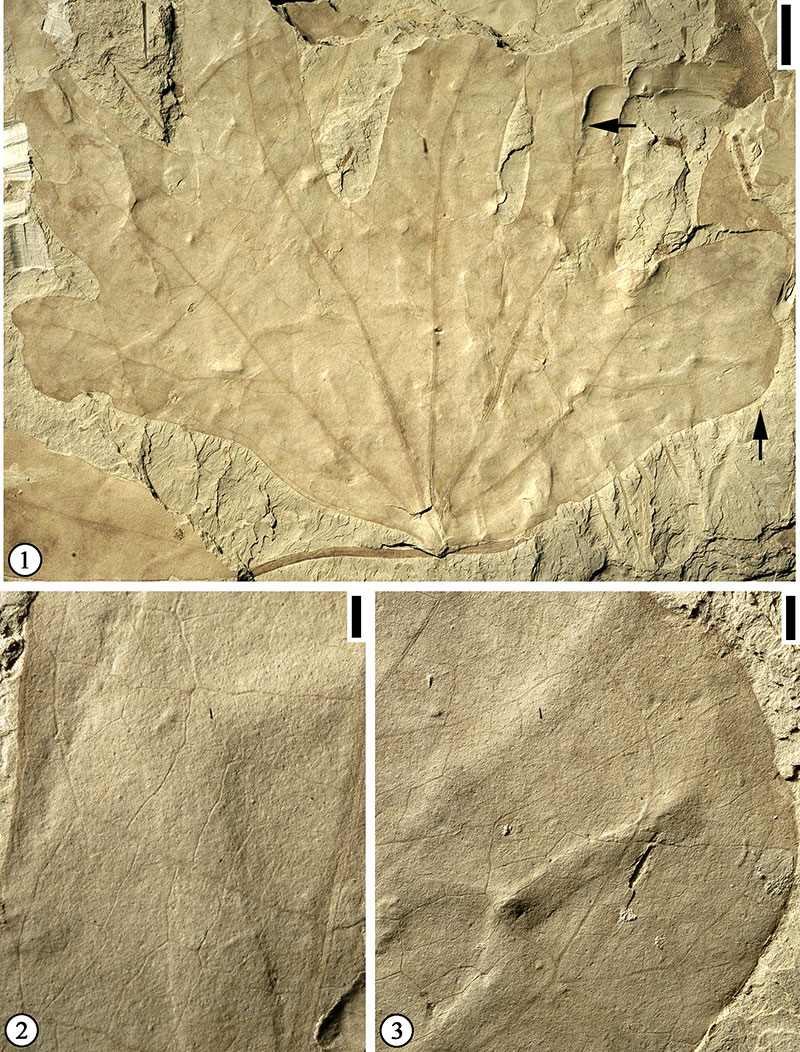
FIGURE 24. Wingia cf. expansolobum (Upchurch and Dilcher) comb. nov. 1, UF15706-14824, specimen showing two complete and two broken lobes. Note thin venation and glandular teeth. Scale bar equals 3 mm. 2, UF15706-24460, specimen showing five lobes. Note that two lobes (indicated by arrows) are broken. Scale bar equals 1 cm. 3, Enlargement of Figure 24.1 (area indicated by arrow) to show glandular teeth. Scale bar equals 1 mm.
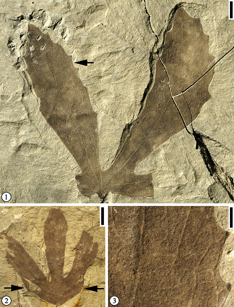
FIGURE 25. Dicotylophyllum skogii sp. nov. 1, UF15706-24573, fragment of leaf to show the petiole and strong primary vein. Scale bar equals 1 cm. 2, Enlargement of Figure 25.1 (area indicated by the upper arrow) to show secondary and intersecondary veins. Scale bar equals 2 mm. 3, Enlargement of the base of the leaf in Figure 25.1 (are indicated by the lower arrow) to show venation. Scale bar equals 2 mm.
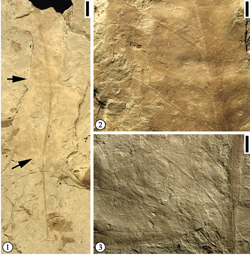
FIGURE 26. Dicotylophyllum leptovenum Wang and Dilcher, 2009. 1, UF15706-24734, incomplete leaf showing strong primary vein and asymmetric lamina base. Scale bar equals 5 mm. 2, Enlargement of Figure 26.1 to show secondary and tertiary veins. Scale bar equals 1 mm.