FIGURE 1. The late Wasatchian paromomyid Phenacolemur citatus. 1: USGS 6573 (original fossil), right maxilla with P3 -M2. 2-4: USGS 21712 (cast), left dentary with P4 -M3 in occlusal (2), buccal (3), and lingual (4) views.
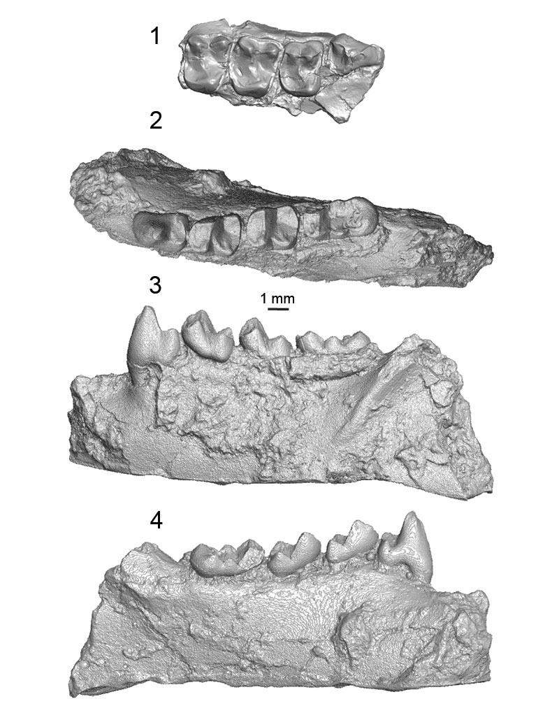
FIGURE 2. Holotype of Trogolemur myodes, AMNH 12599 (Matthew, 1909: plate LII, figure 5). Right dentary with P2 -M3. 1: occlusal view; 2: buccal view; 3: lingual view.
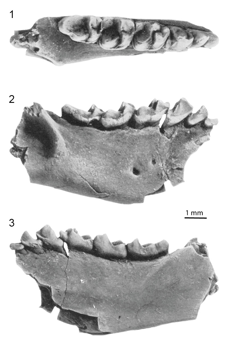
FIGURE 3. Reconstruction of western North America from 40 million years ago. Orange dot indicates the paleoposition of the sites discussed in this paper, whereas the red dot indicates the current relative position of the sites. The map is reproduced with R. Blakey’s permission (Colorado Plateau Geosystems, Inc.).
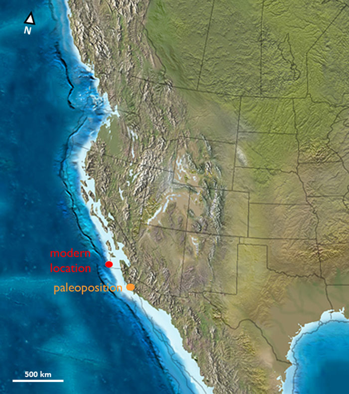
FIGURE 4. Environmental SEM images of four teeth of Walshina esmaraldensis gen. et sp. nov. 1: left M3, SDSNH 76276; 2: right M1, SDSNH 76337; 3: right M2, SDSNH 76338; 4: right M3, SDSNH 72583. All teeth are in occlusal view. Arrow indicates the location of the fovea.
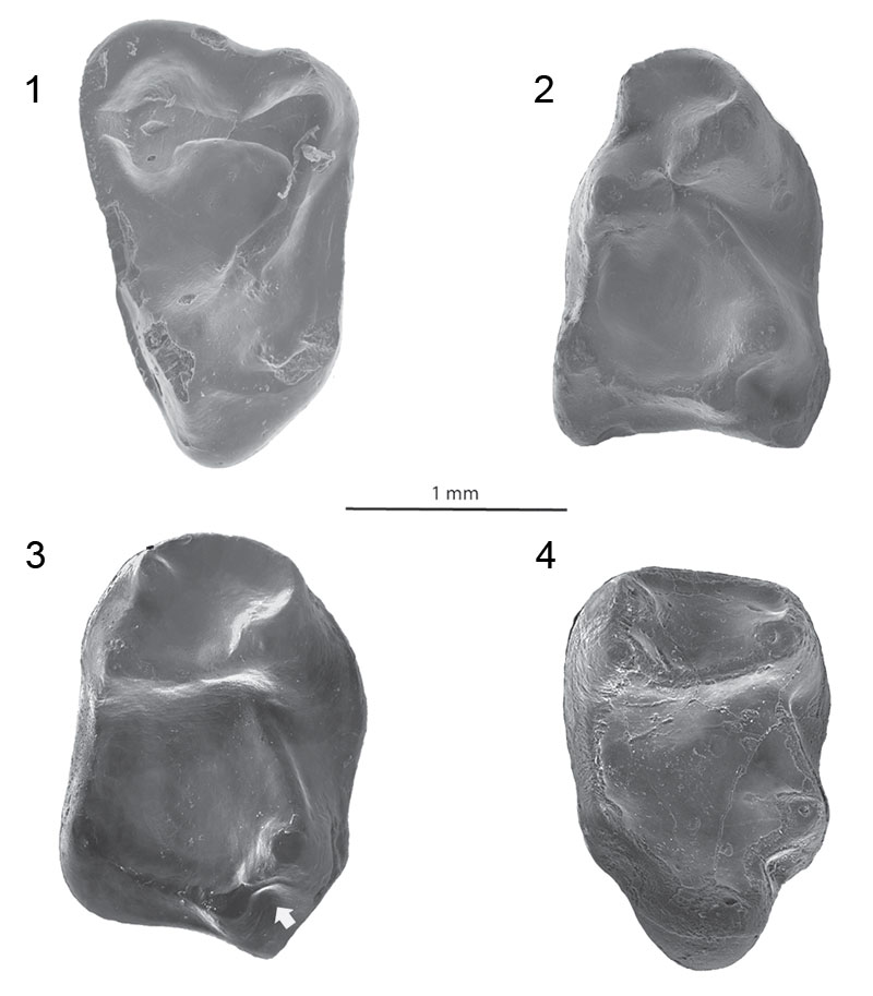
FIGURE 5. Photographs taken with digital camera (1, 4-11) and Micro-CT scan reconstructions generated using Avizo 7 (2, 3). Walshina shifrae comb. nov. (1, 4, 7, 10) - 1: right M1, CM 15797 (holotype; mirrored), in occlusal view; 4: left M2, CM 15103, in occlusal view; 7: left M2, CM 21637, in occlusal view; 10: left M3; CM 15726, in occlusal view. Walshina esmaraldensis gen. et sp. nov. (2, 5) - 2: left M1, LACM 40198 (holotype), in occlusal view; 5: left M2, SDSNH 62850, in occlusal view. Walshina mcgrewi comb. nov. (3, 6, 8, 9, 11) - 3: left M1, CM 15635 (holotype), in occlusal view; 6: left M2, CM15794, in occlusal view; 8, 9, 11: left mandibular fragment with M2, CM 29005, in occlusal (8), buccal (9), and lingual (11) views.
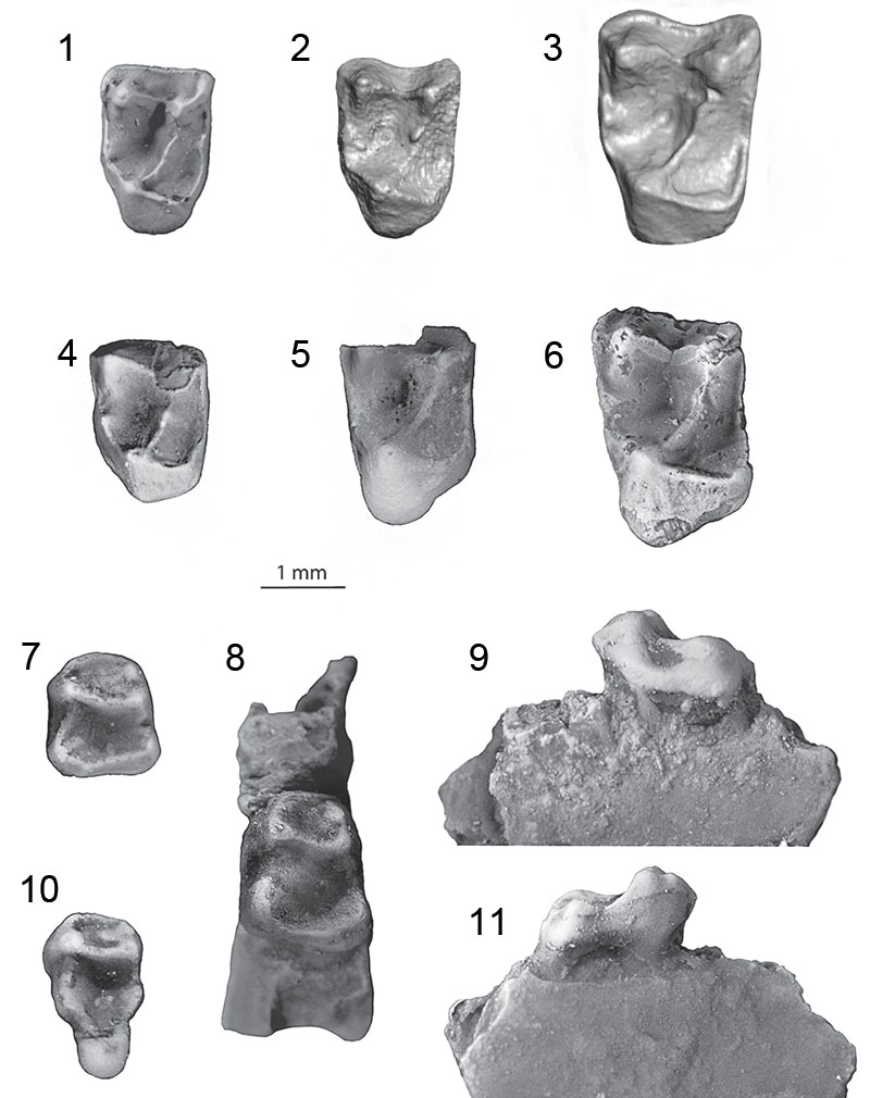
FIGURE 6. Micro-CT scan reconstructions of specimens of Walshina esmaraldensis gen. et sp. nov. generated using Avizo 7: 1: left M1, LACM 40198 (holotype), in occlusal view; 2: left M2, SDSNH 87336, in occlusal view; 3: lingual fragment of a left M2, SDSNH 87337, in occlusal view; 4: lingual fragment of a right M2, SDSNH 42268, in occlusal view; 5, 6, 9, 10: left M2, SDSNH 87332, in occlusal (5), buccal (6), mesial (9) and lingual (10) views; 7, 8, 11, 12: mesial fragment of a left M1, SDSNH 87331, in occlusal (7), buccal (8), mesial (11) and lingual (12) views; 13, 14, 17, 18: left M3, SDSNH 87334, in buccal (13), occlusal (14), lingual (17) and mesial (18) views; 15, 16, 19: distal fragment of a right M3, SDSNH 87335, in buccal (15), occlusal (16) and lingual (19) views.
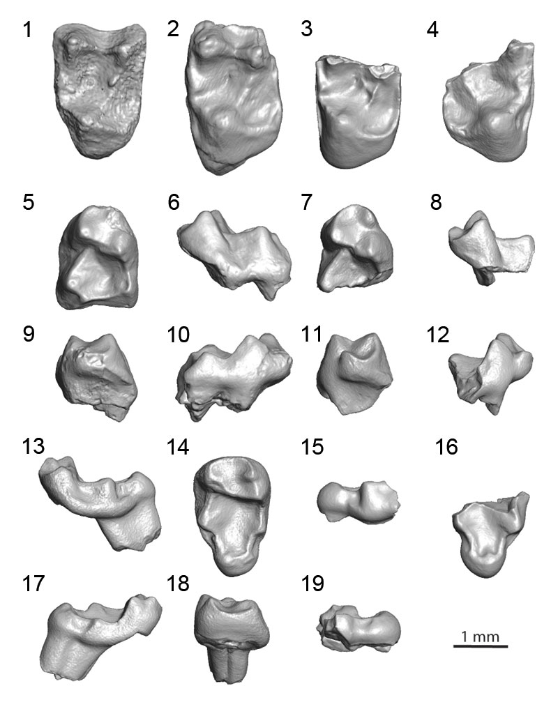
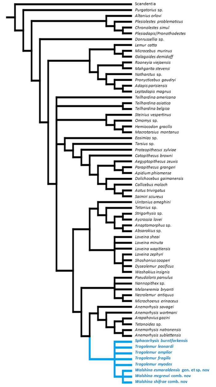
FIGURE 7. Hypothesis of relationships of Walshina gen. nov. in the context of the Order Primates. Strict consensus cladogram based on data modified from Holroyd and Strait (2008), including the addition of seven newly-coded trogolemurins (Trogolemur amplior, Tr. fragilis, Tr. leonardi, Sphacorhysis burntforkensis, Walshina esmaraldensis gen. et sp. nov., W. mcgrewi comb. nov., and W. shifrae comb. nov.), a microchoerine (Melaneremia bryanti), and three anaptomorphins (Anemorhysis sublettensis, An. wortmani, and An. natronensis). Trogolemurins are marked in blue.