FIGURE 1. Map showing the area covered by the Galatian Volcanic Province (GVP) (or Köroglu Volcanics). Sites discussed in this work are marked by green circles: ELM, Elmali Village; SOG, Soguksu National Park; BUG, Bugralar Village; INO, Inozu Valley South Side; INL, Inozu Valley North Side; KAR, Karasar Village; MEN, Menceler Plateau; KIR, Kiraluc Sitenear Nuhhoca Village; AGU, Asagiguney Village; KUZ, Kuzca Village. Yellow circles (PEL, Pelitcik Village; GUD, Gudul; HOC, Hoçaş Village; KOZ, Kozyaka Village) indicate those sites discussed in Akkemik et al. (2009, 2016, 2017). The sites located in the western part (INO, INL, KAR, MEN, KIR, AGU, KUZ, HOC and KUZ) are from early-middle Burdigalian and Hancili Formation (Altun et al., 2002; Akbaş et al., 2002); the sites in the central part (GUD, BUG, ELM, PEL and SOG) are from middle-late Burdigalian and Pazar Formation (Kazancı, 2012; Sen et al., 2017).
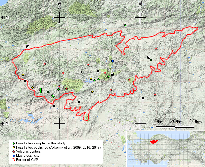
FIGURE 2. Stratigraphic columns of the GVP showing the fossils (dark symbols) originating from the Hançili Formation of the western part (1-2) (Akbaş et al., 2002; Altun et al., 2002) and from within the pyroclastic layers of the Pazar Formation in the central part of the GVP at Çamlıdere-Pelitcik (3) and Kızılcahamam (4). Columns redrawn from figure 3 for Kızılcahamam and figure 6 for Pelitcik of Kazancı (2012) and figure 2 of Şen et al. (2017).
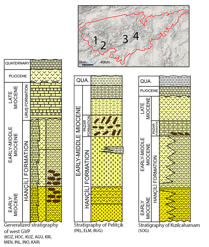
FIGURE 3. Examples of petrified wood specimens. 1, Example of small wood samples found at the Bugralar Fossil Site (BUG), from the central part of GVP, with an age of middle-late Burdigalian. 2, Petrified woods found in their life position in Asagiguney Site (AGU). 3, Material found in Kuzca Village (KUZ) in the west part of the GVP of early-middle Burdigalian age.
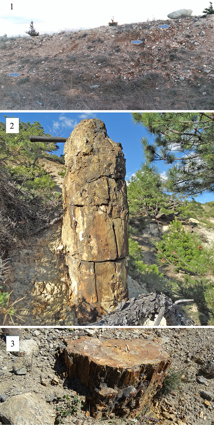
FIGURE 4. Example of allochthonous petrified woods from Kuzca Village (KUZ) in the Galatian Volcanic Province.
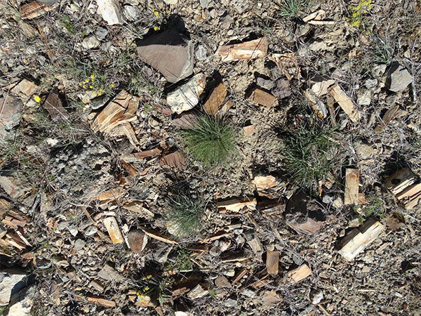
FIGURE 5. Juniperus sp. 1, TS showing growth rings with false rings in summer wood. 2, Close-up of tracheidal arrangement within one growth ring (not inter-tracheidal spaces) and uniseriate rays (TS). 3, RLS showing procumbent cells comprising the ray. 4, RLS showing cupressoid type cross-field pits (RLS). 5, TLS showing axial parenchyma cells with nodular end walls.
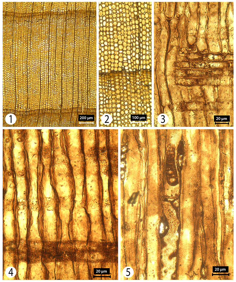
FIGURE 6. Sequoia sp. 1, TS showing distorted (preservational feature) growth rings with clear latewood. 2, TS of one growth ring showing a late wood zone of up to 11 rows of tracheids. 3, TLS showing uniseriate, homogenous rays. 4, TLS showing axial parenchyma cells with smooth end walls and dark contents (preservational feature). 5, Rays with taxodioid type cross-field pits (RLS). 6, Bordered pits in one and two rows with crassulae visible (arrow) (TLS).
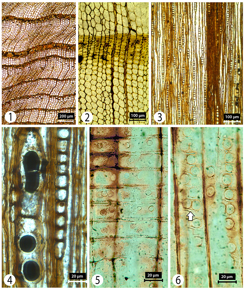
FIGURE 7. Taxodium. 1, TS showing very narrow, indistinct growing rings. 2, TS showing latewood zones of only 2 rows of tracheids. 3-4, TLS showing uniseriate rays. 5, Rays with taxodioid type cross-field pits (RLS).
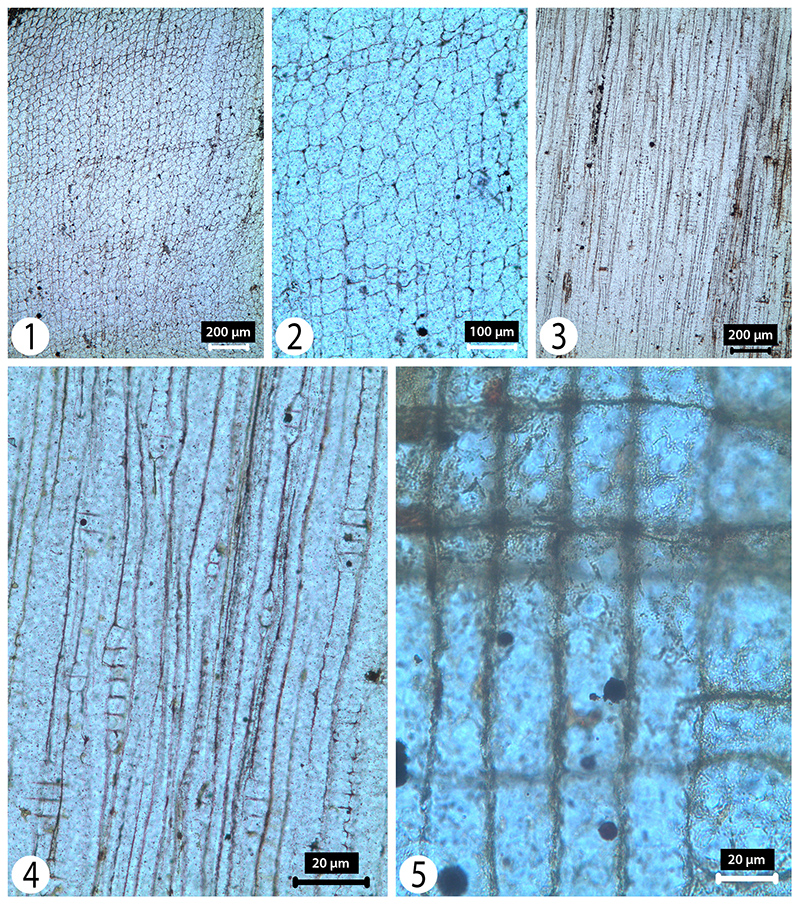
FIGURE 8. Cedrus. 1, TS showing clear growth rings and no (traumatic) resin canals. 2, TS showing growth rings and associated traumatic resin canals. 3, TLS showing uniseriate rays. 4, RLS showing rays with ray tracheids (white arrow) and cupressoid type cross-field pits. 5, Bordered pits with scalloped tori (RLS) (black arrow).
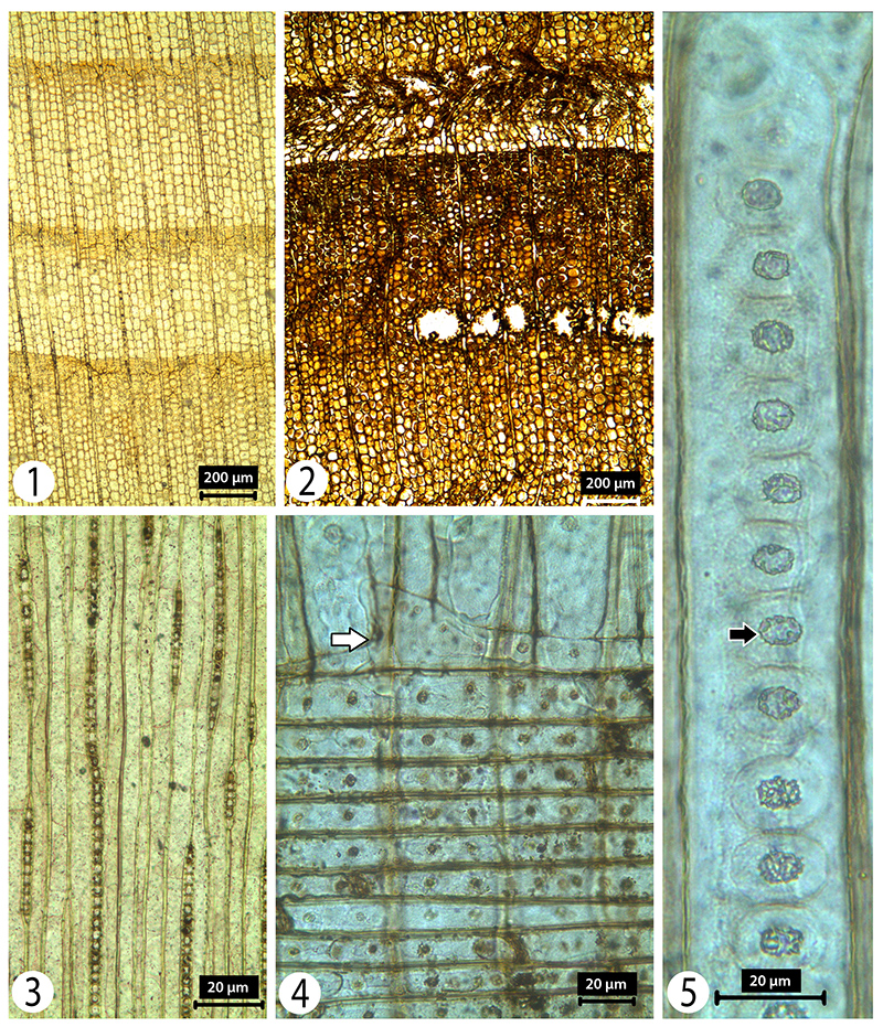
FIGURE 9. Picea. 1, TS showing wide growth rings and resin canal. 2, TS resin canal with no visible epithelial cells (white arrow). 3, TLS showing a ray without any horizontal resin canals and tracheids with helical thickenings. 4, TLS showing a ray with a horizontal resin canal and helical thickenings in the tracheids. 5, Cupressoid type cross-field pits. 6, Ray tracheid (black arrow) with smooth wall and cupressoid type pits in the cross-fields (RLS).
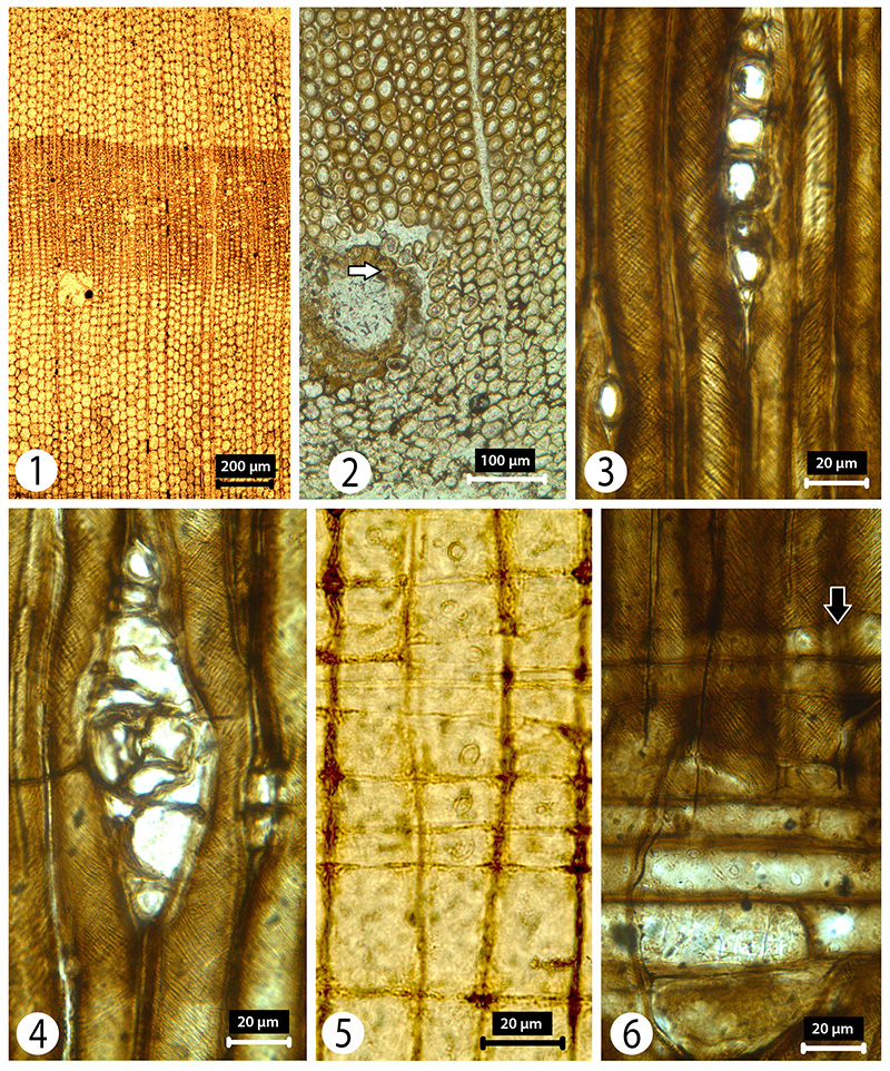
FIGURE 10. Pinus. 1, TS showing wide growth rings and resin canal. 2, TS resin canal with no visible epithelial cells (white arrow). 3-4, Rays with and without horizontal resin canal and tracheids (TLS). 5, RLS showing procumbent cells forming the ray. 6, Pinoid type cross-field pits, and ray tracheids (black arrow) with smooth walls.
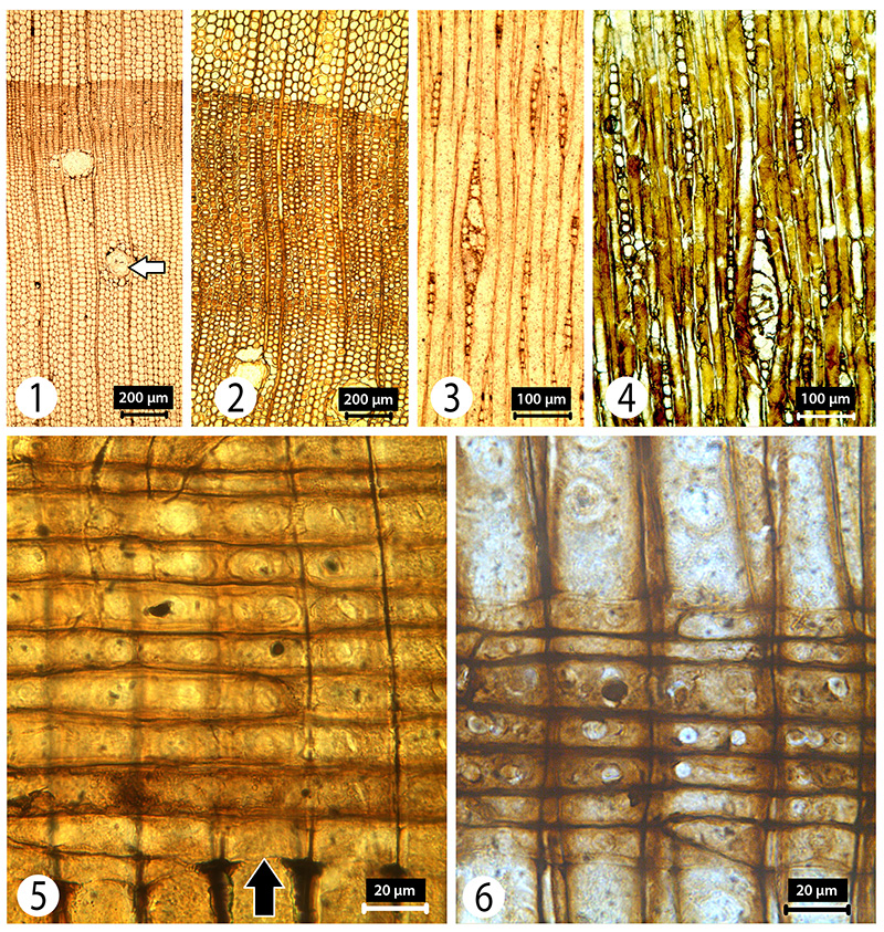
FIGURE 11. Podocarpus. 1, TS across one growth ring with diffuse axial parenchyma cells. 2, Homogenous uniseriate rays (TLS). 3, RLS showing large cupressoid and taxodioid type cross-field pits with two cross-field pits shown in the upper box. Left indicates the borders of pit drawn, right shows the oblique appearance of the pit. 4, RLS showing procumbent nature of the cells forming the ray, and cross-field pits; white arrow indicates the oblique position of the pit; black arrow shows vertical pit. 5, Radial pitting with one row in large (earlywood) tracheids. 6, Axial parenchyma cells with thin end walls (vertical arrow).
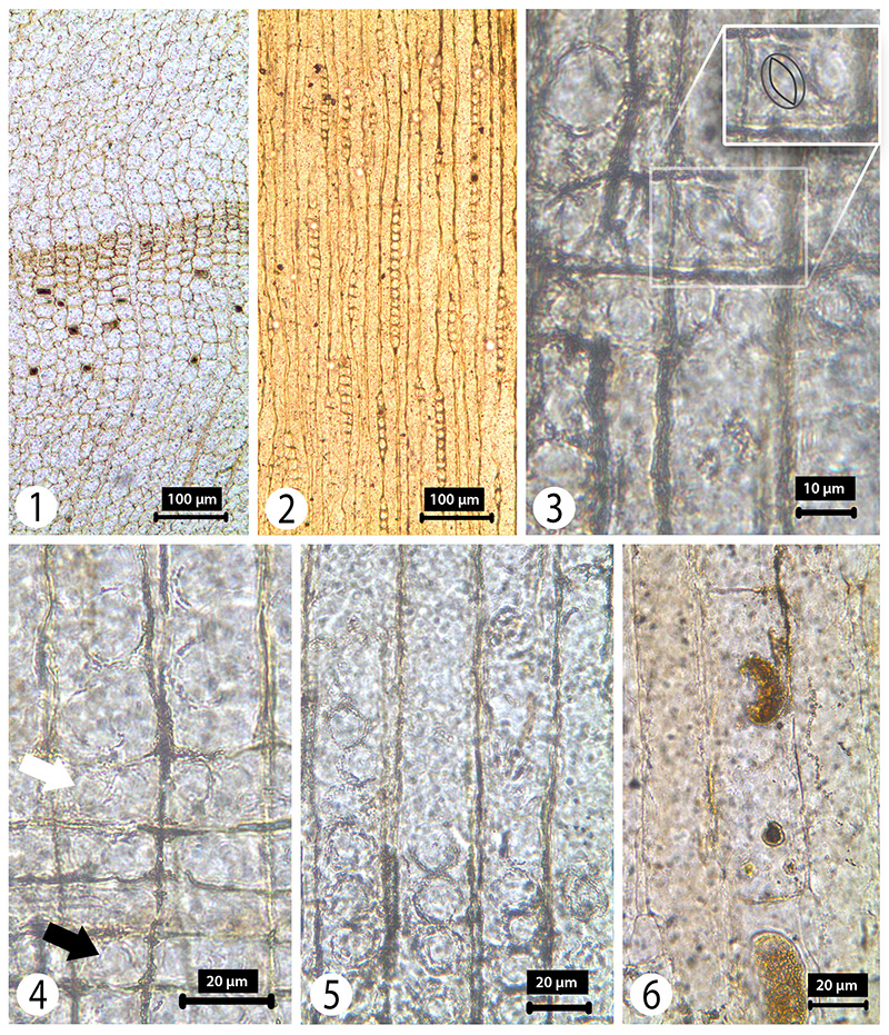
FIGURE 12. Quercus (section Ilex). 1, TS showing solitary vessels with tyloses and one multiseriate ray. 2, TS showing radial alignment of the solitary vessels and uniseriate rays. 3, TLS showing rays of two sizes. 4, RLS of rays with procumbent cells, vessel with a simple perforation plate (black arrow) and filled with tyloses (white arrow).
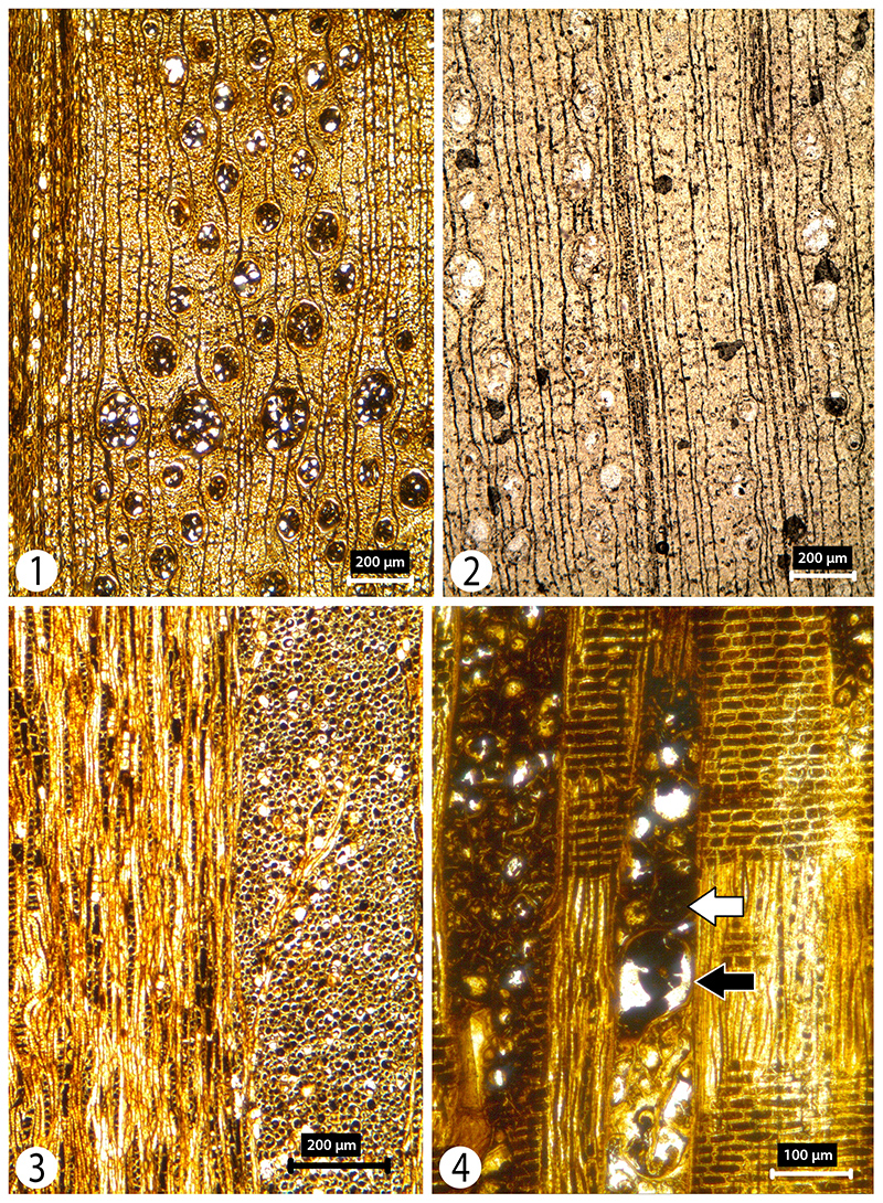
FIGURE 13. Fraxinus. 1, TS showing ring porous nature of the wood. 2, RLS of triseriate rays. 3, TS showing distortion in the early wood caused during preservation. 4, Rays with both procumbent and upright cells (RLS). 5, Axial parenchyma cells and a vessel with alternate bordered pits.
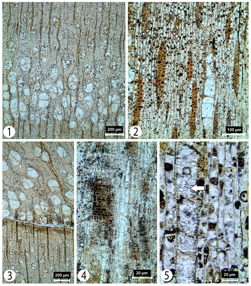
FIGURE 14. Prunus. 1, TS showing relatively unclear preservation caused by distortion during preservation and ring-porous vessels. 2, TLS showing the multiseriate ray arrangement. 3, TLS showing the uniseriate rays between the multiseriate rays. 4, RLS showing the poor preservation of the rays but some cellular detail visible. 5, Better preserved rays with procumbent and upright cells, and simple perforation plate (RLS) (arrow) visible.
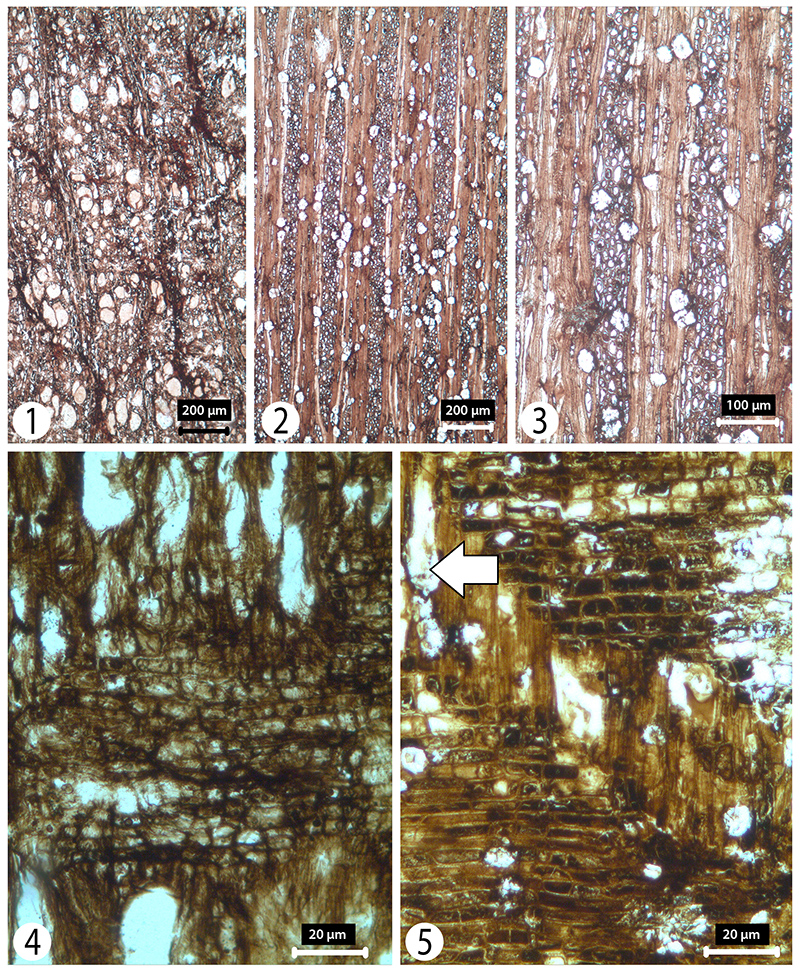
FIGURE 15. Salix. 1, TS showing two growth rings and diffuse porosity of the vessels. 2, TS showing the solitary and paired nature of the vessels. 3, RLS showing ray with both procumbent and upright cells. 4, TLS showing uniseriate rays and vessels.
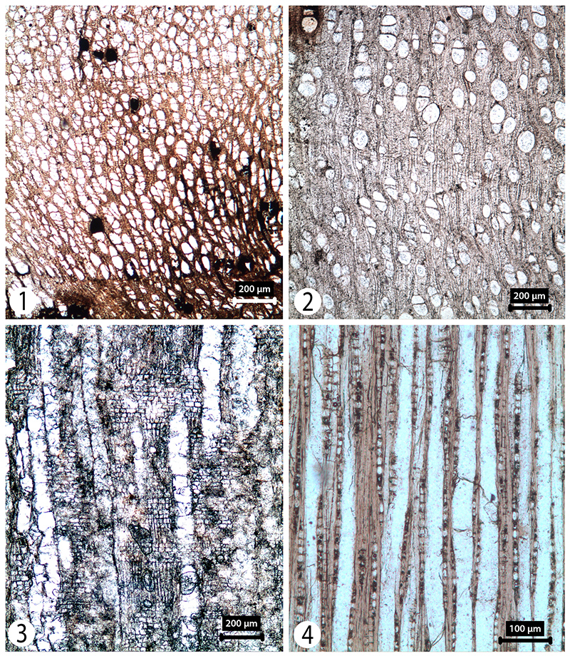
FIGURE 16. Acer. 1, TS showing diffuse porous arrangement of the vessels. 2, TLS showing multiseriate ray arrangement. 3, TLS showing rays of four cells wide and helical thickenings in the vessel wall. 4, RLS showing alternate arrangement of bordered pits on the surface of vessels. 5, RLS showing simple perforation plate and ray with procumbent cells.
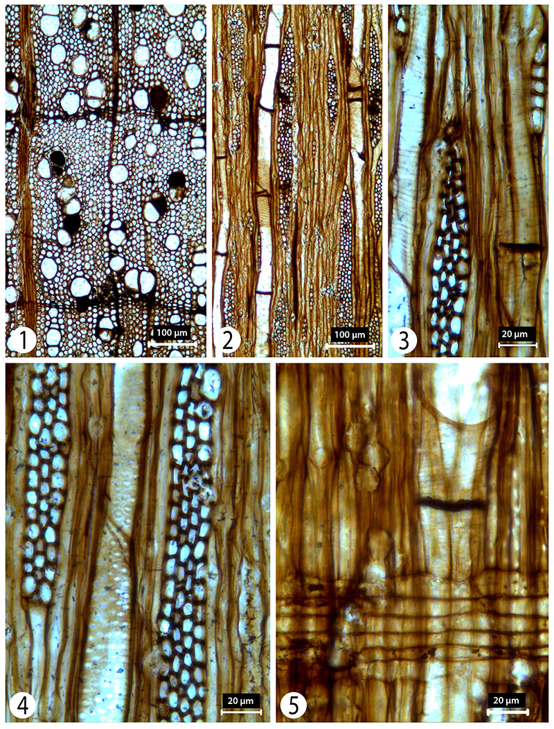
FIGURE 17. Ulmus with poor anatomical preservation. 1-2, TS showing ring porous nature of the earlywood (2) and wavy bands of vessels forming more or less tangential rows in the latewood (1). 3, TLS showing rays of up to 4-5 cells in width. 4, RLS showing rays with procumbent ray cells. 5, RLS showing simple perforation plates (arrows).
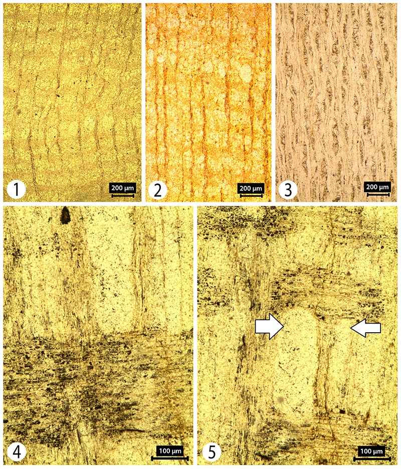
FIGURE 18. Zelkova. 1, TS showing the ring porous nature of the wood. 2, TS showing the vessel arrangement. 3, RLS of crystals in outer two rows of marginal ray cells. 4, RLS showing vessels having obvious helical thickening. 5, TLS showing rays up to 6 cells width and with crystals in their marginal cells (white arrow). 6, RLS of rays with procumbent ray cells and one row of upright marginal cells, simple perforation plates (grey arrow) in vessels and crystals.

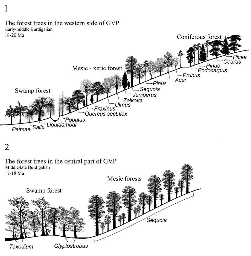
FIGURE 19. 1-2, Summary reconstruction of the two distinct vegetational groups. The elevation line shows possible elevation. The riparian forest vegetation growing on the western side of the GVP at 20-18 Ma based on trees identified from the eight distinct localities (1). Compare this with (2) which summarizes the composition of the more or less contemporaneous swamp forest vegetation (18-17 Ma) growing across the central part of the GVP. (Note that ‘Sequoia’ type wood can include one or more of Sequoia, Sequoiadendron and Metasequoia).