FIGURE 1. Skull of African Lion (Panthera leo, author’s private collection) showing anatomy of vomer and vomerine notch. Turbinates in this specimen are largely missing. 1, anterior view; 2, ventral view; 3, detail of anterior nasal cavity; 4, detail of internal narial region. Abbreviations: IN, internal nares; Ps, presphenoid; V, vomer; VN, vomerine notch, the presumed functional internal nares (N) in all other figures. Rectangles superimposed on upper photographs show areas of detail below. In 3 and 4 the vomer has been lightened for better clarity.
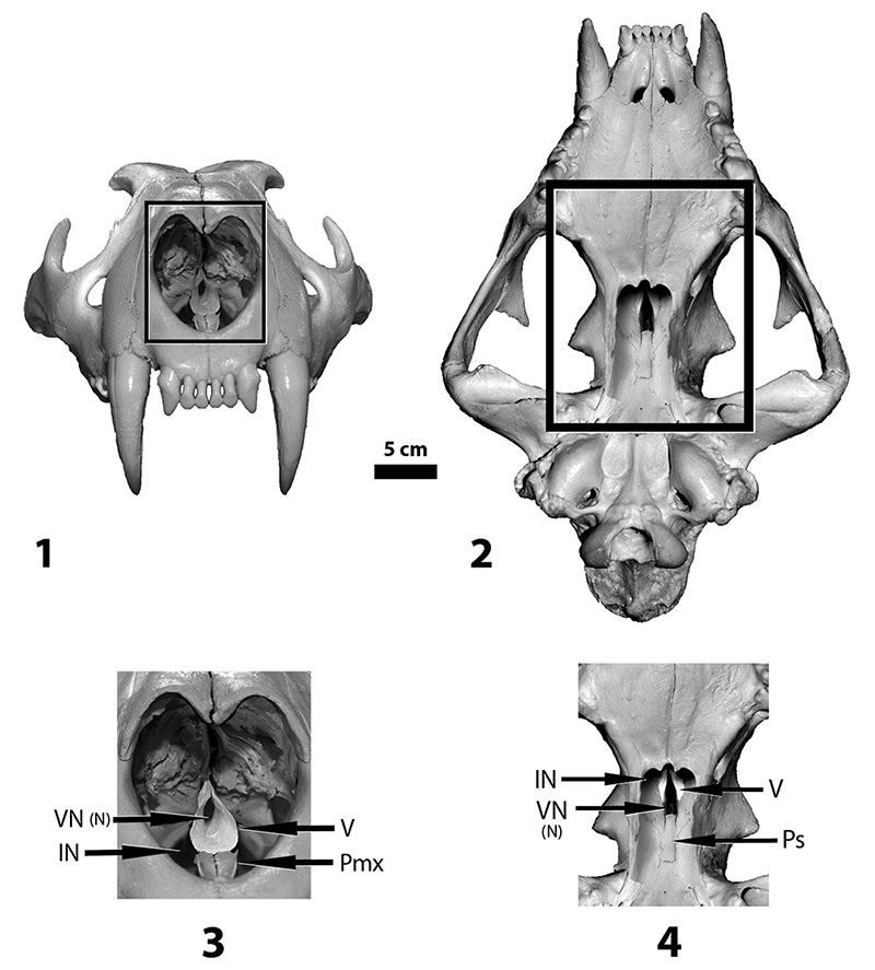
FIGURE 2. Skull of Metarhinus (UCM 44939). 1, entire skull in ventral view; 2, enlargement of the nasopharyngeal region. Rectangle superimposed on left photograph shows area of detail. Abbreviations: IN, internal nares (non-functional); N, functional internal nares (presumably the vomerine notch); PD, palatal depression (secondary palatal plate); V, vomer; VP, vomerine plate.
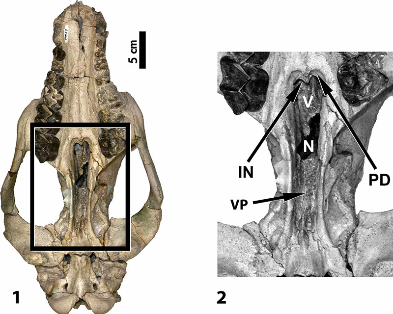
FIGURE 3. Semi-diagrammatic reconstruction of the skull of Metarhinus seen in sagittal section (based on UCM 44939 and FMNH PM 1732). Abbreviations: Bo, basioccipital; Bs, basisphenoid; E, ethmoid; IN, anatomical internal nares (non-functional); Mx, maxilla; N, functional internal nares (vomerine notch); pkt, blind pocket in lateral wall of nasal cavity; Pmx, premaxilla; Ps, presphenoid; V, vomer. Large arrow shows air pathway through the nasal cavity. Dark gray areas with hatch marks indicate cut surfaces.
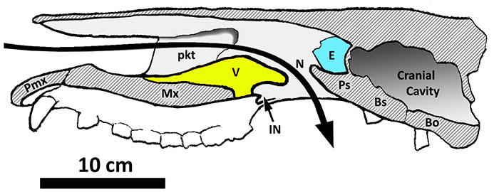
FIGURE 4. AMNH 13164, skull of Sphenocoelus (= Dolichorhinus) in ventral view. 1, Skull as it actually appears; 2, reconstruction of skull showing how it might have appeared when fully intact and prior to taphonomic distortion; 3, detail of reconstructed internal narial region (functional internal nares darkened). Rectangle superimposed on right photograph shows area of detail. Abbreviations: IN, anatomical internal nares (non-functional); N, functional internal nares (vomerine notch); PD, palatal depression (secondary palatal plate); V, vomer; VP, vomerine plate.
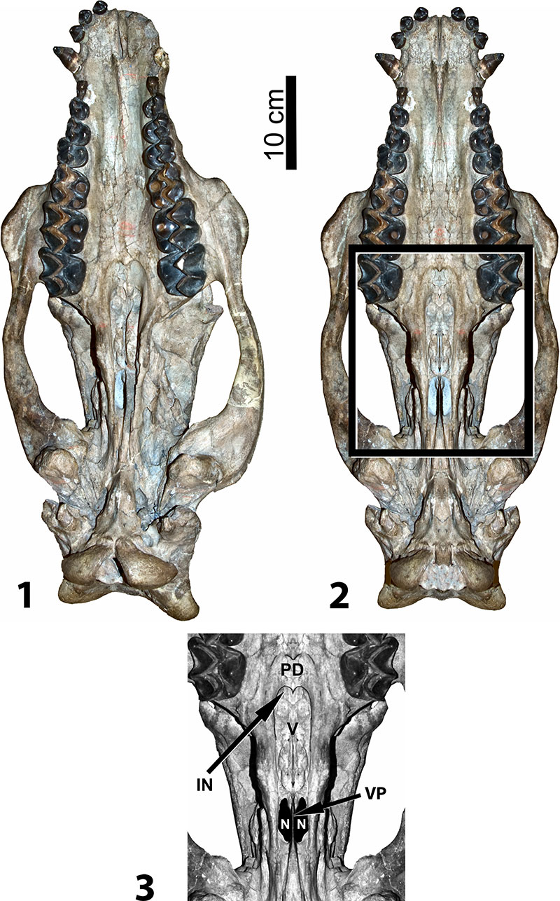
FIGURE 5. AMNH 1851, skull of Sphenocoelus (= Dolichorhinus). 1, left side of skull showing external morphology; 2, right side of skull showing sagittal section (nasal is removed and matrix artificially lightened for better contrast); 3, semi-diagrammatic reconstruction of sagittal section showing presumed air stream. Abbreviations: Bo, basioccipital; Bs, basisphenoid; E, ethmoid; ET, ethmoturbinate; IN, anatomical internal nares (non-functional); Mx, maxilla; Mx (cr), crest of maxilla; N, functional internal nares (vomerine notch); Ps, presphenoid; V, vomer; VNO, area of vomeronasal organ.
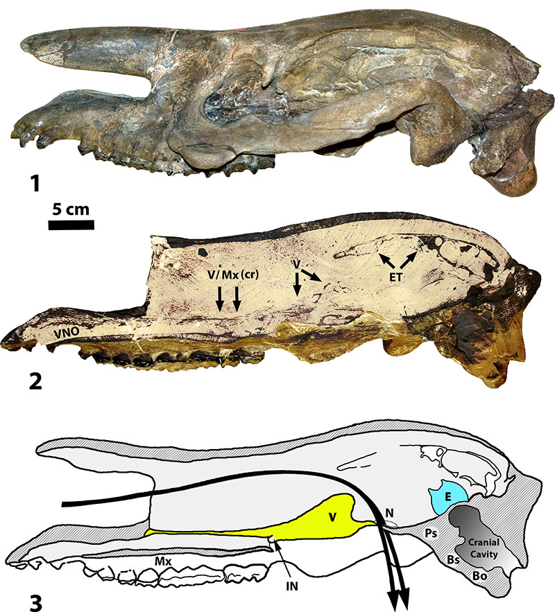
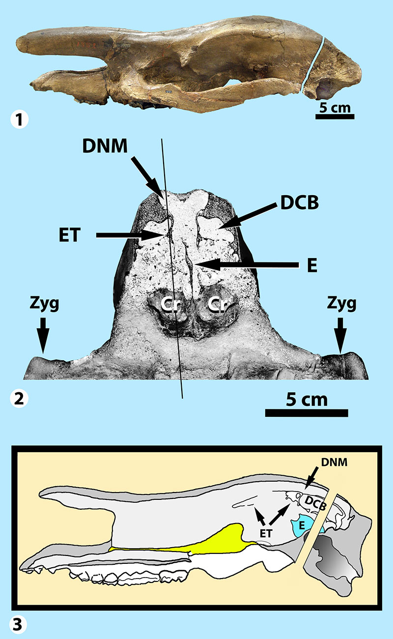
FIGURE 6. Skulls of Sphenocoelus (= Dolichorhinus). 1, AMNH 1852 in lateral view showing location of transverse section near back of skull; 2, transverse section of AMNH 1852 through anterior-most part of braincase looking anteriorly; 3, reconstructed sagittal section of AMNH 1851 (from Figure 5.3) showing the approximate plane of the transverse section in AMNH 1852 relative to internal structures. Abbreviations: Cr, cribriform plate of ethmoid; DCB, dorsal conchal bulla; DNM, dorsal nasal meatus; E, ethmoid (perpendicular plate); ET, ethmoturbinate; Zyg, zygomatic arch. Thin diagonal line in middle image approximates the plane of the sagittal section shown in Figure 5.2.