FIGURE 1. Locality map, showing North Island of New Zealand and the sample sites.
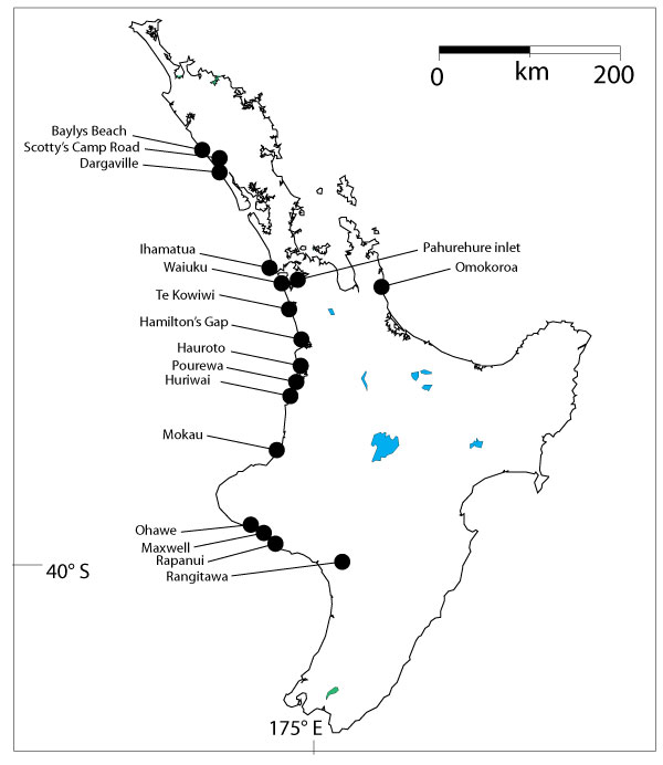
FIGURE 2. Approximate ages of the deposits, with time divisions mostly after Raine et al. (2015), which largely follows Beu (2001). Some of the system of Carter and Naish (1998b) is also included.
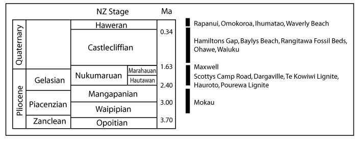
FIGURE 3. Summary of distribution of taxa in the samples. Note some Rangitawa samples have been grouped; Rangitawa-4 and -5 are from float, of unclear origin, Rangitawa-6 is probably Rangitawa Fossil Beds, Rangitawa-7 and -11 are from clear Rangitawa Fossil Beds, and Rangitawa-8, -9 and -10 are from the Rangitawa Pumice.
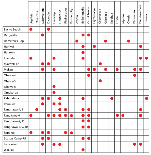
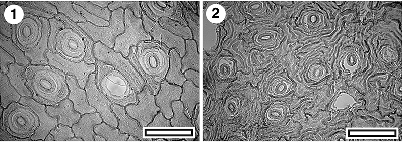
FIGURE 4. Cuticle of Agathis australis and Araucariaceae indet (all TLM); 1. Agathis australis, stomatal rows (SL4401, Baylys Beach, scale bar equals 100 µm); 2. A. australis, detail of stomatal complex showing ‘spoked’ appearance (SL4401, Baylys Beach, scale bar equals 50 µm); 3. Araucariaceae gen. indet. showing stomatal rows (SL4641, Rangitawa-5, scale bar equals 100 µm); 4. Araucariaceae gen. indet. detail of stomatal complex, note lack of ‘spoked’ appearance (SL4641, Rangitawa-5, scale bar equals 100 µm).
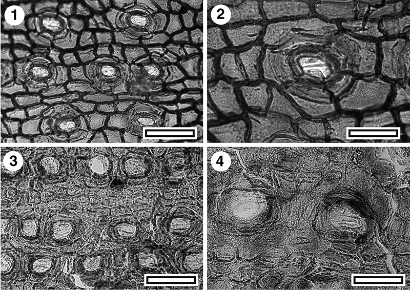
FIGURE 5. Cuticle of Libocedrus sp. (Cupressaceae); 1. SEM view of inner surface, note lateral subsidiary cells from adjacent stomatal complexes touching, and encompassing a shared polar subsidiary cell (S-1651, Rangitawa-6, scale bar equals10 µm); 2. SEM view of outer surface (S-1651, Rangitawa-6, scale bar equals20 µm); 3. TLM view (SL4504, Pahurehure, scale bar equals 50 µm); 4. TLM view, note prominent rim of fused papillae and numerous individual papillae (SL5448, Rangitawa-6, scale bar equals 50 µm); 5. TLM view (SL4503, Pahurehure, scale bar equals 50 µm); 6. TLM view, note some dicylic stomatal complexes (SL4508, Pahurehure, scale bar equals 50 µm).
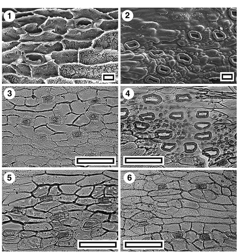
FIGURE 6. Cuticle of Dacrycapus and Dacrydium (Podocarpaceae, all TLM); 1. Dacrycapus dacrydioides, note very smooth and relatively thin anticlinal cell walls (SL2345, Mokau, scale bar equals 50 µm); 2. D.acrycapus dacrydioides, detail of stomatal complex (SL2345, Mokau, scale bar equals 20 µm); 3. Dacrydium cupressinum, note relatively robust and rough anticlinal cell walls (SL4511, Pahurehure, scale bar equals 50 µm); 4. D. cupressinum, detail of stomatal complex (SL4511, Pahurehure, scale bar equals 20 µm).
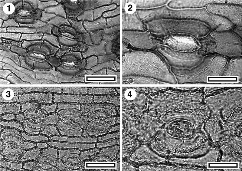
FIGURE 7. Cuticle of Halocarpus bidwilli ( Podocarpaceae, all Rangitawa-6); 1. TLM view of stomatal complexes, note essentially random orientation (SL5454, scale bar equals 50 µm); 2. TLM view, detail of stomatal complex (SL46621, scale bar equals 20 µm); 3. SEM view of outer surface showing sunken stomatal complexes (S-1650, scale bar equals 20 µm); 4. SEM view of inner surface (S-1650, scale bar equals 10 µm).
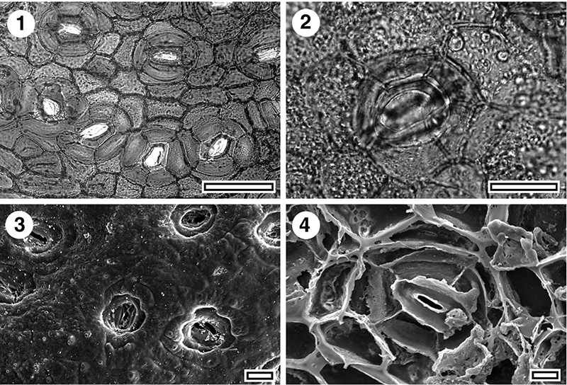
FIGURE 8. Cuticle of Phyllocladus. sp. (Podocarpaceae, Rangitawa-6, all TLM); 1. Note multiple rows of stomatal complexes (SL4650, scale bar equals 100 µm); 2. Detail of stomatal complexes, note ring of thin cuticle around the guard cells (SL4650, scale bar equals 50 µm).
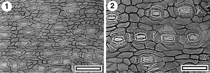
FIGURE 9. Cuticle of Prumnopitys taxifolia (Podocarpaceae, all Rangitawa-6) and indet, conifer (all Mokau); 1. P. taxifolia, TLM view (SL4981, Rangitawa-6, scale bar equals 50 µm); 2. P. taxifolia, TLM view, detail of stomatal complex (SL4981, Rangitawa-6, scale bar equals 20 µm); 3. P. taxifolia, SEM view of inner surface, note marked ‘bulging’ of stomatal complex outline (S-1656, Rangitawa-6, scale bar equals 10 µm); 4. P. taxifolia, SEM view of outer surface (S-1656, Rangitawa-6, scale bar equals 10 µm); 5. Indet. conifer, TLM view (SL2355, Mokau, scale bar equals 50 µm); 6. Indet. conifer, TLM view (SL2355, Mokau, scale bar equals 50 µm); 7. Indet. conifer, SEM view of inner surface, note clear buttressing (S-1657, Mokau, scale bar equals 20 µm); 8. Indet. conifer, SEM view of outer surface (S-1657, Mokau, scale bar equals 20 µm).
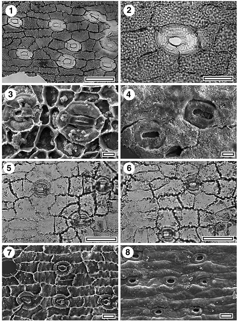
FIGURE 10. Cuticle of Phormium tenax (Asphodelaceae); 1. TLM view (SL4983 Rangitawa-6, scale bar equals 200 µm); 2. TLM view (SL4983 Rangitawa-6, scale bar equals 100 µm); 3. TLM view (SL4649, Rangitawa-6, scale bar equals 50 µm); 4. TLM view, detail of stomatal complexes (SL4649, Rangitawa-6, scale bar equals 20 µm); 5. SEM view of inner surface (S-1655, Rangitawa-6, scale bar equals 10 µm); 6. SEM view of outer surface, note marked alternation of stomatal complexes (S-1655, Rangitawa-6, scale bar equals 10 µm); 7. TLM view (SL2353, Mokau, scale bar equals 50 µm); 8. TLM view, detail of stomatal complexes (SL2353, Mokau, scale bar equals 20 µm).
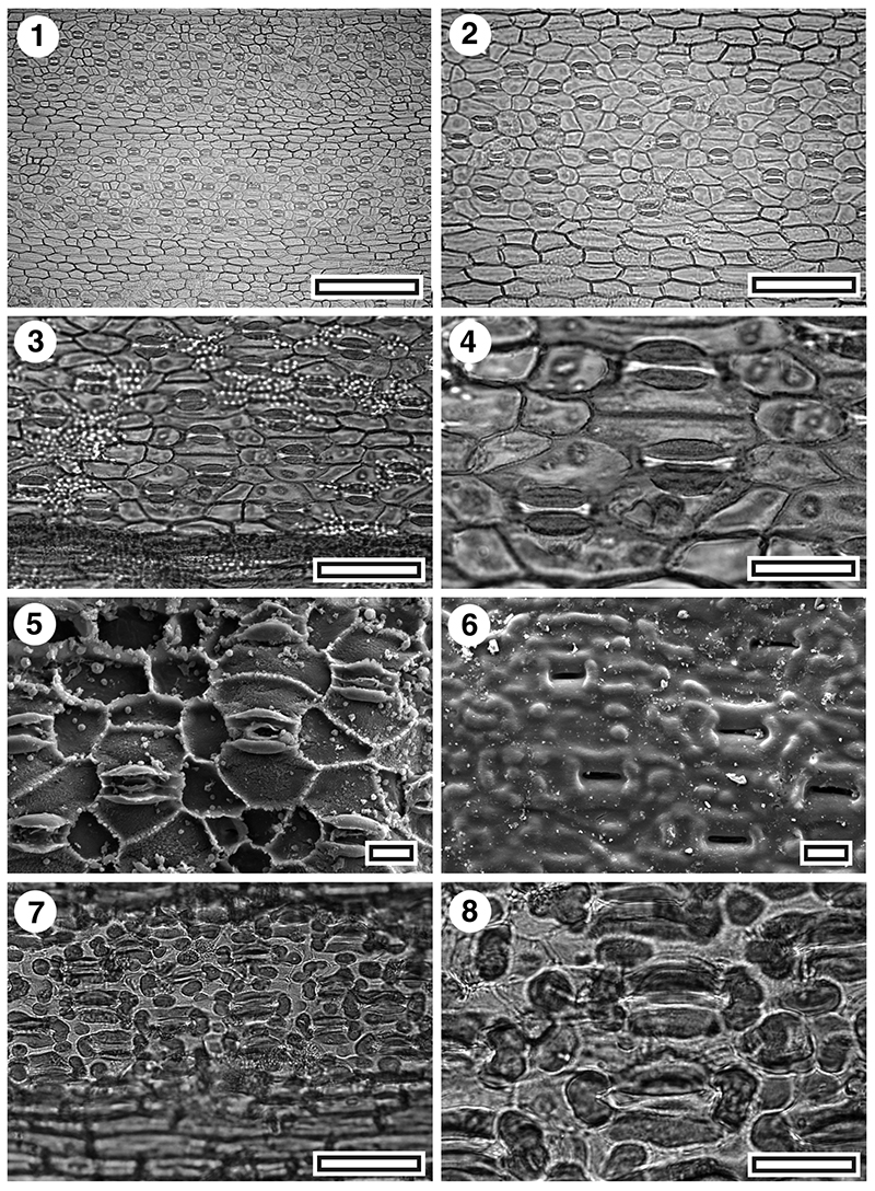
FIGURE 11. Cuticle of Astelia (Asteliaceae), Cyperaceae and Arecaceae; 1. Astelia sp., TLM view (SL5436, Hamiltons Gap, scale bar equals 50 µm); 2. Astelia sp., TLM view (SL5436, Hamiltons Gap, scale bar equals 20 µm); 3. Astelia sp., SEM view of inner surface (S-1661, Hamiltons Gap, scale bar equals 50 µm); 4. Astelia sp., SEM view of outer surface (S-1661, Hamiltons Gap, scale bar equals 50 µm); 5. Cyperaceae gen. et sp. indet., TLM view (SL2376, Maxwell-07, scale bar equals 50 µm); 6. Cyperaceae gen. et sp. Indet., TLM view, detail of stomatal complexes (SL2376, Maxwell-07, scale bar equals 20 µm); 7. Arecaceae gen. et sp. indet., TLM view (SL4671, Rapanui, scale bar equals 100 µm); 8. Extant Rhopalostylus sapida, TLM view (OPH2638, scale bar equals 50 µm).
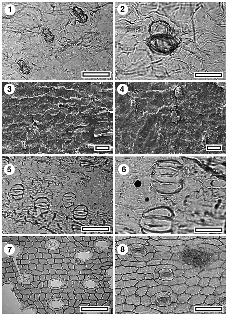
FIGURE 12. Cuticle of Pseudowintera sp. (Winteraceae, all Rangitawa-6); 1. TLM view with several stomatal complexes, note granular texture (SL5452, scale bar equals 50 µm); 2. TLM view, detail of a stomatal complex (SL5452, scale bar equals 20 µm); 3. SEM view of inner surface (S-1652, scale bar equals 10 µm); 4. SEM view of outer surface showing two stomatal pores, obscured by cutin (S-1652, scale bar equals 10 µm).
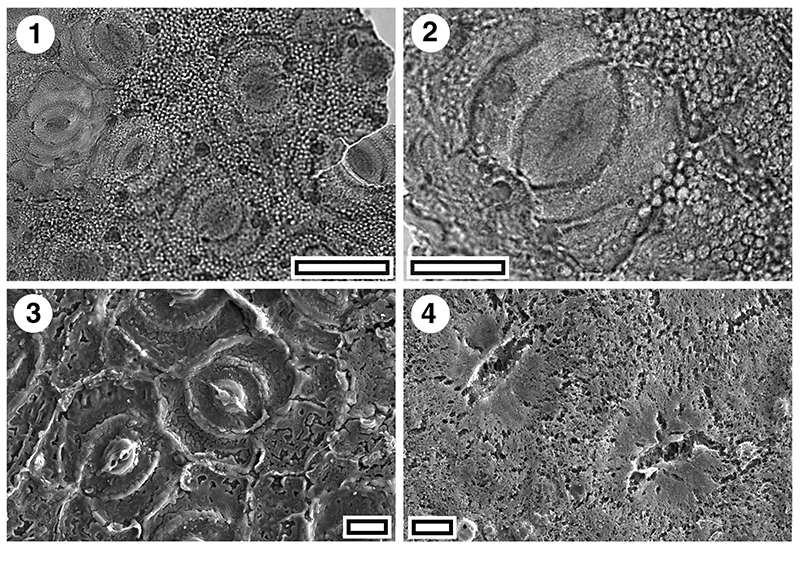
FIGURE 13. Cuticle of extant Beilschmiedia (Lauraceae); 1. Beilschmiedia tawaroa, TLM view (CHR367126, scale bar equals 50 µm); 2. B. tawaroa, TLM view, detail of stomatal complex (left) and trichome attachment site (right) (CHR367126, scale bar equals 20 µm); 3. B. tawaroa, SEM view of outer surface (S-1784, CHR367126, scale bar equals 10 µm); 4. B. tawaroa, SEM of inner surface SEM view (S-1784, CHR367126, scale bar equals 10 µm); 5. B. tawa, TLM view (OTA01439, scale bar equals 50 µm); 6. B. tawa, TLM view, detail of stomatal complexes (OTA01439, scale bar equals 20 µm); 7. B. tawa, SEM view of outer surface with clearly defined stomatal complexes and slit-like apertures (S-1719, OPH2627, scale bar equals 10 µm); 8. B. tawa, SEM view of inner surface with two stomatal complexes (S-1719, OPH2627, scale bar equals 10 µm).
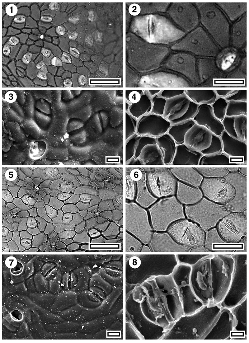
FIGURE 14. Cuticle of extant Beilschmiedia and Litsea (Lauraceae); 1. Beilschmiedia tarairi, TLM view (OTA46493, scale bar equals 50 µm); 2. B. tarairi, TLM view, detail of two stomatal complexes (OTA46493, scale bar equals 20 µm); 3. B. tarairi, SEM view of outer surface (S-1718, OPH2628, scale bar equals 10 µm); 4. B. tarairi, SEM view of inner surface (S-1718 OPH2628, scale bar equals 10 µm); 5. Litsea calicaris, TLM view (OTA04767, scale bar equals 50 µm); 6. L. calicaris, TLM view, detail of stomatal complex, note very rounded outline (OTA04767, scale bar equals 20 µm); 7. L calicaris, SEM view of outer surface (S-1153, OTA04767, scale bar equals 10 µm); 8. L. calicaris, SEM view of inner surface (S-1153, OTA04767, scale bar equals 10 µm).
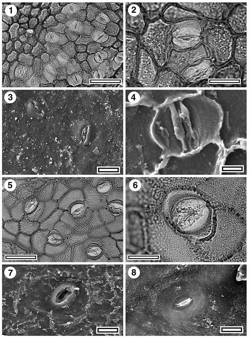
FIGURE 15. Cuticle of fossil Litsea and Beilschmiedia (Lauraceae, all Rangitawa-6); 1. Beilschmedia sp., TLM view (SL4979, scale bar equals 50 µm); 2. Beilschmedia sp., TLM view (SL4979, scale bar equals 20 µm); 3. Beilschmedia sp., SEM view of outer surface (S-1654, scale bar equals 10 µm); 4. Beilschmedia sp., SEM view of inner surface (S-1654, scale bar equals 10 µm); 5. Litsea sp., TLM view (SL4982, scale bar equals 50 µm); 6. Litsea sp., TLM view, detail of stomatal complexes (SL4982, scale bar equals 20 µm); 7. Litsea sp., SEM view of outer surface with two stomatal complexes. Note distinctive raised aspect of the subsidiary cells (SEM3957, scale bar equals 10 µm); 8. Litsea sp., SEM view of inner surface (SEM3959, scale bar equals 10 µm).
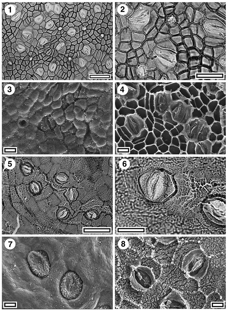
FIGURE 16. Cuticle of fossil Cryptocarya sp. (Lauraceae, all Huriwai); 1. TLM view with several stomatal complexes and trichome attachment site (SL4977, scale bar equals 50 µm); 2. TLM view, detail of stomatal complexes (upper) and trichome attachment site (lower right) (SL4977, scale bar equals 20 µm); 3. SEM view of outer surface with almost cryptic stomatal complexes obscured by cutin growth (S-1670, scale bar equals 10 µm); 4. SEM view of inner surface of a stomatal complex, note the ‘butterfly’ cuticular scales (S-1670, scale bar equals 10 µm).
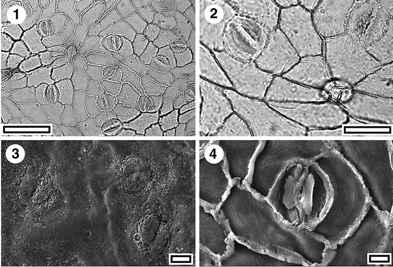
FIGURE 17. Cuticle of Toronia toru and Knightia excelsa (Proteaceae, all Pahurehure, all TLM); 1. Toronia toru, stomatal complexes, and, at upper left, a trichome attachment (SL4500, scale bar equals 100 µm); 2. T. toru, detail of stomatal complex (SL4500, scale bar equals 50 µm); 3. Knightia excelsa, stomatal complexes and trichome attachments (SL4514, scale bar equals 50 µm); 4. K. excelsa, detail of stomatal complex (SL4514, scale bar equals 20 µm).
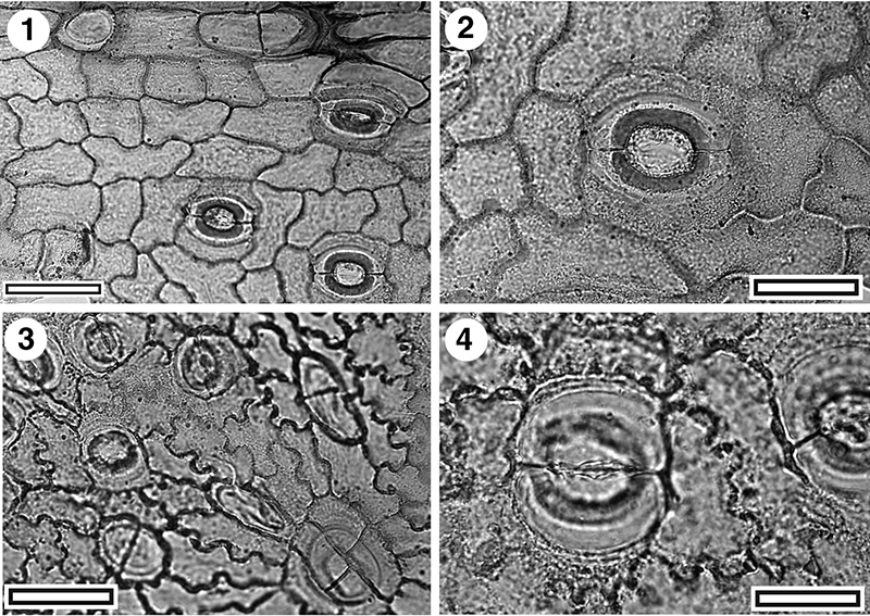
FIGURE 18. Cuticle of Carmichaelia sp. (Fabaceae); 1. TLM view showing stomatal complexes. The plant axis in the image is horizontal and the stomatal orientation is transverse (SL2365, Mokau, scale bar equals 100 µm); 2. TLM view, detail of stomatal complex (SL2365, Mokau, scale bar equals 20 µm); 3. SEM view of inner surface of a stomata complex (S-1648, Rangitawa-6, scale bar equals 10 µm); 4. SEM view of outer surface of a stomatal complex (S-1648, Rangitawa-6, scale bar equals 10 µm); 5. TLM view of stomatal complexes and trichome bases near upper and lower margins (SL2379, Maxwell-07, scale bar equals 50 µm); 6. TLM view of stomatal complexes and a trichome base at lower left (SL2379, Maxwell-07, scale bar equals 50 µm).
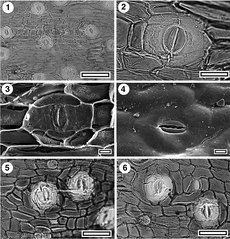
FIGURE 19. Cuticle of Grisellinia sp., (Griseliniaceae, all Hamilton’s Gap); 1. TLM view of three stomatal complexes (SL5478, scale bar equals 50 µm); 2. TLM view, detail of stomatal complex (SL5478, scale bar equals 20 µm); 3. SEM view of outer surface of a stomatal complex, note the prominent, wide peristomatal rim (S-1660, scale bar equals 10 µm); 4. SEM view of inner surface of a stomatal complex (S-1660, scale bar equals 10 µm).
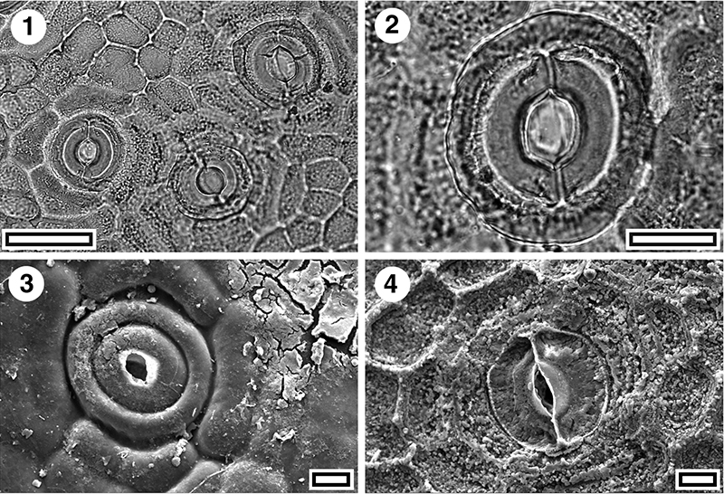
FIGURE 20. Cuticle of fossil and extant Olearia sp. (Asteraceae); 1. TLM view of stomatal complexes, with papillae and trichomes between (SL5480, Hamiltons Gap, scale bar equals 50 µm); 2. TLM view, detail of two stomatal complexes, note the prominent peristomatal rims (SL5480, Hamiltons Gap, scale bar equals 20 µm); 3. SEM view of inner surface, note hollow papillae (S-1659, Hamiltons Gap, scale bar equals 10 µm); 4. SEM view of outer surface, note common papillae and trichomes (S-1659, Hamiltons Gap, scale bar equals 20 µm); 5. Extant Olearia arborescens, TLM view of stomatal complexes, papillae and trichomes (OTA43559, scale bar equals 100 µm); 6. Extant Olearia rani, TLM view of stomatal complexes, papillae and trichomes (OTA23174, scale bar equals 50 µm).
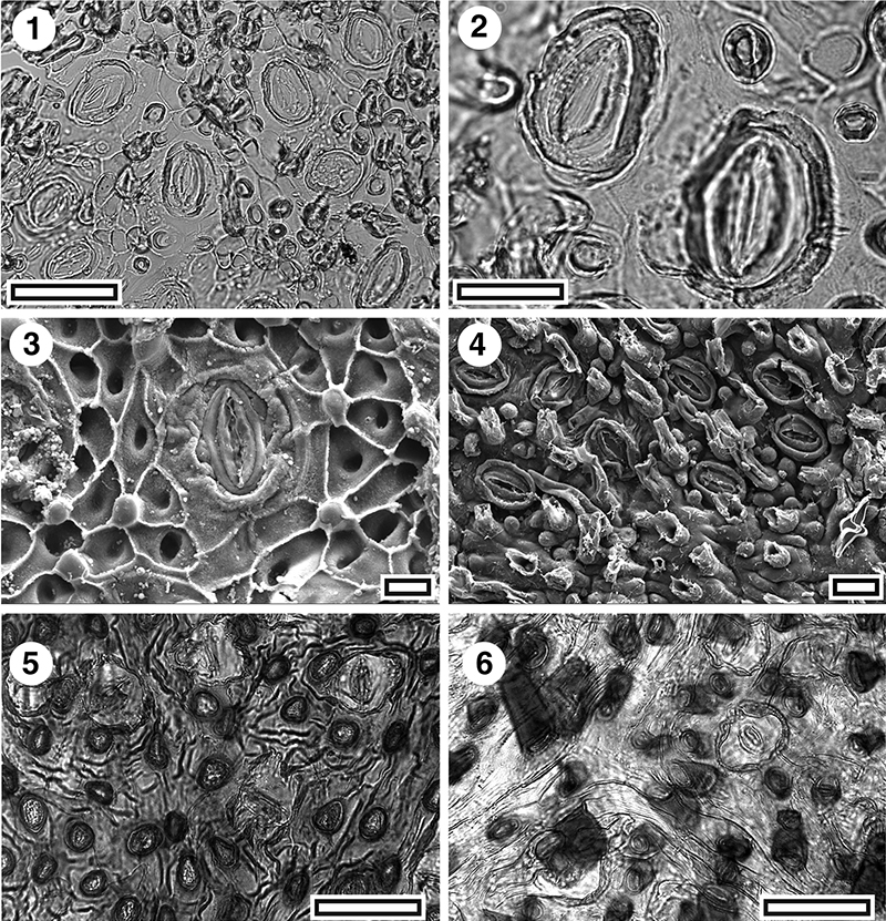
FIGURE 21. Cuticle of Myrsine sp. (Myrsinaceae, both TLM); 1. Group of stomatal complexes, note slight ridging on subsidiary cells (SL4658 Rangitawa-6, scale bar equals 100 µm); 2. Stomatal complexes, note prominent ridging (SL4655 Rangitawa-6, scale bar equals 100 µm).