FIGURE 1. Survey photo of a field crew at Buckner Ranch Site 1 in 1939 (41BE2-27). The majority of these workers had no geologic or paleontologic experience and were paid $0.20/hour through the WPA.
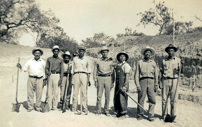
FIGURE 2. Removing overburden from Site 1, Buckner Ranch, April 1939 (41BE2-94). The tractor and Fresno scraper were hired from a local rancher who was paid $0.60/hour rental fees and $0.30/hour to drive.
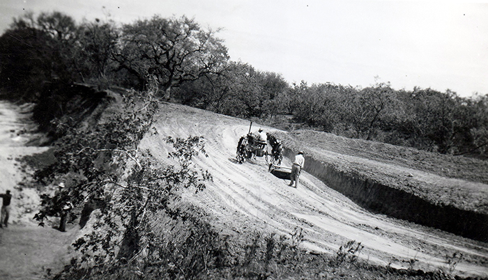
FIGURE 3. Quarry at Site 1, Buckner Ranch along south side of Blanco Creek, Bee County, Texas (TMM 30896) (41BE2-132). Stairway cut into Pleistocene sedimentary strata of the Berclair Terrace. Workers on left standing near top of the Goliad Formation. Workers in lower right kneeling with proboscidean skulls in the Goliad Formation.
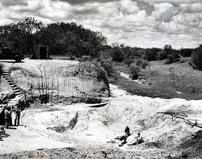
FIGURE 4. Gomphothere skull with tusks (TMM 30896-390) at Buckner Ranch Site 1 (41BE2-112). This image illustrates the collection technique employed on the project where nearly all matrix surrounding individual specimens was removed in the field prior to jacketing.
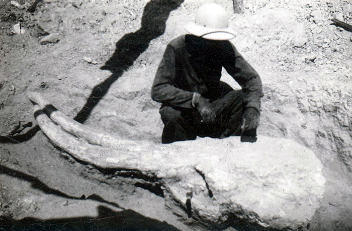
FIGURE 5. Ten Mile Waterhole Creek quarry in 1939, Live Oak County, Texas. (Survey photo from archives at the Vertebrate Paleontology Collections.)
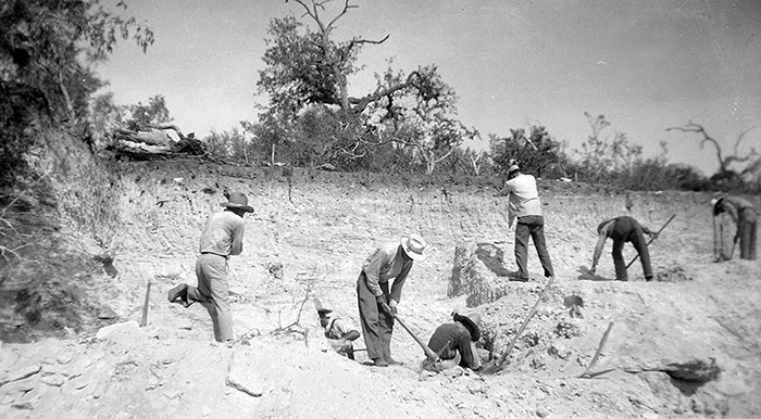
FIGURE 6. Site 15, Farish Ranch along Medio Creek in Bee County, Texas (41BE2-21). The contact is clear between the darker Berclair Terrace deposits above and the lighter Goliad Formation below.
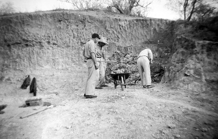
FIGURE 7. Bridge Ranch localities. 1. Original label states: “2 mi. east of Normanna-hammer at contact of conglomerate and underlying sandstone. Contact is conformable, vertebrates from conglomerates.” (41BE2-29). 2, “Section in Goliad Formation at fossil excavation east of Normanna.” This photo illustrates interbedded sandstones and carbonate-pebble conglomerate, hammer for scale (41BE2-31).
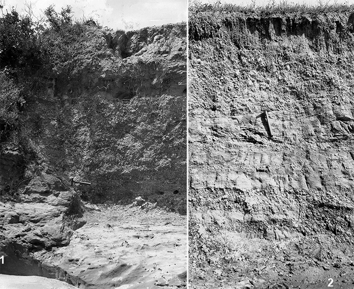
FIGURE 8. Geologic Time Scale (Gradstein et al., 2012) with Magnetic Polarity Time Scale (MPTS) and North American Land Mammal Ages (NALMA). Young et al. (2010) incorporated subsurface data and marine biostratigraphy for their correlation of the Goliad Formation to the time scale. The column on the right shows age constraints and correlation for the updip outcrop of the Goliad Formation. Although Young et al. (2010) followed Plummer (1932) in the usage of the Lagarto Formation as a unit within the Fleming Group, the current paper follows Wilson (1956) and others in the use of Fleming as a formation name. The Cold Spring Fauna from the Fleming Formation is late Barstovian in age (Tedford et al., 2004).
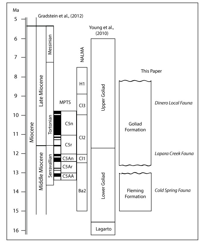
FIGURE 9. Geologic map showing extent of the Goliad Formation from Texas State Geologic Map (Barnes, 1992), major fluvial fairways based on Hoel (1982), approximate location of shoreline facies in the upper Goliad Formation (Young et al., 2010), and major vertebrate localities shown as stars labeled 1-4.
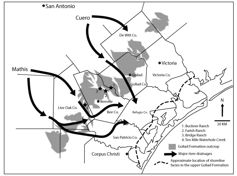
FIGURE 10. Outcrop view of pedogenic carbonate nodules in Goliad Formation gray mudstone within Blanco Creek near TMM 30898. Nodules are concentrated in upper 10 cm of bed below exposure surface and appear to display weak inverse grading.
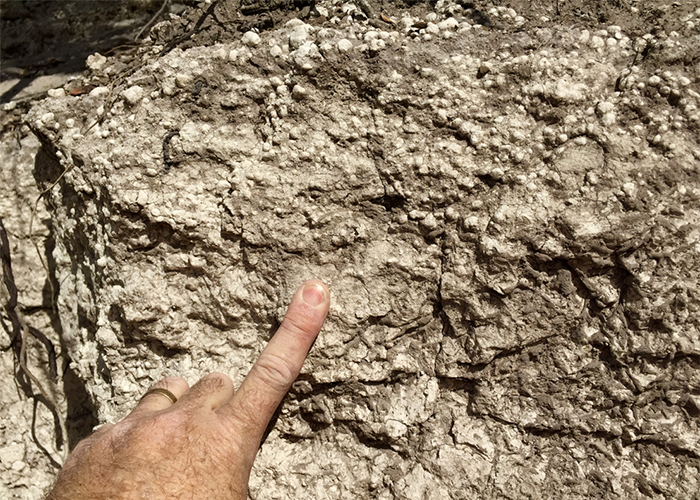
FIGURE 11. Goliad Formation exposed along Medio Creek southeast of Normanna, Texas. Black bag is approximately 25 cm long. Light gray sandstone bed overlying bone-bearing, carbonate-nodule conglomerate was sampled for detrital zircon geochronology (JR1601).
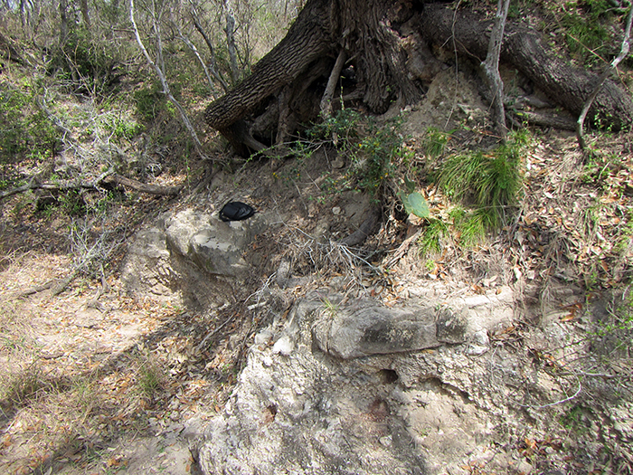
FIGURE 12. Map of the Bee County area with major vertebrate localities comprising the Lapara Creek Fauna shown by stars. Dashed line is the boundary of Bee County, defined by Blanco Creek along the northeast margin.
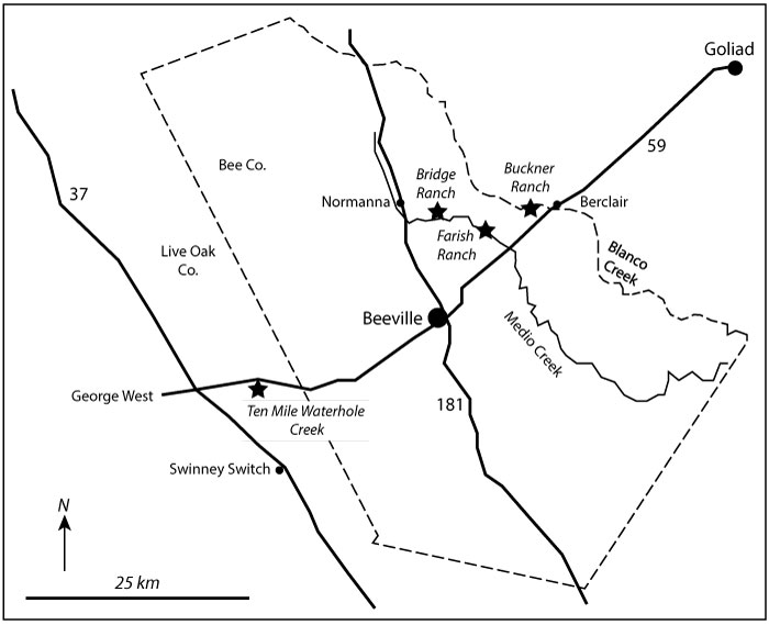
FIGURE 13. Field photos of four main locality areas. 1, Outcrop of Goliad Formation along Blanco Creek near TMM 30898. 2, Farish Ranch (TMM 31081) locality in 2015 along the south side of Medio Creek, Bee County, TX. The locality is now extremely overgrown with vegetation. 3, Ten Mile Waterhole Creek locality in 2015. The original excavation is now represented by the small depression in the foreground. Full size pick-up truck is on the skyline for scale. 4, Bridge Ranch locality TMM 31132. In 2016, the site was overgrown and the only evidence of Goliad Formation strata was observed in float.
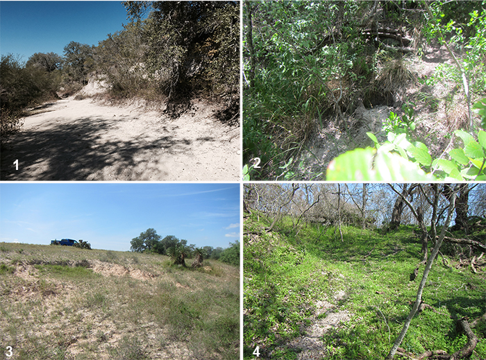
FIGURE 14. Excavation in Berclair Terrace sediments with top of the Goliad Formation evidenced by light gray sediments to the right of the small excavator.
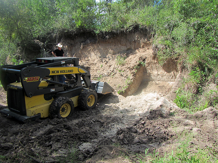
FIGURE 15. Wall of 2016 excavation at Site 1 (TMM 30896) with 0.5 m rock-pick for scale in shadow. Approximately 1.5-2 m of gray mudstone of the Goliad Formation is overlain by brownish conglomerate and sandstone of the Berclair Terrace. The original back-wall of Site 1 can be seen in the center and material to the right of the sharp boundary is backfill from the original Survey excavation. Bone fragments were found near the Brunton Compass (lower right) at the contact between the mudstones and underlying muddy sandstone. This is the horizon that yielded the majority of fossil vertebrates during the Survey excavation. Samples for paleomagnetic analysis were collected from this interval. Just south of this photo, the Berclair Terrace deposits are 4-5 m thick.
FIGURE 16. Mudstone casts of pelecypods. 1, TMM 30896, mudstone molds of two specimens. 2, TMM 30898 mudstone molds of multiple specimens in carbonate nodule conglomerate matrix.
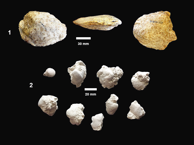
FIGURE 17. Unidentified elements showing both sides of five different specimens illustrating the range of morphologies and sizes. One side with bumpy texture and the other with longitudinal ridges. 1, TMM 30898-34. 2, TMM 30898-35. 3, TMM 30898-36. 4, TMM 30898-37. 5, TMM 30898-38.
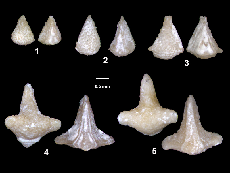
FIGURE 18. TMM 31081-1284, Ictalurus cf. lambda, pectoral spine, anterior, posterior and ventral views (top to bottom).
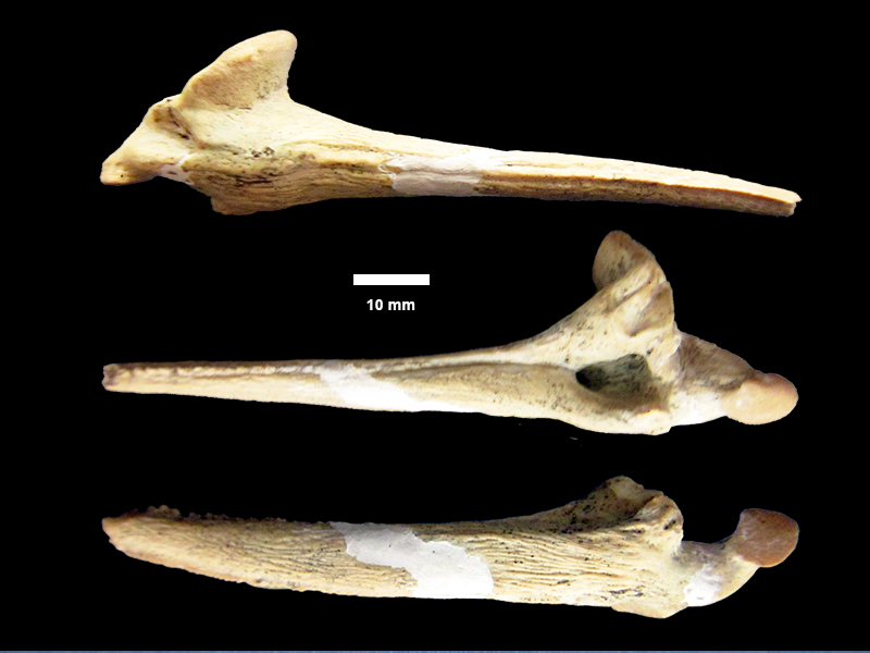
FIGURE 19. Actinopterygii, indeterminate. 1, TMM 30898-29, blade-like tooth with striated roots. 2, TMM 30898-30, blade-like tooth with striated roots. 3, TMM 30898-31, hook-like tooth. 4, TMM 30898-32, peg-like tooth with sub-circular to ovoid occlusal shape and enamel cap. 5, TMM 30898-33, peg-like tooth, lateral view.
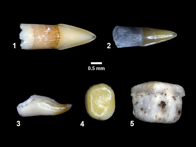
FIGURE 20. TMM 31081-280, cf. Trachemys sp., partial carapace. 1, dorsal view (bones outlined in black, scutes in white). 2, lateral view. 3, ventral view. 4, carapace right posterolateral view (bones outlined in black, scutes in white). Note double-notched peripherals, sculpting on costals and suprapygals.
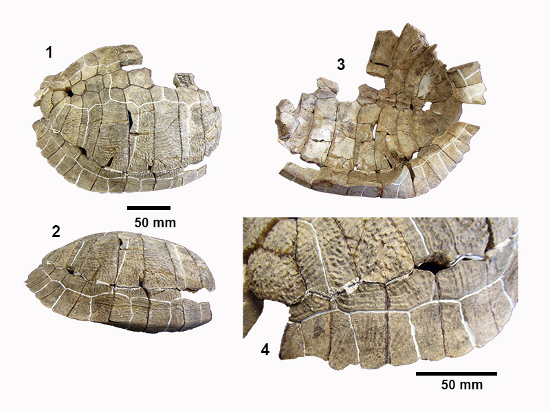
FIGURE 21. TMM 31081-280, cf. Trachemys sp. 1, nuchal bone. 2, right hypoplastron. 3, right and left xiphiplastra. 4, right hyoplastron. Specimens have been previously marked with black ink at bone edges and white ink at scute edges.
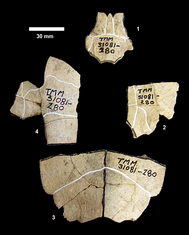
FIGURE 22. Emydidae indet. 1, TMM 31081-163, partial plastron, dorsal view. 2, TMM 3081-14 partial plastron ventral view. 3, TMM 3081-14 lateral view.
FIGURE 23. TMM 31081-145, Apalone sp., carapace fragment with neurals five-seven (N5-N7) and costals five-seven (C5-C7). Sutures shown by red lines.
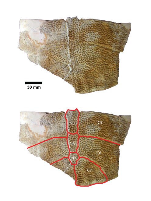
FIGURE 24. Apalone sp. 1, TMM 31132-769, plastron fragment illustrating texture (thickness ~ 8.8 mm). 2, TMM 31081-1488, hypoplastron, maximum length = 209 mm, width = 92 mm. 3, TMM 31081-139, partial xiphiplastron. 4, TMM 31081-1562, carapace fragment with four adjacent costals (co2-co5, left to right), co3 maximum dimensions: 177 mm x 58 mm.
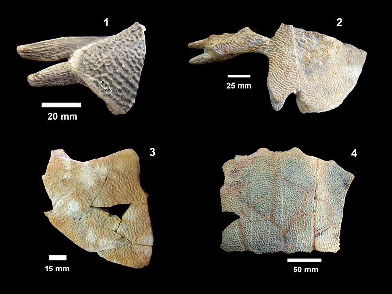
FIGURE 25. TMM 31081-1314, cf. Gopherus sp. 1, carapace lateral view. 2, plastron, oblique ventral view.
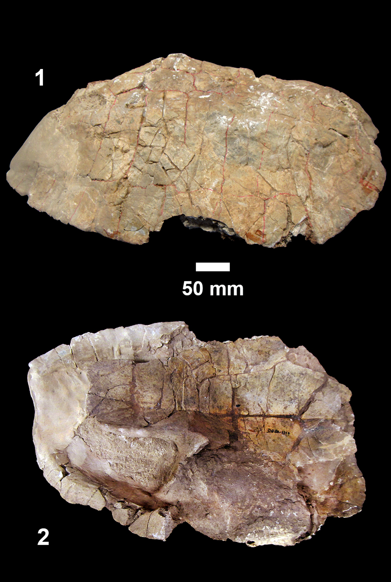
FIGURE 26. TMM 31081-1314, cf. Gopherus sp., anterior end of carapace with individual elements outlined in red and labeled (nu-nuchal, n-neural, c-costal, p-peripheral).
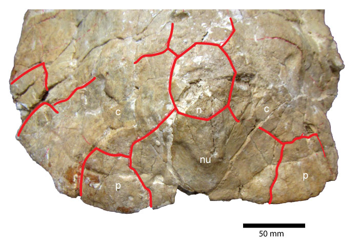
FIGURE 27. Hesperotestudo sp., 1-2, TMM 31081-600 peripheral. 3, TMM 31081-82, caudal vertebra. 4-5, dermal ossicles.
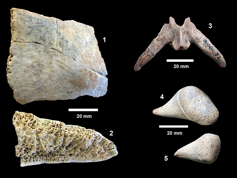
FIGURE 28. TMM 31081-584, Alligator cf. mississippiensis, skull fragment showing broad flat snout, centerline between nasals is evident, with partial premaxilla, maxilla and nasals (anterior to the left).
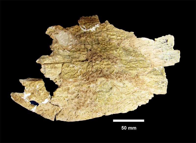
FIGURE 29. TMM 31081-40, Alligator cf. mississippiensis, 1, mesial mandible fragment-lingual view. Dorsal portion of the rostral tip of the splenial is missing along linear break. The ventral portion is very thin and not clearly broken. The Meckelian groove and the foramen intermandibularis oralis are clear between the splenial and the symphysis. 2, left mandible fragment-lingual view. S-splenial, SS-splenial suture.
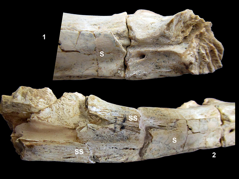
FIGURE 30. TMM 31081-40, Alligator cf. mississippiensis, mandible fragment. 1, Lingual view. 2, labial view. 3, anterior fragment, dorsal view, anterior to the left.
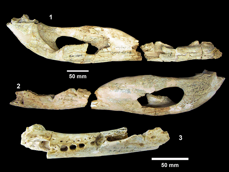
FIGURE 31. Alligator cf. mississippiensis, skull fragments. 1, TMM 30896-280, dorsal view, slightly crushed, snout missing. Specimen was embedded in plaster by Survey preparators. 2, TMM 30896-280, oblique view of left orbit with slightly elevated ridge along interior edge of orbit, little to no ridge around the supratemporal fenestrae, and gentle slope between frontal and prefrontals. 3, TMM 31081-810, partial right premaxillary, ventral view showing dorsally oriented nares at top. (anterior to the left).
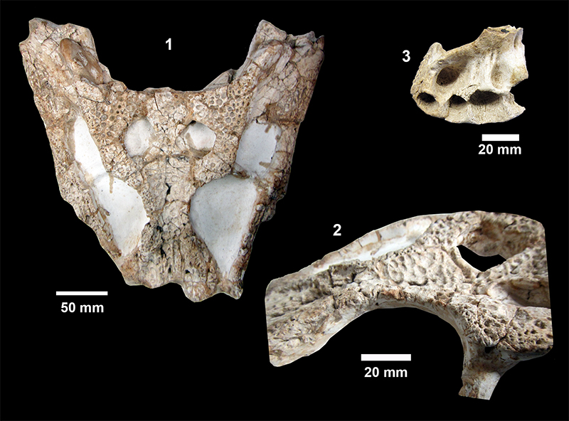
FIGURE 32. 1, TMM 31081-1263, cf. Anserinae, ulna. 2, TMM 31132-347, cf. Mycteria sp., partial tarsometatarsus, anterior and posterior views.
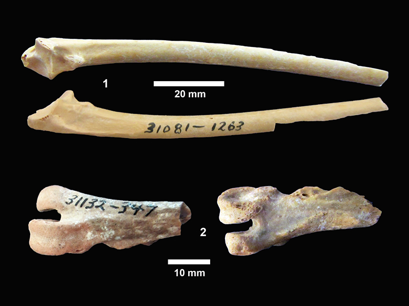
FIGURE 33. cf. Pronotolagus apachensis, line drawing of occlusal views. 1, TMM 30898-24 P2. 2, TMM 30898-25 partial p or m.
FIGURE 34. TMM 30898-21, Perognathus cf. minutus, partial M2, posterior edge broken. 1, occlusal. 2, anterior. 3, lateral view.
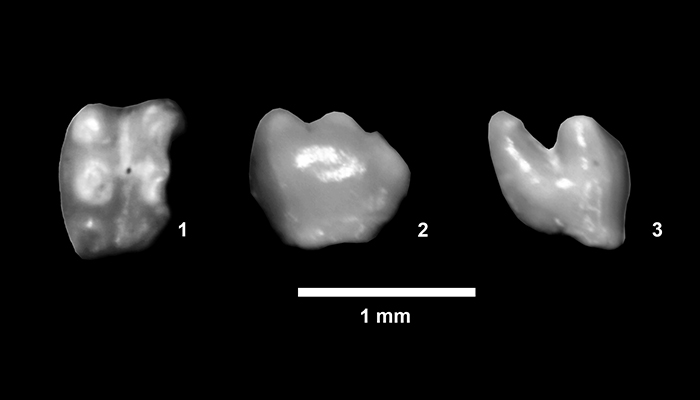
FIGURE 35. Ceratogaulus cf. rhinoceros, line drawings of occlusal surface. 1, TMM 31132-143 left P4. 2, TMM 31170-67 left P4. 3, TMM 31132-120 right p4.
FIGURE 36. TMM 31132-458, Aelurodon taxoides, left dentary with c, p2-m2. 1, labial view. 2, lingual view. 3, ventral view showing weak sypmphyseal flange.
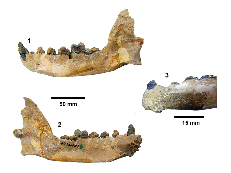
FIGURE 37. Aelurodon taxoides. 1, TMM 31170-65, right M2, occlusal view. 2, TMM 30936-106, m1, lingual view. 3, TMM 30936-40, partial right dentary, labial view, anterior to the right.
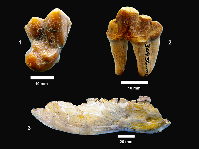
FIGURE 38. TMM 31081-1255, cf. Eucyon sp., partial right maxilla with P4-M2. 1, occlusal view. 2, labial view, anterior to the right.
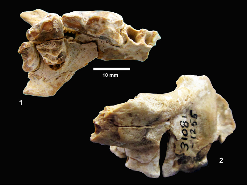
FIGURE 39. TMM 30896-241, Leptocyon vafer, partial right dentary with p2-m2. 1, lingual view, anterior to left. 2, occlusal view of p4-m2, anterior to right.
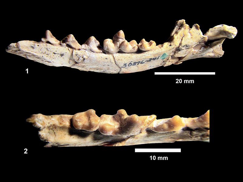
FIGURE 40. Ischyrocyon gidleyi. 1, TMM 31184-2, R M1, 24.7 mm x 34.7 mm, paracone and metacone are worn nearly flat. (Note: this specimen was figured by Wilson (1960), but incorrectly labeled as 31132-29.) 2, TMM 31132-29, R M2, 18.9 mm x 26.4 mm. 3, TMM 31081-1243, canine, maximum length: 112 mm.
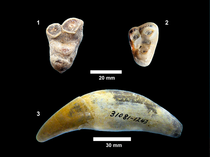
FIGURE 41. Ischyrocyon gidleyi. 1, TMM 30936-236, m2, length: 24.9 mm, width: 17.0 mm, taller trigonid is composed of a large protoconid and very reduced metaconid, the talonid is dominated by a hypoconid. 4, TMM 31132-412, P4, length: 26.3 mm, width: 15.1 mm, labial view. 5, occlusal view.
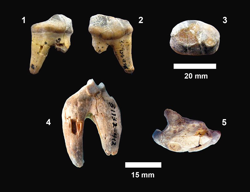
FIGURE 42. TMM 31081-465, Ischyrocyon gidleyi, humerus, maximum length: 303 mm.
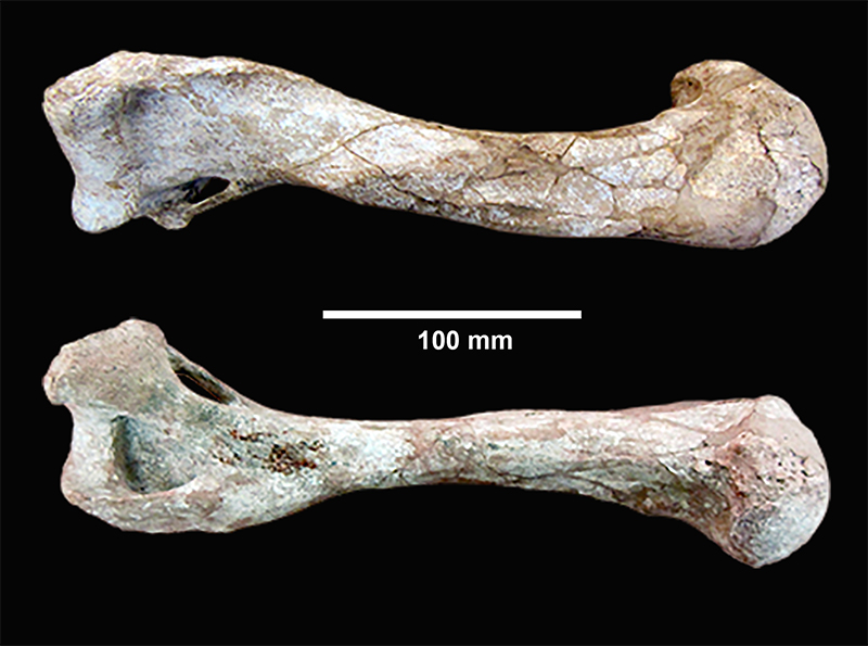
FIGURE 43. Chart of dimensions for upper and lower third molars of B. buckneri specimens. Lower third molars are slightly narrower than uppers. Lower third molars from mandibles with lower tusk alveoli span the length range of all third molars, but are all only moderately worn.
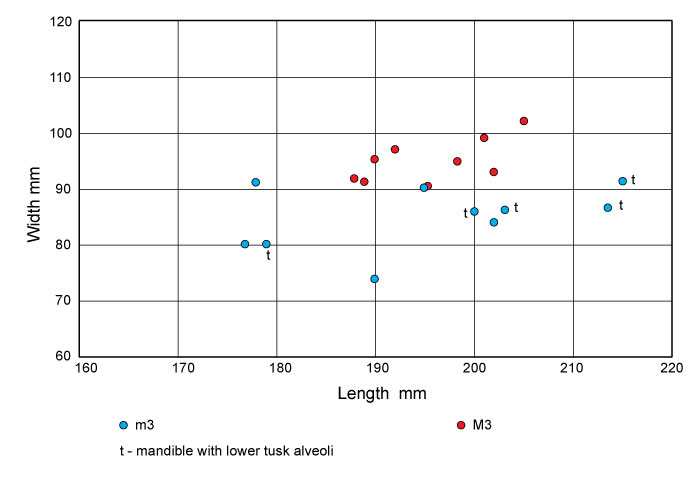
FIGURE 44. TMM 30896-390, Blancotherium buckneri, holotype, left side of skull and mandible. Plaster reconstruction of the cranium, ascending ramus and condyle, and distal upper tusks have been removed digitally. There are still some areas with plaster reconstruction such as toward the anterior margin of the symphysis.
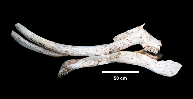
FIGURE 45. TMM 30896-570, Blancotherium buckneri, labial view of right dentary with down-curved symphysis and m2-m3.
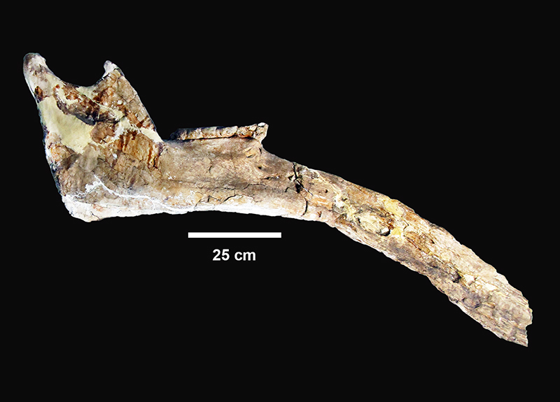
FIGURE 46. TMM 30896-570, Blancotherium buckneri, occlusal view of right m3 showing advanced state of wear on four main lophids with pretrite trefoils, a smaller fifth lophid, and a small posterior heel and, (anterior to the left).
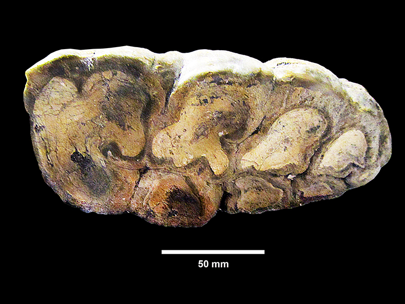
FIGURE 47. TMM 30896-278, Blancotherium buckneri, partial right dentary with m2-m3. 1, labial view. 2, occlusal view, anterior to the left. Very worn m2 and moderately worn m3 with well-developed pretrite trefoils evident on the first and second lophids. The fourth lophid exhibits slight wear and the fifth is unworn indicating that this was a mature adult. 3, anterior view of broken symphysis showing matrix filled alveoli for lower tusks (a). Although the specimen is deformed, presumably the result of burial compaction, the alveoli are clear and dorsoventrally elongate roughly parallel to the labial margins of the symphysis. The left alveolus (right side of photo) closely approximates the size and shape of the isolated lower tusks (TMM 30896-520, 526).
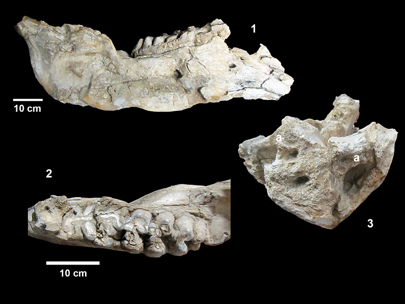
FIGURE 48. TMM 30896-311, Blancotherium buckneri mandible. 1, right and left dentaries crushed flat with anterior to the right. Both right and left m3s are visible. One cm squares on scale bar. The specimen is imbedded in a plaster cradle. 2, occlusal view of left m3 anterior to the right illustrating four well developed lophids with pretrite trefoils, a smaller fifth lophid and a posterior conulid. The valleys between lophids contain secondary conulids resulting in a dense packing of enamel and complex occlusal pattern. 3, anterior view of symphysis illustrating spatulate geometry as well as an apparent alveolus for a lower right tusk.
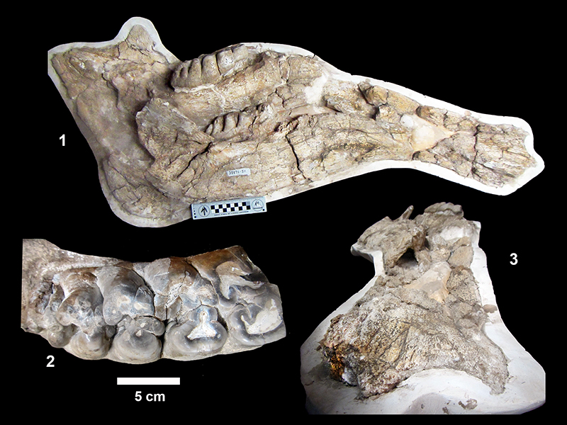
FIGURE 49. TMM 30896-64, Blancotherium buckneri. 1, mandible, occlusal view, alveoli for m1, m2 with moderate wear, m3 just erupting. 2, left and right m2-m3, anterior to the right.
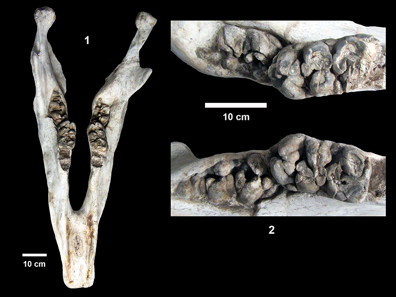
FIGURE 50. TMM 30896-271 Blancotherium buckneri, mandible with short symphysis. 1, view from right labial side with m3’s unerupted, m2’s, and m1 in left mandible (alveoli in right). 2, view from left showing very worn m1 and collapsed bone associated with possible lower tusk alveolus. 3, occlusal view of right dentary showing alveoli for m1, m2 and unerupted m3. 4, anterior view of symphysis showing plaster-filled alveoli for right lower tusk. (Scale bar applies to 1-3.)
FIGURE 51. TMM 30896-657, Blancotherium buckneri, mandible. 1, occlusal view showing right and left m3, open alveoli for m2, and short narrow symphysis (anterior end broken). Although mesial portions of both dentaries are crushed, it is clear that the molar is m3 and moderate wear suggests an early mature adult. The left m3 exhibits 4 main lophids with well-developed pretrite trefoils, a smaller fifth lophid and a small posterior heel. The alveoli anterior to m3 suggest that m2 was lost not long before death. 2, lateral view from right side showing slight downward deflection of symphysis and possible collapsed lower tusk alveolus along anterior labial margin.
FIGURE 52. TMM 30896-318 Blancotherium buckneri. 1, anterior-oblique view of U-shaped symphysis. 2, cross-section of possible lower tusk alveolus exposed along break in symphysis. 3, left m3 showing four main lophids, a smaller fifth lophid and a small posterior heel, anterior to the left.
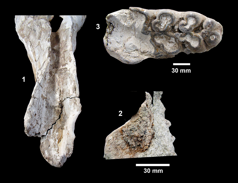
FIGURE 53. TMM 30896-108 Blancotherium buckneri, mandible. 1, oblique view of symphysis with “lower tusk” sculpted in plaster by Survey preparators. 2, anterior view of symphysis after plaster tusk was removed showing potential alveolus for lower tusk.
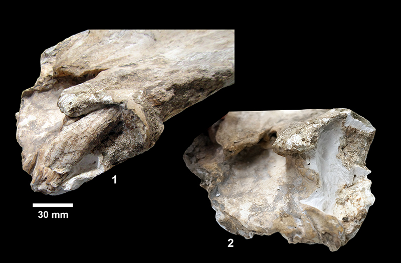
FIGURE 54. TMM 30896-325 Blancotherium buckneri, partial palate with right and left M2 and M3. 1, occlusal view, one cm squares on scale bar, anterior to the right. 2, left M3 occlusal view, 4 main lophs with pretrite trefoils, smaller fifth loph and posterior conules, anterior to the left. 3, labial view of left M3 showing relative crown height, anterior to the right.
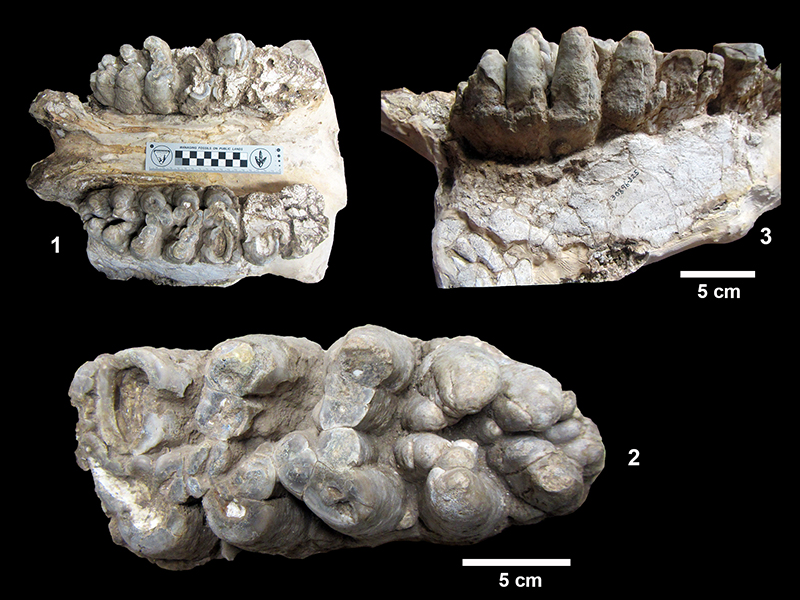
FIGURE 55. TMM 30896-269, Blancotherium buckneri. 1, partial palate, anterior to left with left M2-M3 and right M3. This specimen is imbedded in a block of plaster which was digitally removed. 2, left M3 with moderate wear showing 4 main lophs and a posterior heel, occlusal view.
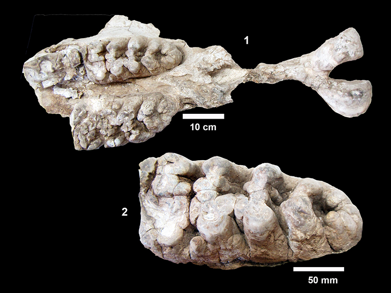
FIGURE 56. TMM 30896-381, Blancotherium buckneri, slightly worn right M3. 1, occlusal view, (201 x 99 mm). 2, lingual view, anterior to the right.
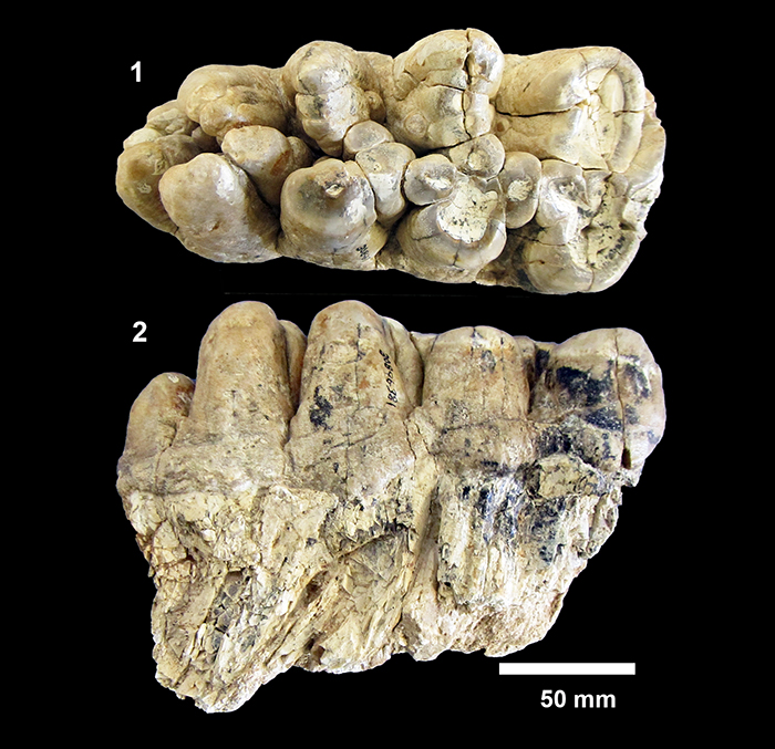
FIGURE 57. TMM 30896-389, Blancotherium buckneri, Survey photo of skull showing palate with very worn M2, slightly worn M3, and an unworn aberrant “tooth” posterior to M3 (41BE2-49).
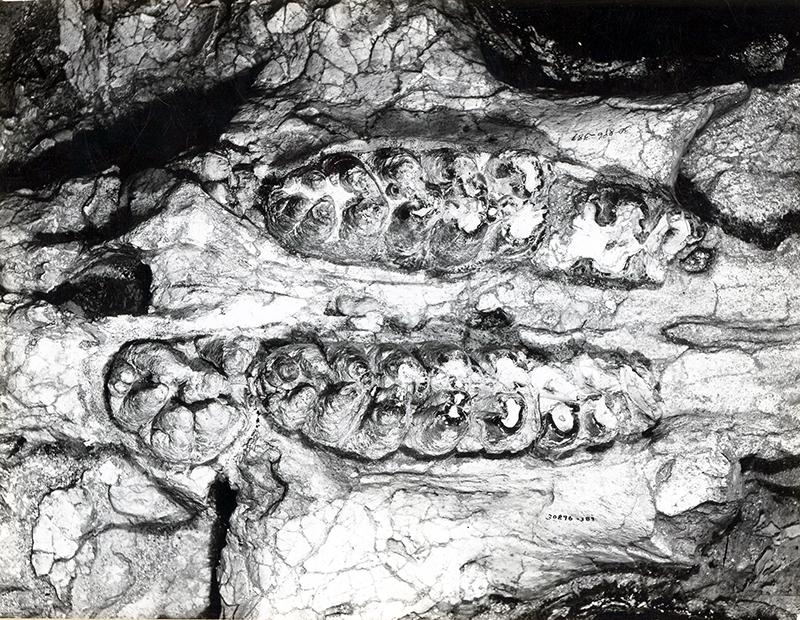
FIGURE 58. TMM 30896-656 Blancotherium buckneri, skull. Lateral view of right side showing the jugal (J), right M2-M3, left M3, and the proximal end of the left upper tusk. The remainder of the tusk is shown in Figure 60. Also visible is a first rib (R) that was wedged under the zygomatic arch.
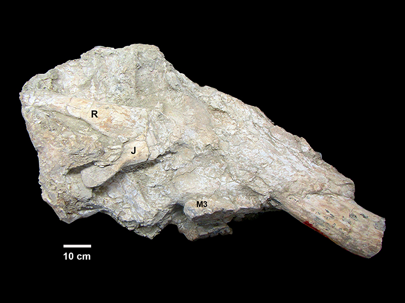
FIGURE 59. TMM 30896-656, Blancotherium buckneri, left M3 showing advanced state of wear with four major lophs and a smaller fifth loph, anterior to the left. Pretrite trefoil pattern is evident. This is how the fossil appeared after removal of the plaster jacket, illustrating the amount of field preparation done by the Survey collectors. 1 cm squares on scale.
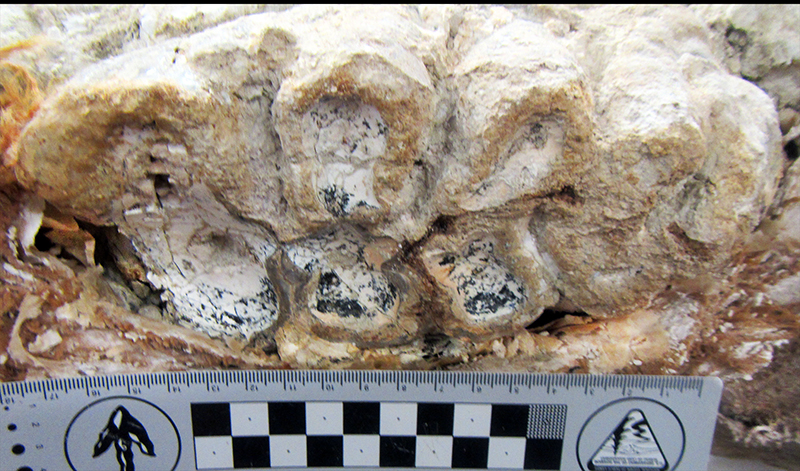
FIGURE 60. TMM 30896-656, Blancotherium buckneri. 1, cross-section of left upper tusk illustrating alternating dark and light bands interpreted as annual growth rings. The tusk was sawed off by Survey collectors prior to jacketing (see Figure 58). 2, Lingual view showing red markings that match the tusk in the skull. The tusk would have been slightly up-curved and angled outward from the midline. No significant helical spiral was observed in the longitudinal ridges.
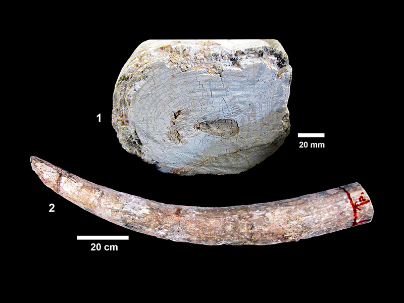
FIGURE 61. Maximum diameter vs. distance along left upper tusk of Blancotherium buckneri (TMM 30896-656). The original specimen was cut at ~ 40 cm in the field for collection and transport.
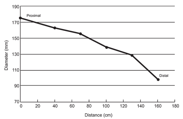
FIGURE 62. Survey quarry at Buckner Ranch Site 1 (TMM 30896) with three Blancotherium buckneri skulls, one of which preserves the upper tusks nearly in place suggesting upward and outward curvature (41BE2-131). The tusk on the left side of the photo was not associated with a particular skull. All skulls are oriented with palate up, and the top skull was in close association with a number of disarticulated post-cranial elements including pelvis, scapula, vertebra and ribs. This photo illustrates the significant amount of specimen preparation done in the field.
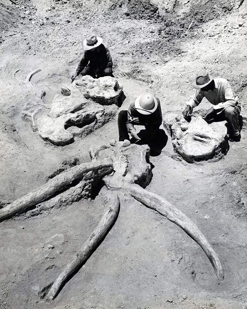
FIGURE 63. Blancotherium buckneri, lower tusks. 1, TMM 30896-520, 305 mm x 43 mm maximum width. 2, TMM 30896-526, 310 mm x 47 mm maximum width, both showing fragments of enamel band on one side. Plaster reinforcement was added by the Survey preparators.
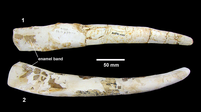
FIGURE 64. TMM 30896-298, Blancotherium buckneri, stylohyoid bone.
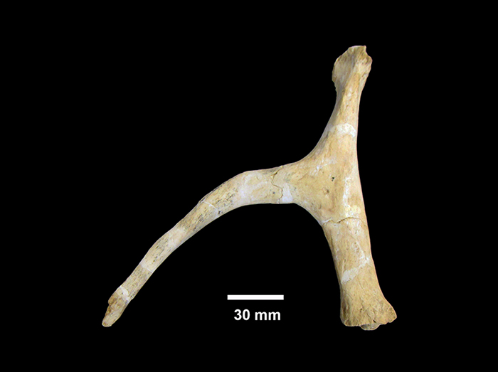
FIGURE 65. Blancotherium buckneri, post cranial elements. 1, TMM 30896-254 left humerus. 2, TMM 30896-299 ulna. 3, TMM 30896-85 left femur. (This specimen is somewhat deformed presumably due to burial compaction.) 4, TMM 30896-197 tibia.
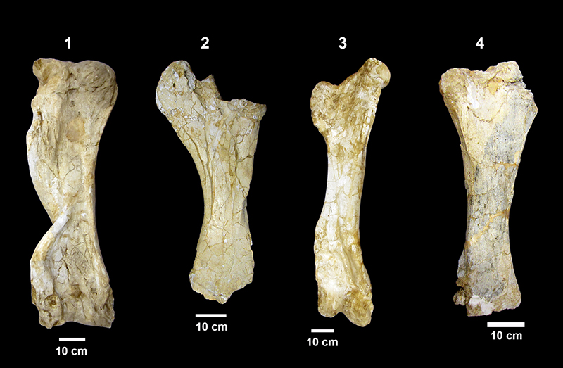
FIGURE 66. Blancotherium buckneri, post-cranial elements. 1, TMM 30896-51 right scapula, maximum length parallel to scapular spine: 730 mm, maximum width: 650 mm, significant plaster reconstruction of the scapular spine and dorsal edge. 2, TMM 30896-101 left pelvis, length: 860 mm, height: 810 mm, plaster reconstruction around the edge of the bone and on the back of the specimen for support. 3, TMM 30896-255 sacrum, length: 400 mm, proximal width: 284 mm, distal width: 180 mm. 4, TMM 30896-12 atlas, 494 mm maximum width, 239 mm maximum height. 5, TMM 30896-324 thoracic vertebra, spinous process is 605 mm tall, centrum is ~170 mm wide and 148 mm tall.
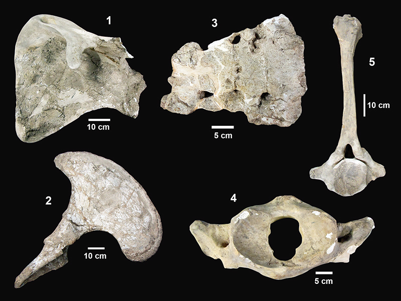
FIGURE 67. Comparison of Blancotherium buckneri size to other fossil proboscideans and estimations of body mass based on data and calculations in Christiansen (2004). Colored circles are from Christiansen (2004); black bar shows range of measured humerus lengths for B. buckneri. Mammuthus data are from multiple species.
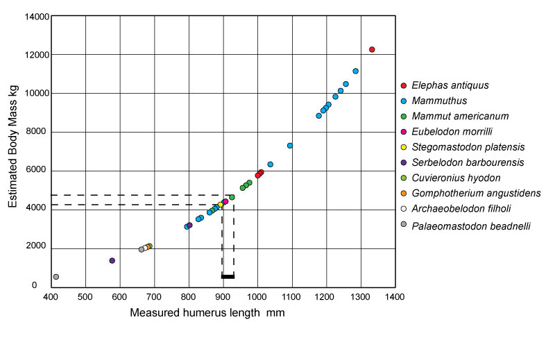
FIGURE 68. cf. Gomphotherium sp. 1, TMM 30936-90 upper tusk fragment with enamel band. 2, same specimen, cross-sectional view with the enamel band on left side. Note the wear-flattened dorsal surface making nearly a right angle with the enamel band. 3, TMM 31170-47 upper tusk fragment with enamel band and dorsal wear surface.
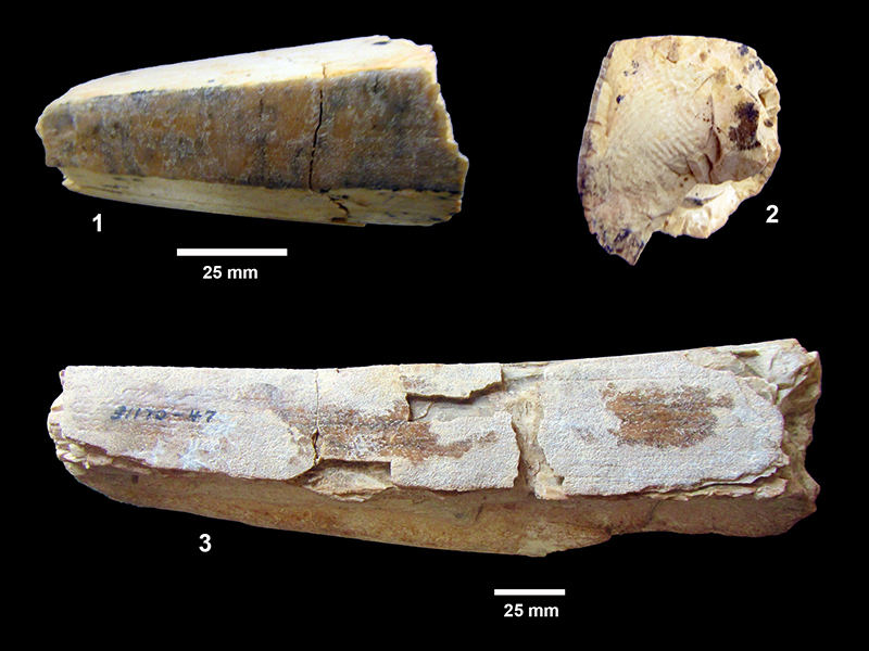
FIGURE 69. TMM 31132-365, cf. Gomphotherium sp., lower tusk fragment. 1, dorsal view. 2, cross-sectional view showing concentric rings and “engine-turning” pattern.
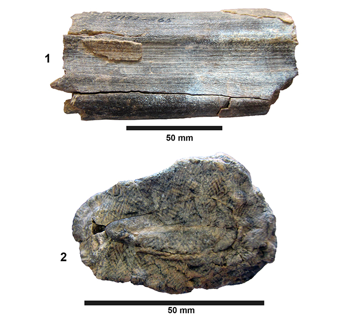
FIGURE 70. cf. Gomphotherium sp., isolated molars. 1, TMM 30936-289 left m3, occlusal view illustrating four main lophids and a posterior heel with relatively open valleys and pretrite trefoils. 2, TMM 30936-289 left m3, oblique view. 3, TMM 30936-97 right M3. 4, TMM 30936-331 second molar with three main lophs and two posterior conules. 5, TMM 31132-274 m3? Occlusal view of four main lophids with small posterior heel. Although the advanced state of wear makes identification difficult, this specimen compares well with the smaller morphotype with relatively clean valleys.
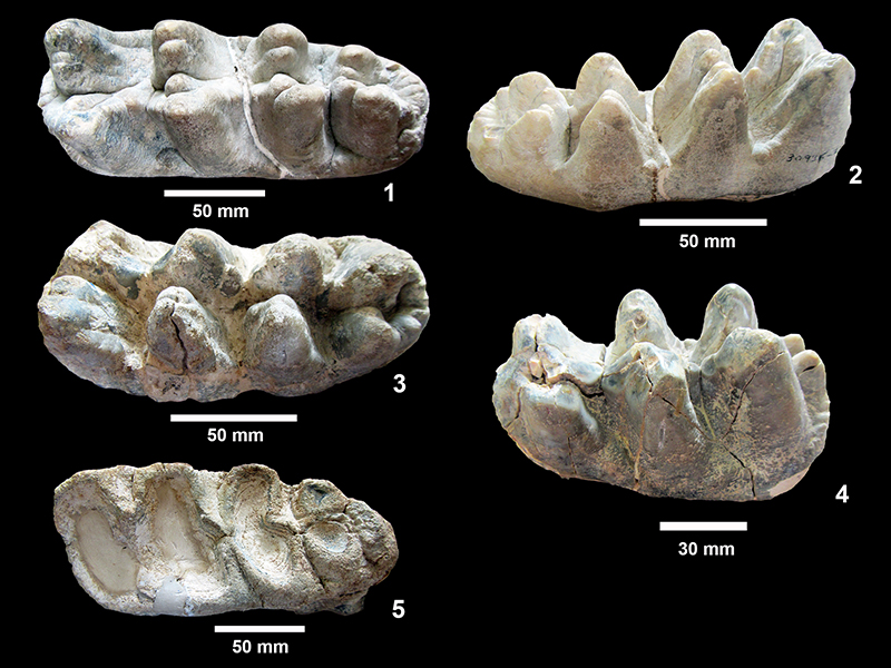
FIGURE 71. TMM 31081-1 cf. Gomphotherium sp., partial right dentary with dp2, alveoli for dp3, and unerupted dp4. The dp2 is 22.3 x 12.9 mm and exhibits moderate wear, while dp4 is 70.6 mm in length and trilophodont with well-developed anterior and posterior cingula. 1, lingual view, anterior to the left. 2, dorsal view.
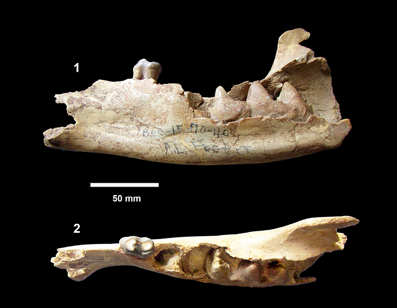
FIGURE 72. TMM 31081-685, Ustatochoerus cf. medius, right partial maxilla. 1, lateral view, anterior to the right. Note the steepening of the profile above P2 and well-developed labial cingula on P2-P4. (Significant plaster reinforcement in posterior portions.) 2, occlusal view of P1-M3 (anterior portion of M3 is broken).

FIGURE 73. TMM 30896-496, Pseudoceras skinneri, partial right dentary with p3-m3, labial view, anterior to the right.
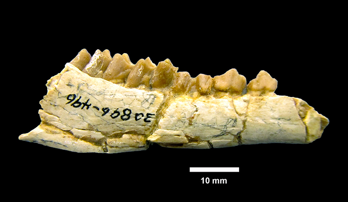
FIGURE 74. Ramoceros ramosus. 1, TMM 31132-73 partial horn, lateral view. 2, posterior view. 3, dorsal view showing the arrangement of the tines, labeled Tine 1-4. 4, TMM 30936-294, partial horn, distal tine.
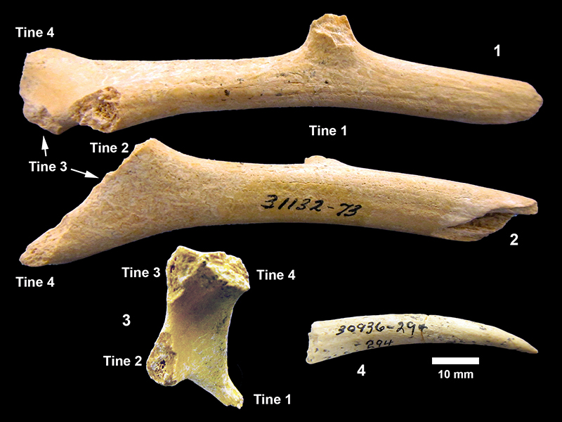
FIGURE 75. Synthetoceras tricornatus. 1, TMM 30936-23 nasal horn. (String in photo is tied to specimen card that was removed digitally.) 2, TMM 30936-506, frontal horn fragment. The horn displays a nob on the end as is common is Synthetoceras, but is very short. 3, TMM 30936-280 M2. 4, TMM 31132-563 M2. 5, TMM 31081-1471 M2.
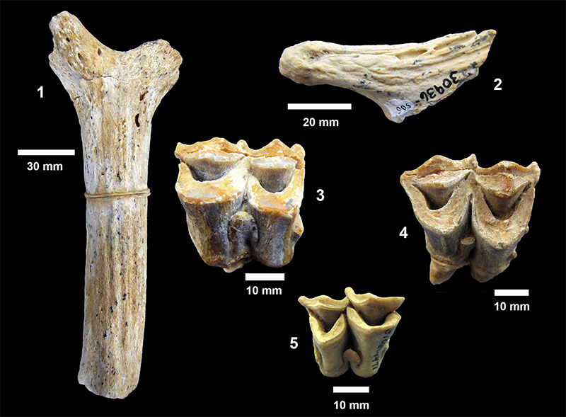
FIGURE 76. Cranioceras teres. 1 and 2, TMM 31132-455, partial skull with proximal horn, (two views of same specimen). 3, TMM 31132-300 M2.
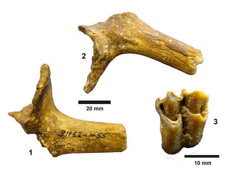
FIGURE 77. Nothotylopus camptognathus dentaries. 1, TMM 30896-26 left dentary, labial view-holotype described by Patton (1969). 2, TMM 30896-198 right partial dentary with p4, m1 roots, partial m2 and m3, labial view. 3, TMM 30896-198 occlusal view with p1 roots, p3 roots, p4, m1 roots, partial m2 and m3, anterior to the left.
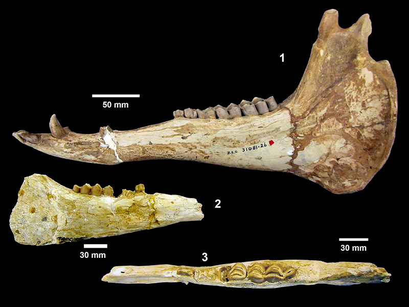
FIGURE 78. TMM 31081-460, Aepycamelus sp., left dentary. 1, labial view with p1 (broken) above foramen, p2 (alveolus only), p3-m3. 2, occlusal view of p2 alveolus, p3 and p4 (unworn), m1, m2 (anterior).
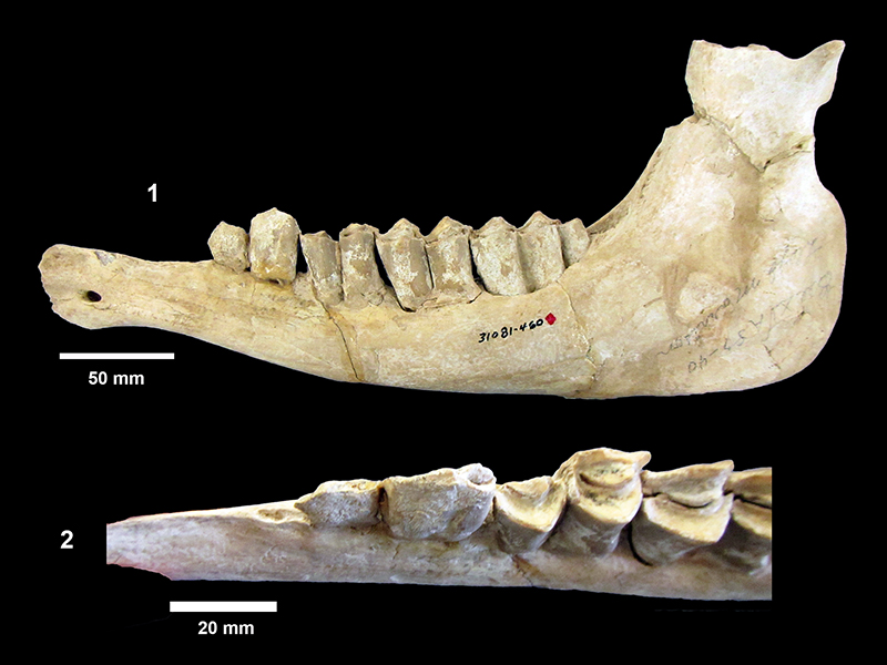
FIGURE 79. TMM 31081-460, Aepycamelus sp., left dentary. 1, showing suspicious attachment of p3 and p4 to the ramus in conventional photo, anterior to the left. 2, rendered isosurface from CT scan showing double rooted alveolus for p2 and complex nature of the “join” between p3, p4 and the ramus. 3, arbitrary slice through CT scan showing roots for p3 and p4 as well as m1-3. No roots are observed beneath alveolus of p2.
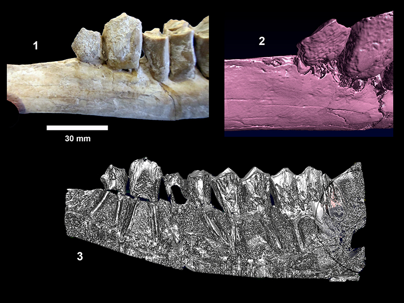
FIGURE 80. TMM 31081-18, Aepycamelus sp. 1, partial right dentary with p3-m3, labial view anterior to the right. 2, occlusal view p3-m1, anterior to the right.
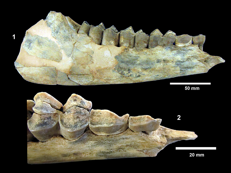
FIGURE 81. TMM 31081-532, 682, 578 Aepycamelus sp., cervical vertebrae.
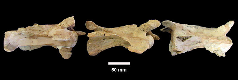
FIGURE 82. Procamelus. 1, TMM 31081-665, Procamelus occidentalis, left dentary with i1-m3, labial view, anterior to the left. 2, TMM 31081-429, Procamelus grandis, left dentary with partial m1, m2, m3. (Note extensive plaster reconstruction with multiple colors of brown and white plaster.)
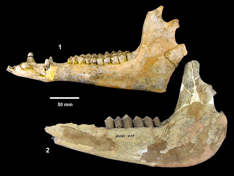
FIGURE 83. TMM 31132-332, Protolabis cf. coartatus, mandible. 1, left dentary labial view. 2, occlusal view. 3, angular flare on left dentary (inverted posterior view).
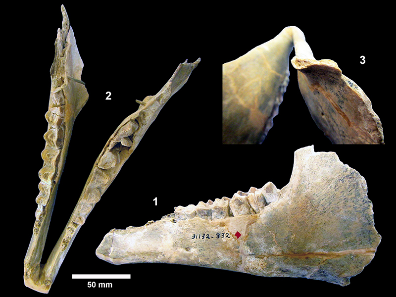
FIGURE 84. TMM 31081-28, Protolabis cf. yavapaiensis, partial left dentary with roots of p3, p4-m2, m3 (broken). 1, labial view, anterior to the left. 2, occlusal view.
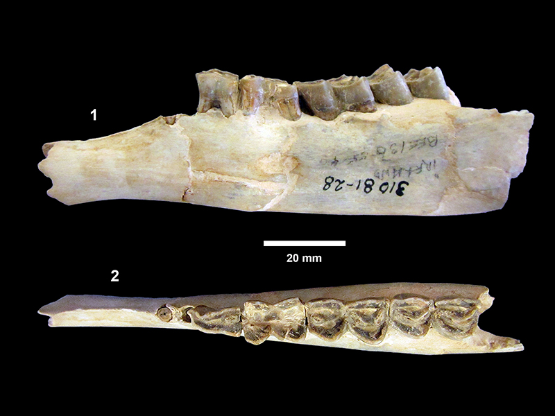
FIGURE 85. TMM 31132-202, “Prosthennops” xiphidonticus, partial mandible. 1, labial view, anterior to the left. 2, dorsal view with C (left and right), dp3, dp4, m1, m2.
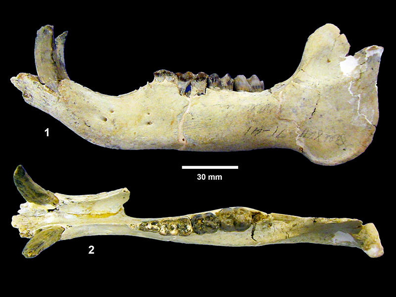
FIGURE 86. TMM 31132-202, “Prosthennops” xiphidonticus, mandible, CT scan, exterior surface. 1, lingual view, anterior to the right. 2, labial view, anterior to the left. 3, occlusal view dp3, dp4 m1, m2, anterior to the left.
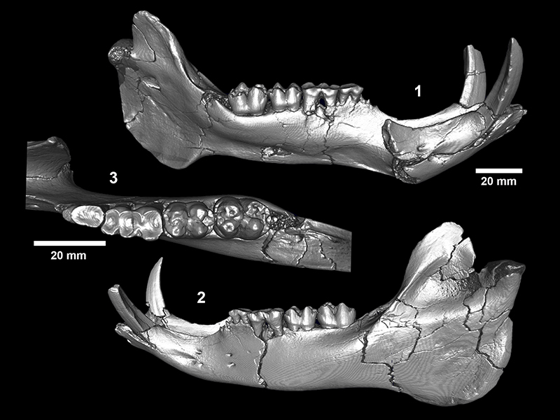
FIGURE 87. TMM 31132-202, “Prosthennops” xiphidonticus, partial mandible. 1, arbitrary section through CT volume illustrating canine, unerupted p2-p4 and m3, along with dp3, dp4, m1, m2, anterior to the right. 2, unerupted p2-p4 extracted from CT volume, oblique labial view, anterior to the left. (Metal rod inserted during Survey preparation is also visible.) 3, Unerupted p2-p4 extracted from CT volume, occlusal view, anterior to the left. Both second and third premolars have a single trigonid cusp, while p4 has both protoconid and metaconid.
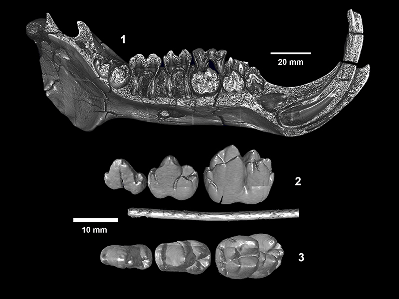
FIGURE 88. TMM 31081-673, “Prosthennops” xiphidonticus. 1, partial mandible with very worn p3-m3 and alveoli for p2, occlusal view, anterior to the left, brown plaster reconstruction near symphysis. 2, ventral view. 3, TMM 31081-363, partial right dentary, occlusal view dp4-m1, anterior to the right.
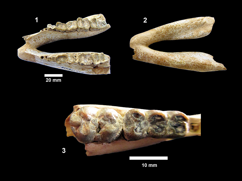
FIGURE 89. TMM 30181-131,2 Teleoceras major. 1, partial skull illustrating brachycephalic morphology, broad zygomatic arch (with significant plaster repair) and narrow, slightly upturned nasals, right side, anterior to the right. 2, left side, anterior to the left. 3, left side tooth row, P3-M3 top to bottom, occlusal view illustrating well developed antecrochets on M1 and M2 and cristae on M1.
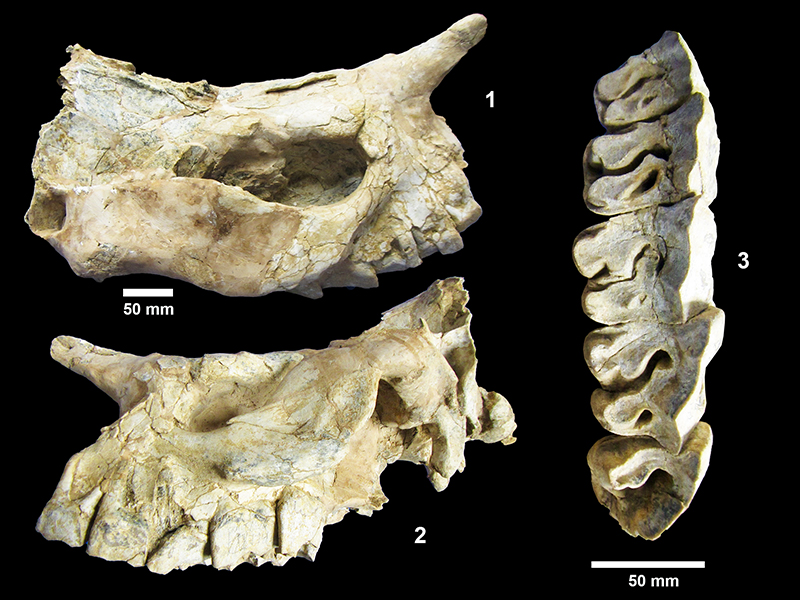
FIGURE 90. Teleoceras major. 1, TMM 31081-29, partial skull illustrating left zygomatic arch, and lambdoidal crest. 2, posterior view illustrating occipital condyles and lambdoidal crest. Skull is partially crushed with top-to-the right shear. 3, TMM 31081-1153, partial right dentary showing mental foramen below p4 and i2. 4, TMM 31081-1084, left dentary with p3-m3, labial view, anterior to the left. 5, TMM 31081-435, right dentary with p3-m3, labial view, anterior to the right.
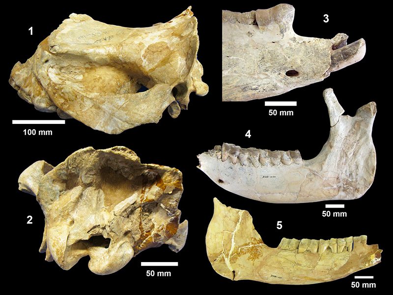
FIGURE 91. Teleoceras major post-cranial elements. 1, TMM 31081-568 left femur. 2, TMM 31081-577 left humerus. 3, TMM 31081-667 right radius.
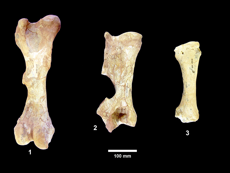
FIGURE 92. Pseudhipparion curtivallum. 1, TMM 30896-196, lingual view of P. curtivallum holotype with anterior to the right. 2, occlusal view. 3, TMM 31132-250 left dentary with p2-m3, occlusal view, anterior to the left. 4, TMM 31132-236, left P4-M3 occlusal view, anterior to the left, note isolated protocone; hypoconal groove open on M2 and closed fossette on M1. 5, TMM 31081-371, R P3, 15.9 x 16.7 mm length x width at occlusal surface, MCH = 30 mm, 6.3 x 3.6 mm protocone length x width.
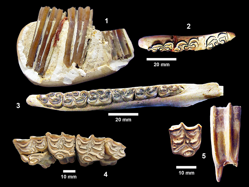
FIGURE 93. Nannippus sp. 1, TMM 31081-660A, right P3, occlusal view showing isolated oval protocone, small pli caballin, moderately complex plications on fossette borders, strong hypoconal groove, strong parastyle. This specimen is moderately well worn: 17.9 x 18.3 mm, PL = 6.6 mm, MCH = 23.4 mm. 2, TMM 30896-450A&B, associated L M1-M2, occlusal view anterior to the left (M1: 18.2 x 16.2 mm, PL = 6.7 mm, MCH = 27.6 mm; M2:18.4 x 17.7 mm, PL = 7.3 mm, MCH = 25.6 mm). 3, TMM 31081-16, mandible with p2-m3. Labial view, TRL = 109.1 mm anterior to the right, darker brown areas are plaster. 4, TMM 31081-16, mandible with p2-m3, anterior to the right.
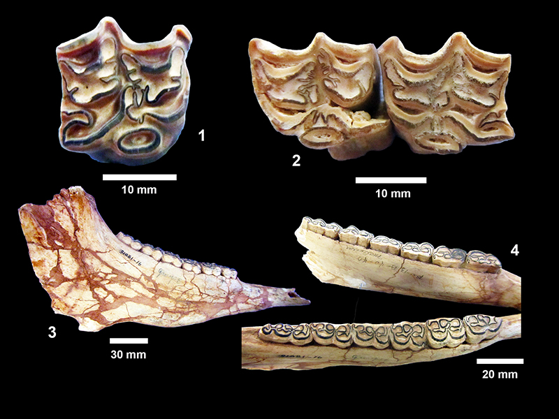
FIGURE 94. TMM 31081-735, Nannippus sp., mandible with p2-m3, occlusal view, left TRL ~112 mm, right TRL ~110 mm, anterior to the right.
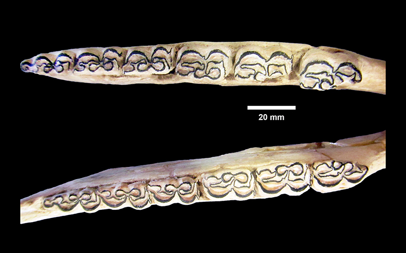
FIGURE 95. Hipparion tehonense. 1, TMM 31081-1554, P4, lateral view and occlusal view (18.7 x 19.0 mm, PL = 7.2 mm, PW = 3.8 mm). 2, TMM 31081-1555 M2, lateral and occlusal views. 1 and 2 illustrate the reduction of plication complexity on fossette borders with wear. 3, TMM 31170-90 (originally 90A), left M1/M2, occlusal view, (21.0 x 18.9 mm, MCH = 36.9 mm). 4, TMM 31170-288 (originally 92C), right P3/P4, occlusal view, (19.4 x 16.3 mm, MCH = 32.3 mm).
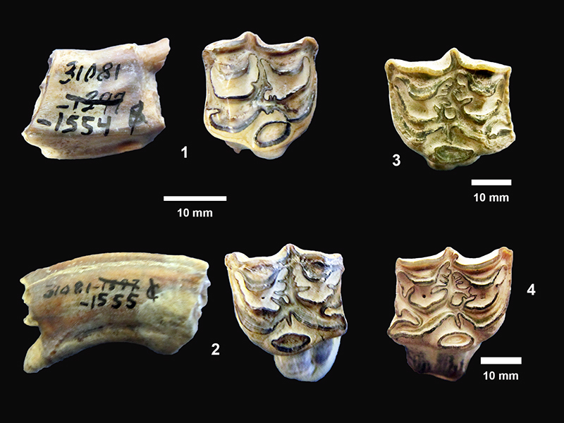
FIGURE 96. TMM 31081-501, Cormohipparion ingenuum. 1, partial right dentary with p2-p4 occlusal view, anterior to right, p2: 21.9 x 10.6 mm, p3: 20.0 x 11.8 mm, p4: 20.9 x 11.5 mm. 2, labial view.

FIGURE 97. TMM 31081-844, Cormohipparion occidentale. 1, right side of skull, anterior to the right, showing facial fossa. 2, right tooth row with P2-M3, occlusal view, illustrating isolated protocone, complex fossette borders, double pli caballins on P3-M3 and open hypoconal groove.
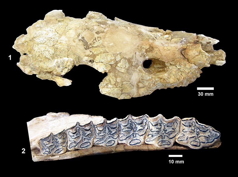
FIGURE 98. TMM 31081-1076, Cormohipparion occidentale. 1, right P2-M3, anterior to right, TRL = 135 mm, left P4-M3 (P4: 22.3 x 21.0 mm, M1: 21.3 x 20.0 mm, M2: 21.4 x 19.2 mm, M3: 19.7 x 13.9 mm), 2, labial view of left P4-M3, anterior to the right.
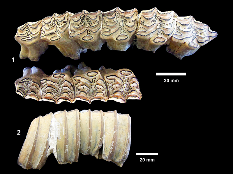
FIGURE 99. Cormohipparion occidentale. 1, TMM 31081-7 partial right dentary showing unerupted p2-4 with dp2-4 above, anterior to the right. 2, TMM 31081-7, occlusal view dp2-4, m1, m2, anterior to the right. M1 with slight wear, m2 just erupted, dp2: 27.0 x 12.0 mm, dp3: 23.6 x 13.0 mm, dp4: 25.3 x 10.6 mm, m1: 23.8 x 8.8 mm. 3, TMM 31081-637, left dentary with p2-m3 (unworn), occlusal view, anterior to the right, p2: 25.5 x 11.0 mm, p3: 24.5 x 11.6 mm, p4: 22.2 x 16.7 mm, m1: 22.0 x 10.4 mm, m2: 24.3 x 9.0 mm, m3: ~24 x ~7.5 mm (unworn). 4, TMM 31081-780, right dentary with p2-m3, occlusal view, anterior to the right. 5, TMM 31081-780, right dentary, labial view, plaster reconstruction near specimen number.
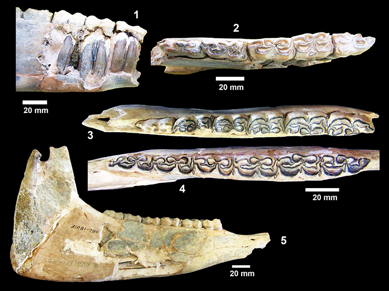
FIGURE 100. Neohipparion affine. 1, TMM 30936-466, left M1/M2, occlusal view. 2, TMM 30936-440, left p3/p4, occlusal view.
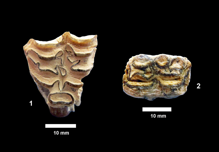
FIGURE 101. Calippus placidus. 1, TMM 30896-530 (figured holotype by Quinn (1955) as type specimen of Calippus optimus), included in Calippus placidus by Hulbert (1988a), occlusal view. 2, labial view anterior to the left. 3, TMM 30896-528, left P2-M3 in partial skull, occlusal view, anterior to the left. TRL = 100 mm. (Quinn (1955) designated this specimen and the associated TMM 30896-530 as the holotype of C. optimus.) 4, TMM 30896-528, crushed partial skull, anterior to the right, (significant plaster repair especially towards anterior and posterior portions of specimen.
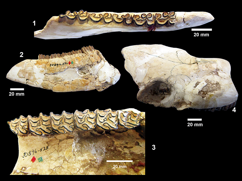
FIGURE 102. Calippus regulus. 1, TMM 30936-153, partial right dentary with p2-m3, occlusal view, anterior to the right, TRL: 88.5 mm. 2, TMM 31081-711, left P2-M3, occlusal view, anterior to the left. 3, labial view, anterior to the right. (Note: this specimen also includes right series P2-M3.) 4, TMM 31081-171, M1/M2, occlusal view, 12.7 mm x 13.4 mm. 5, labial view. 6, TMM 30896-610, P4 sectioned view (14.3 x 14.0 mm, PL = 5.2 mm). 7, oblique lateral view.
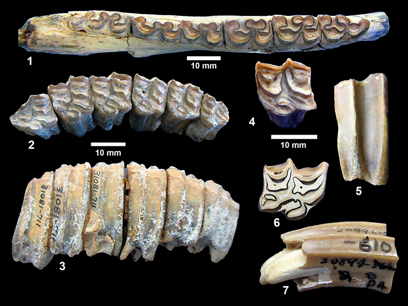
FIGURE 103. TMM 30896-173, Calippus regulus, juvenile partial skull with right and left DP2-M1. As shown on the right side, M1 is just erupting and shows no wear. The M1 on the left side has been sectioned. (Note this skull was designated the holotype of Calippus anatinus by Quinn, 1955.)
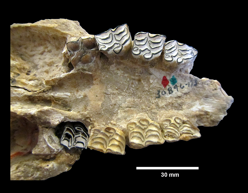
FIGURE 104. Calippus martini. 1, TMM 30896-127 right P2-M3 (broken), occlusal view, anterior to the right. [Figured by Quinn (1955) as Protohippus perditus ]. Measurements in Table 17. 2, TMM 31170-73 right P3/P4, 21.5 x 21.3 mm, MCH = 41.7 mm, protocone = 7.3 x 4.3 mm. 3, TMM 31081-169 right M1/M2, 20.3 x 20.0 mm, MCH = 38.0, protocone = 7.1 x 3.9 mm. 4, TMM 30896-533 left p2, 21.9 x 10.8 mm, figured by Quinn (1955) as Equus laparensis.
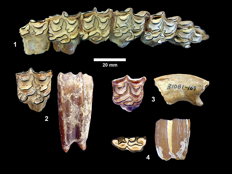
FIGURE 105. Calippus martini. 1, TMM 30896-569 partial skull, oblique dorsal view, anterior to the right. 2, TMM 30896-569 partial skull with P4-M3, very worn, P4: 18.3 x 21.4 mm, M1: 17.9 x 21.9 mm, M2: 17.9 x 20.3 mm, M3: 21.1 x 18.3 mm.
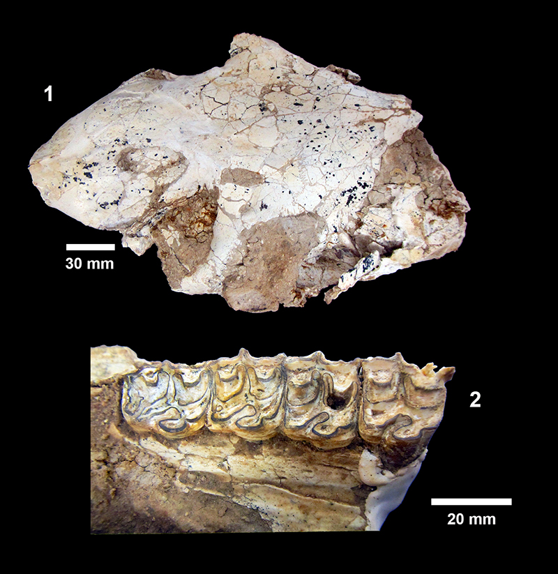
FIGURE 106. Protohippus supremus. 1, TMM 31081-664 partial right dentary with p2-m3, occlusal view, anterior to the left, TRL = 142 mm. 2, TMM 31081-541 partial left dentary with p2-m3, anterior to the left, TRL = 137.8 mm. 3, TMM 30896-503 right P2-M3, occlusal view, anterior to the right. TRL = 134 mm. [Figured by Quinn (1955) as type of Hippotigris sellardsi.] 4, labial view, anterior to the left. P4 MCH = 45 mm.
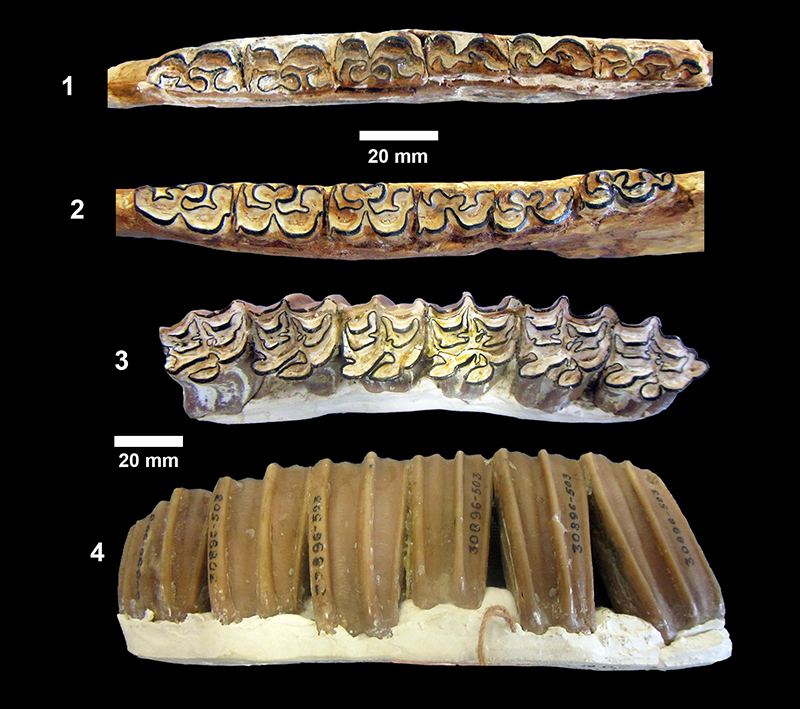
FIGURE 107. Pliohippus pernix. 1, TMM 31132-333 partial right dentary with p2-m3, occlusal view, anterior to the left. 2, lingual view, anterior to the left. TRL = 155 mm. [Quinn (1955) designated this specimen as the holotype of Dinohippus subvenus.] 3, TMM 31081-625 left P3/P4, occlusal view 26.5 x 25.9 mm. 4, posterior view showing high curvature, MCH = 52.8 mm.
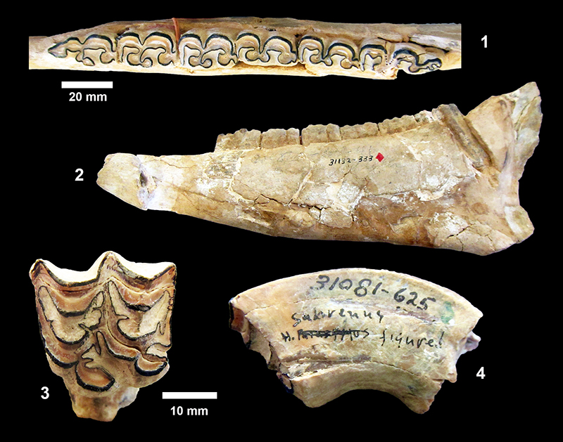
FIGURE 108. TMM 30936-348, Hypohippus cf. affinis, P/M, occlusal view, 32.31 x 24.6 mm, MCH = 16.7 mm.
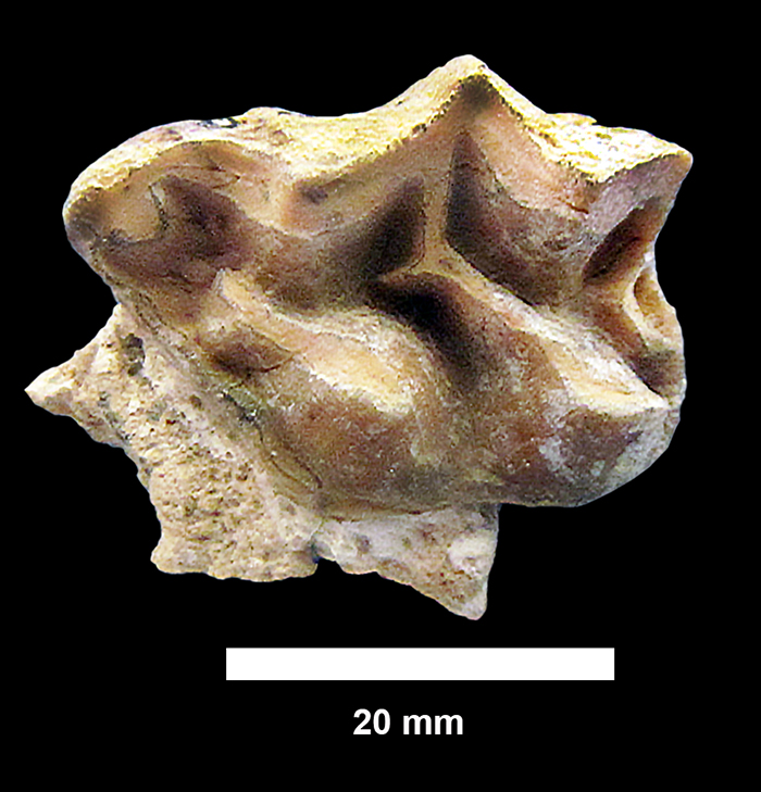
FIGURE 109. Composition of each of the four local faunas. Open squares = questionable occurrence. Bu R = Buckner Ranch, F R = Farish Ranch, Br R = Bridge Ranch, TMC = Ten Mile Waterhole Creek.
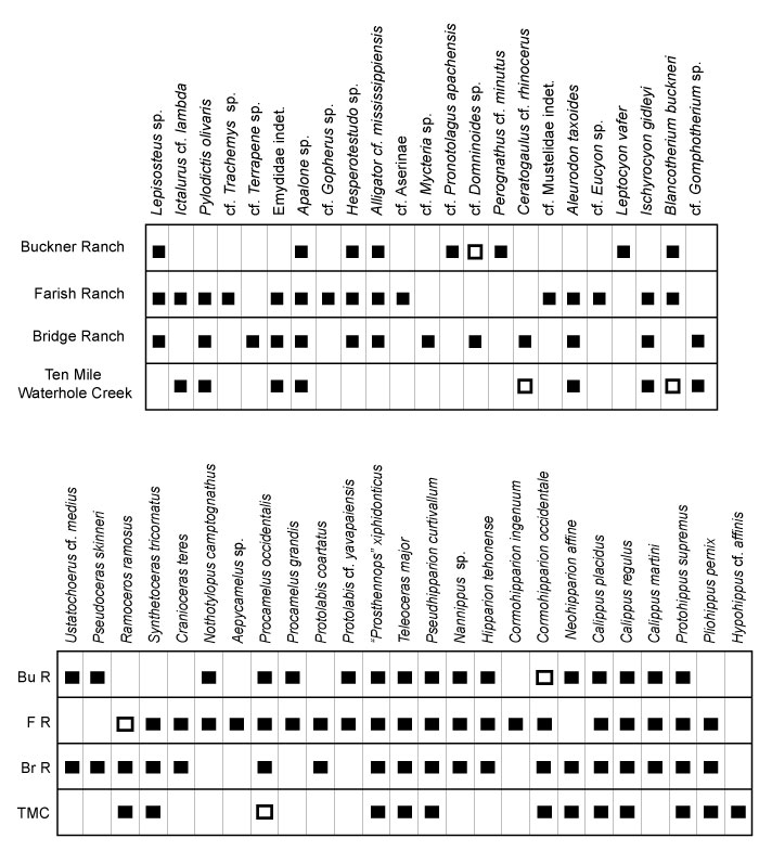
FIGURE 110. Temporal ranges (Hemingfordian - Hemphillian) for taxa from the Lapara Creek Fauna. Gray band illustrates interpreted age of the Lapara Creek Fauna. Ages of “early Clarendonian” from older literature have been interpreted to reflect the first half of the Clarendonian rather than strictly Cl1. Ranges are shown for explicit taxa even though some identifications are conferred.
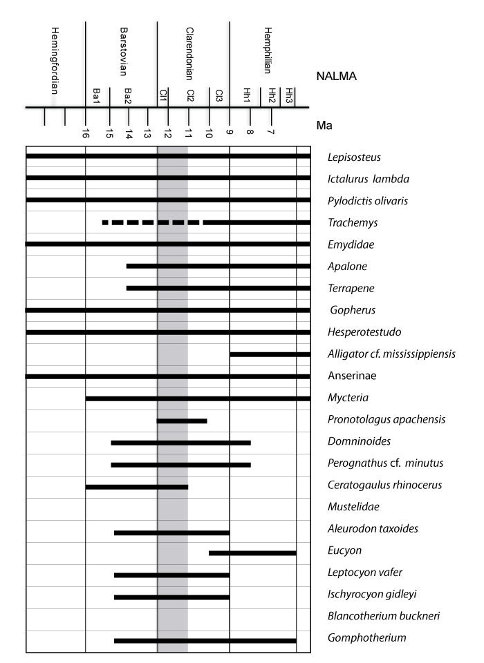
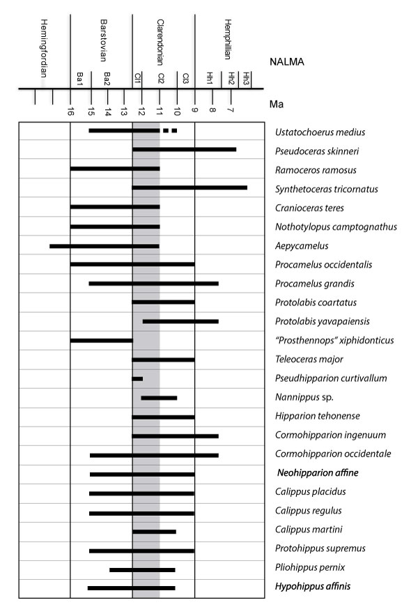
FIGURE 111. Concordia diagram for detrital zircon U/Pb results from JR1601.
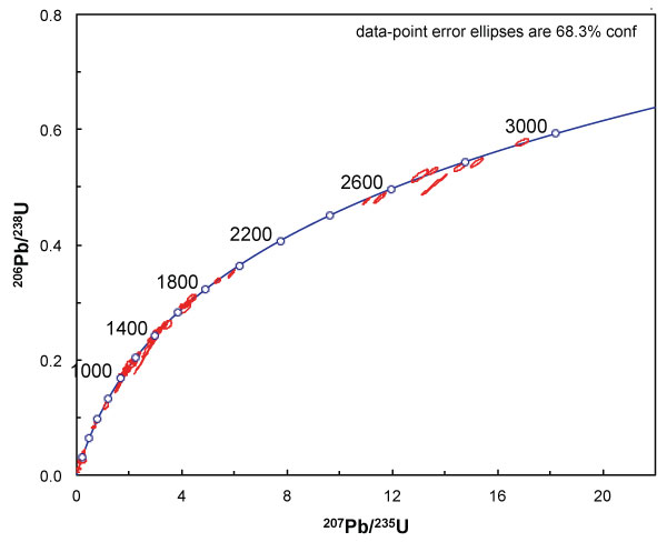
FIGURE 112. Age probability distribution for detrital zircon results from the lower Goliad Formation sample JR1601. 1, complete age spectrum. 2, all grains younger than 300 Ma. Statistically significant age peaks annotated with calculated age.
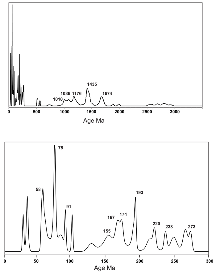
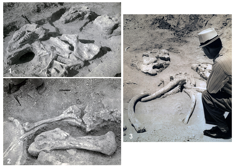
FIGURE 113. Buckner Ranch quarry (TMM 30896). 1, Three gomphothere skulls, all palate up (41BE2-56). Skull in left foreground associated with disarticulated, post-cranial material including pelvis, vertebrae, and ribs. Although the long axis of the three skulls appear to be roughly aligned, the post-cranial material appears disorganized although closely packed. 2, Same three skulls, different view with tusks, two associated with skull and two isolated (41BE2-130). 3, Disarticulated, post-cranial material including femur, vertebra and ribs (41BE2-57).