FIGURE 1. Thylacine skeleton, WAM M195 (M3318).
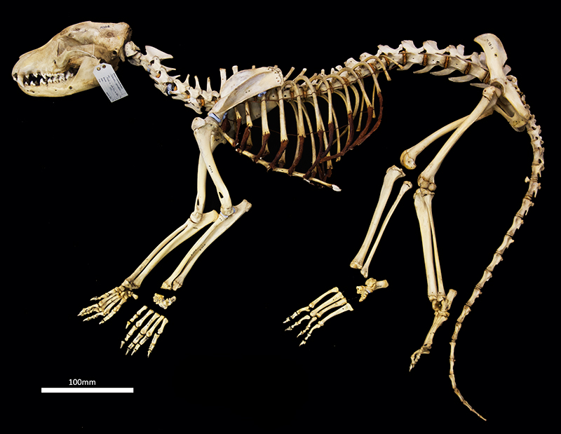
FIGURE 2. Skull of WAM M195 (M3318). 1, dorsal view; 2, ventral view. Scale bar equalsequals 20 mm.
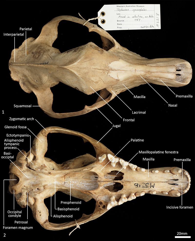
FIGURE 3. Juvenile damaged skull of WAM M17189. 1, dorsal view; 2, ventral view. Scale bar equals 20 mm.
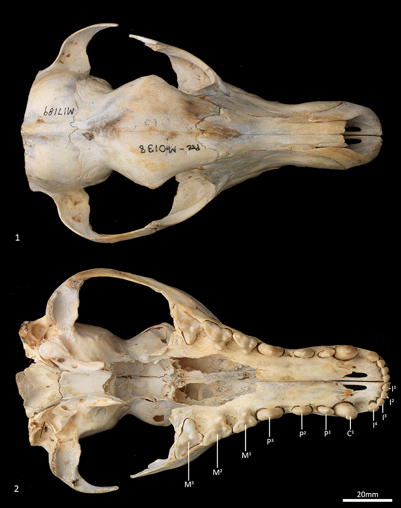
FIGURE 4. Skull of WAM M195 (M3318). 1, right lateral view; 2, left lateral view. Scale bar equals 20 mm.
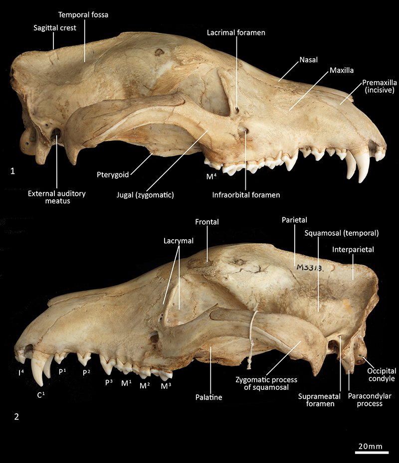
FIGURE 5. Juvenile damaged skull of WAM M17189. 1, right lateral view; 2, left lateral view. Scale bar equals 20 mm.
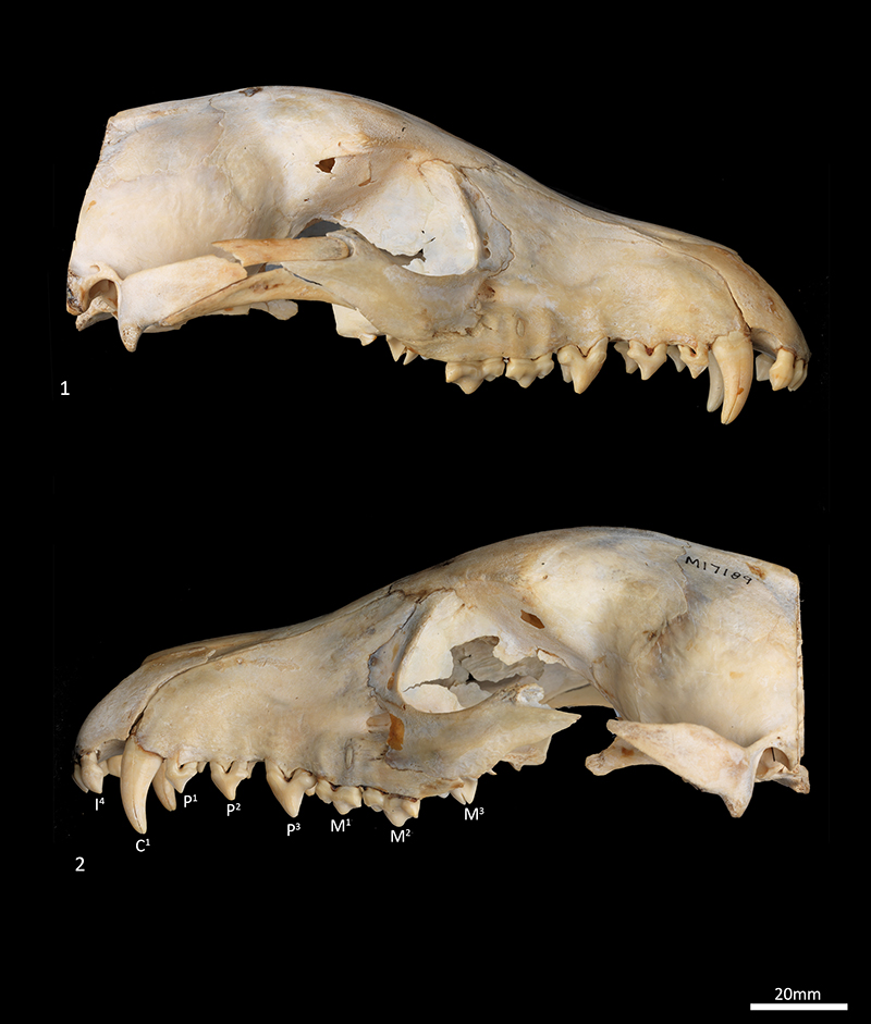
FIGURE 6. Skull of WAM M195 (M3318). 1, rostral view; 2, caudal view. Scale bar equals 20 mm.
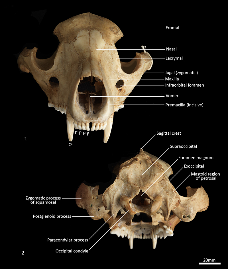
FIGURE 7. Features and foramina of skull WAM M195 (M3318). 1, ventral view; 2, caudoventral view. Scale bar equals 20 mm.
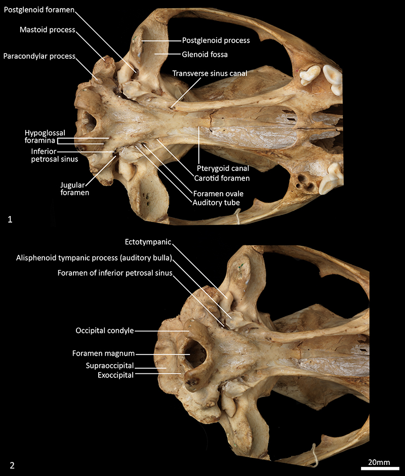
FIGURE 8. Features and foramina of skull WAM M195 (M3318). 1, ventrolateral view; 2, dorsolateral view of rostral orbit; 3, lateral view. Scale bar equals 20 mm.
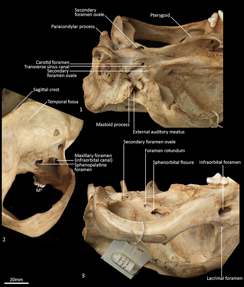
FIGURE 9. Upper dentition of WAM M195 (M3318). 1, ventral view; 2, ventrolateral view. Scale bar equals 20 mm. C1, upper canine; I1-4, upper incisor 1 to 4; M1-4, upper molar 1 to 4; P1-3, upper premolar 1 to 3.
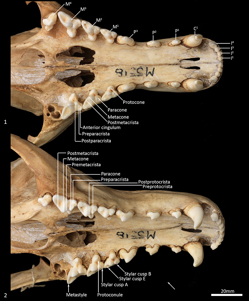
FIGURE 10. Mandible of WAM M195 (M3318). 1, left lateral view; 2, right lateral view; 3, left medial view; 4, right medial view. Scale bar equals 20 mm.
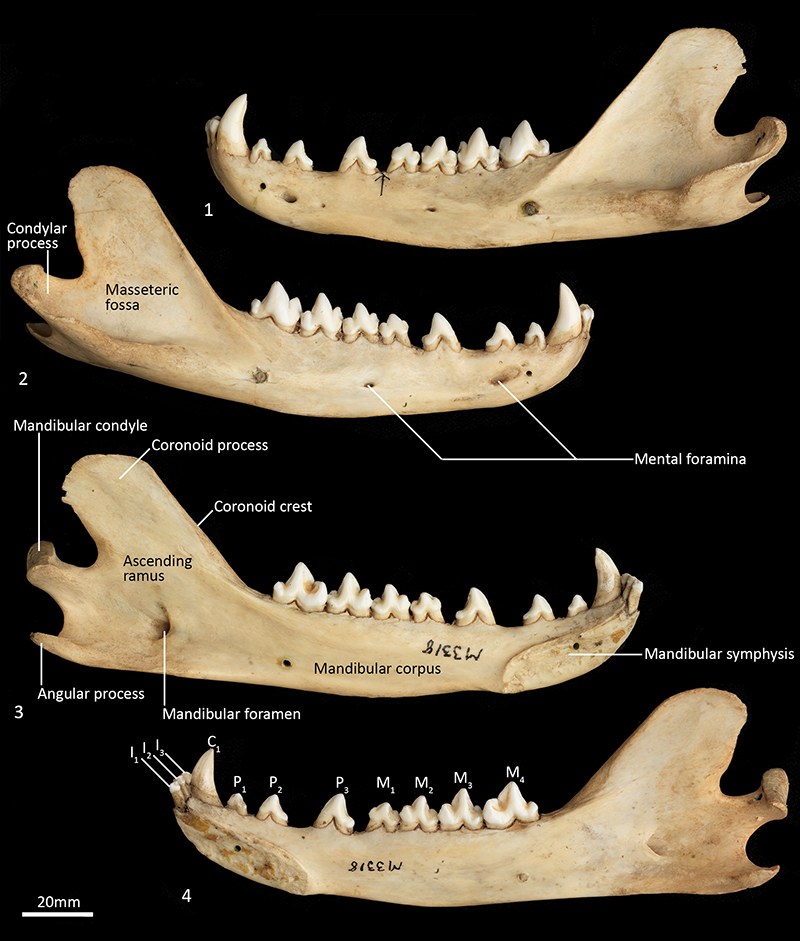
FIGURE 11. Mandible of WAM M195 (M3318). 1, dorsal view; 2, ventral view. Scale bar equals 20 mm.
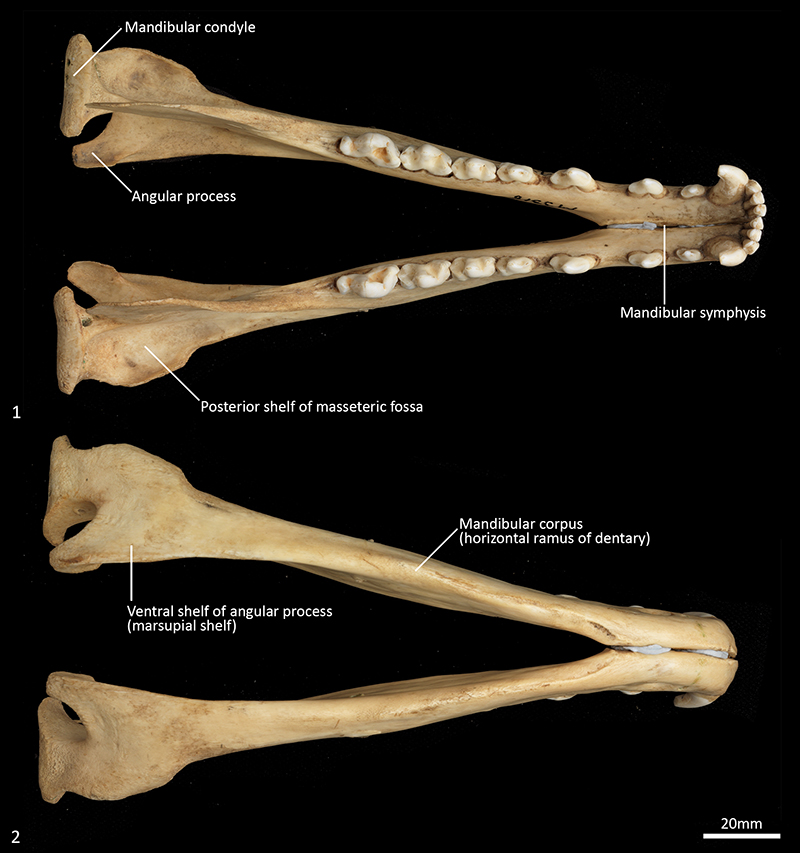
FIGURE 12. Lower dentition of WAM M195 (M3318). 1, dorsal view; 2, dorsolateral view. Scale bar equals 20 mm. C1, lower canine; I1-3, lower incisor 1 to 3; M1-4, lower molar 1 to 4; P1-3, lower premolar 1 to 3.
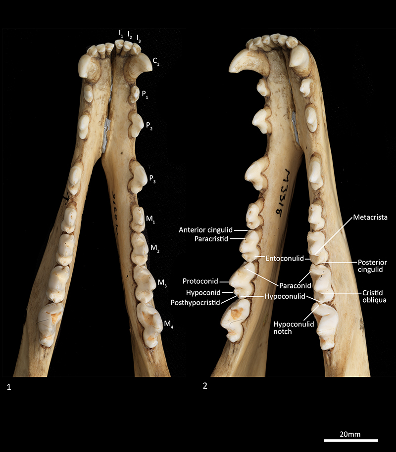
FIGURE 13. Mandible of WAM M17189. 1, left lateral view; 2, right lateral view; 3, right medial view; 4, left medial view; 5, occlusal view. Scale bar equals 20 mm.
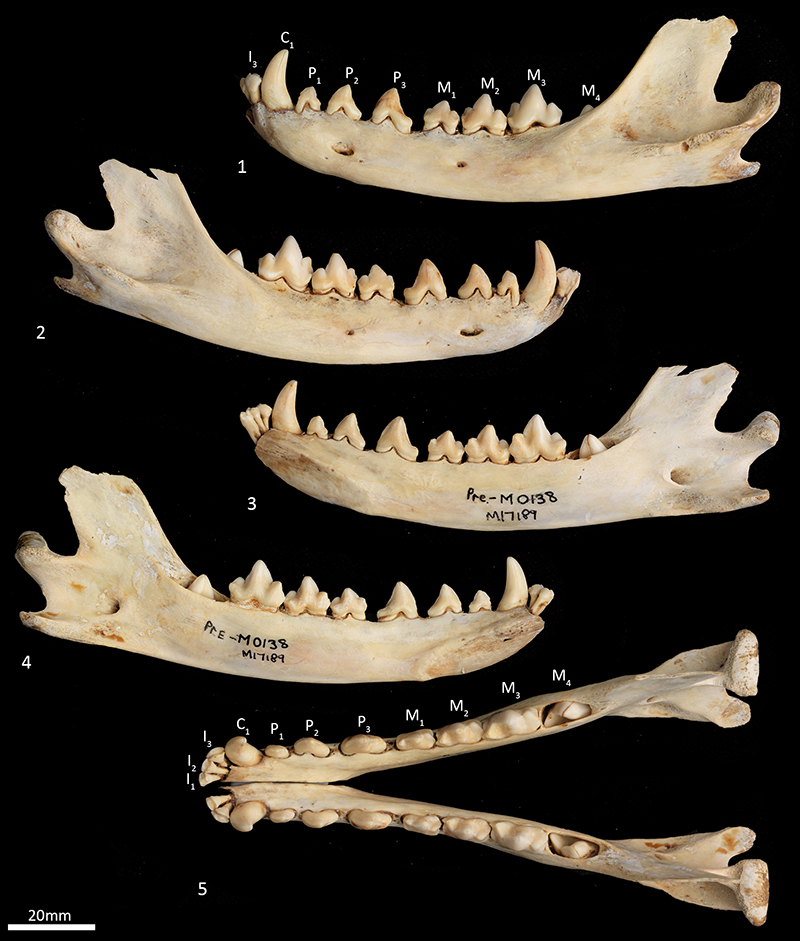
FIGURE 14. Cervical vertebrae SAMA M1960. 1, articulated cervical vertebrae, dorsal view; 2, atlas (C1) cranial view; 3, atlas caudal view; 4, atlas dorsal view; 5, atlas ventral view; 6, axis (C2) dorsal view; 7, axis ventral view; 8, axis lateral view; 9, axis caudal view; 10, third cervical vertebrae (C3) cranial view; 11, articulated cervical vertebrae lateral view; 12, C3 ventral view. Scale bar equals 20 mm.
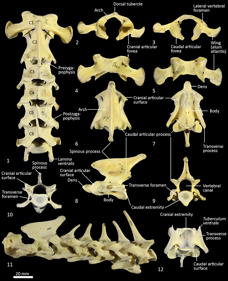
FIGURE 15. Thoracic vertebrae SAMA M95. 1, complete thoracic series (T1-T13) lateral view; 2, dorsal view; 3-5, fourth thoracic vertebra lateral (3), cranial (4) and dorsal (5) view; 6-8, twelfth thoracic vertebra lateral (6), cranial (7) and dorsal (8) view. Scale bar equals 20 mm.
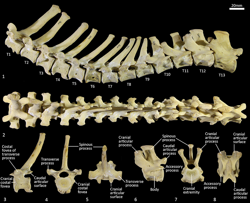
FIGURE 16. Lumbar vertebrae SAMA M1960. 1, complete lumbar series (L1-L6), dorsal view; 2, lateral view; 3, third lumbar vertebra (L3) cranial view; 4, L3 dorsal view; 5, L3 lateral view. Scale bar equals 20 mm.
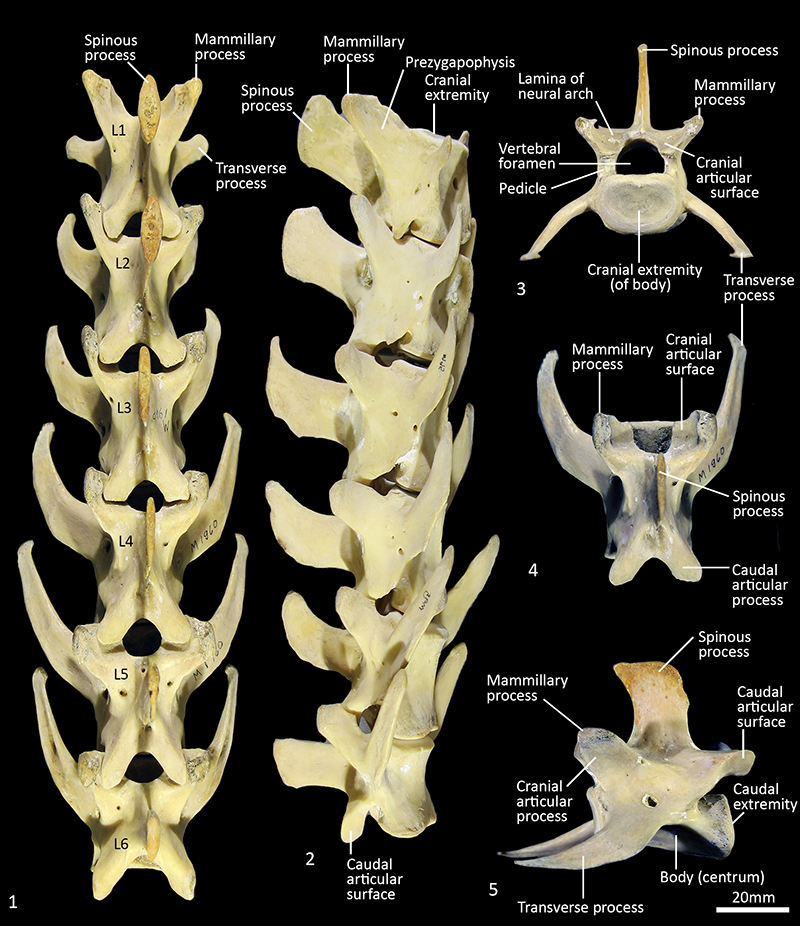
FIGURE 17. Sacral and caudal vertebrae SAMA M1960. 1, complete sacro-caudal series, dorsal view; 2, sacrum and proximal caudal series, dorsal view; 3, first caudal vertebra (Ca1) cranial view; 4, Ca1 dorsal view; 5, Ca1 lateral view; 6, eighth caudal vertebra (Ca8) ventral view; 7, Ca8 cranial view; 8, Ca8 lateral view; 9, examples of proximal, middle and distal chevron bones from ventral and dorsal views. Scale bar equals 20 mm.
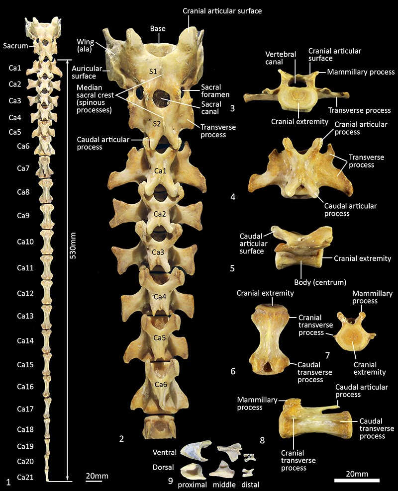
FIGURE 18. Forelimb (right) topography WAM M195 (M3318). 1, Proximal; 2, distal elements. Scale bar equals 20 mm.
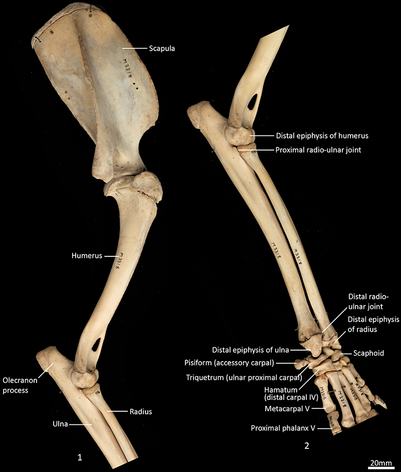
FIGURE 19. Scapula (right) SAMA M1960. 1, lateral view; 2, medial view; 3, dorsal view; 4, caudal view; 5, cranial view; 6, glenoid view. Scale bar equals 20 mm; NB 5 and 6 not to scale.
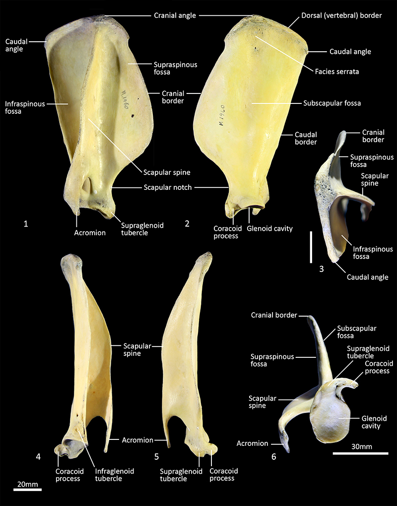
FIGURE 20. Scapula (left) WAM M195 (M3318). 1, lateral view; 2, medial view; 3, caudal view. Scale bar equals 20 mm.
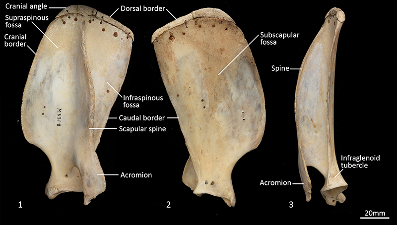
FIGURE 21. Clavicles SAMA M1960. 1, left lateral view; 2, left medial view; 3, right lateral view; 4, right medial view. Scale bar equals 20 mm.
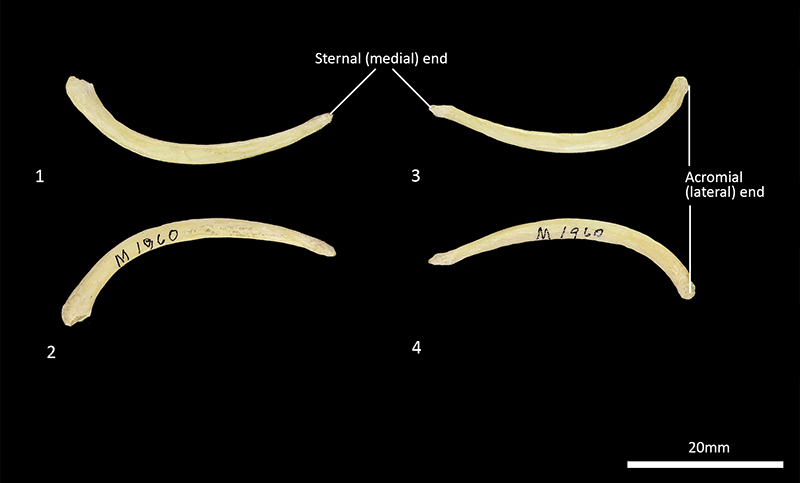
FIGURE 22. Right Humerus SAMA M1960. 1, cranial view; 2, caudal view; 3, lateral view; 4, medial view; 5, proximal view; 6, distal view. Scale bar equals 20 mm; NB 5 and 6 not to scale.
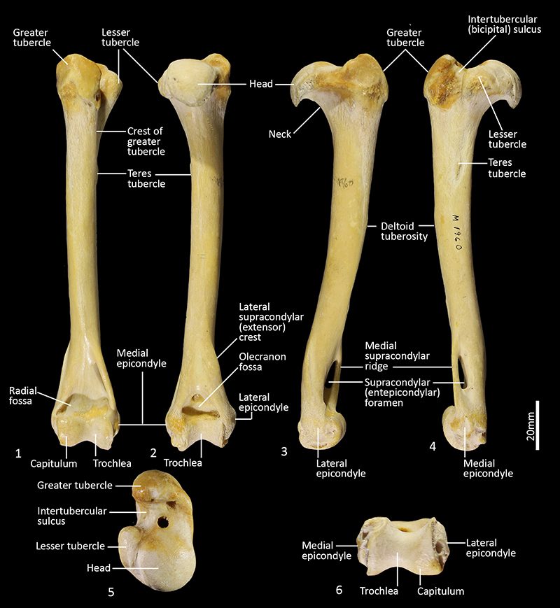
FIGURE 23. Humerus (left) WAM M195 (M3318). 1, cranial view; 2, caudal view; 3, lateral view; 4, medial view; 5, proximal view; 6, distal view. Scale bar equals 20 mm; NB 5 and 6 not to scale.
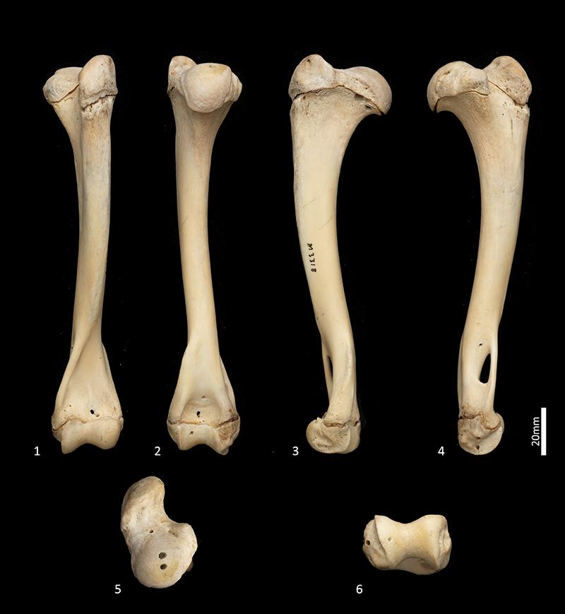
FIGURE 24. Ulna and radius (right) SAMA M1960. 1, radius cranial view; 2, ulna cranial view; 3, ulna caudal view; 4, radius caudal view; 5, ulna lateral view; 6, radius lateral view; 7, radius medial view; 8, ulna medial view; 9, radius proximal view; 10, radius distal view. Scale bar equals 20 mm; NB 9 and 10 not to scale.
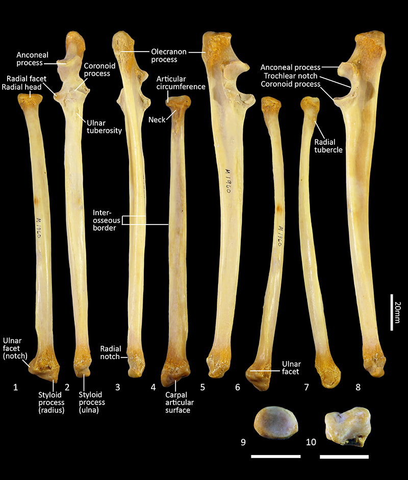
FIGURE 25. Ulna and radius (left) WAM M195 (M3318). 1, ulna cranial view; 2, ulna medial view; 3, ulna lateral view; 4, radius lateral view; 5, radius cranial view; 6, radius medial view; 7, radius proximal view; 8, radius distal view. Scale bar equals 20 mm; NB 7 and 8 not to scale.
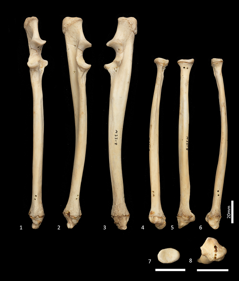
FIGURE 26. Manus (left) SAMA M1960. 1, proximal view (digitigrade posture); 2, lateral view (digitigrade posture); 3, dorsal view (plantigrade posture); 4, palmar/ventral view (plantigrade posture); 5, medial view (digitigrade posture). Scale bar equals 20 mm.
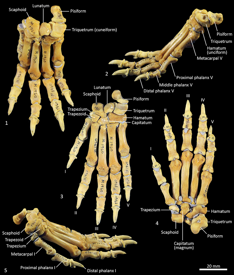
FIGURE 27. Carpal bones (left) SAMA M1960, dorsal, ventral, proximal, distal, medial and lateral views. 1-6, Scaphoid; 7-12, lunatum; 13-18, triquetrum; 19-24, pisiforme; 25-30, trapezium; 31-36, trapezoid; 37-42, capitatum; 43-48, hamatum. Scale bar equals 10 mm.
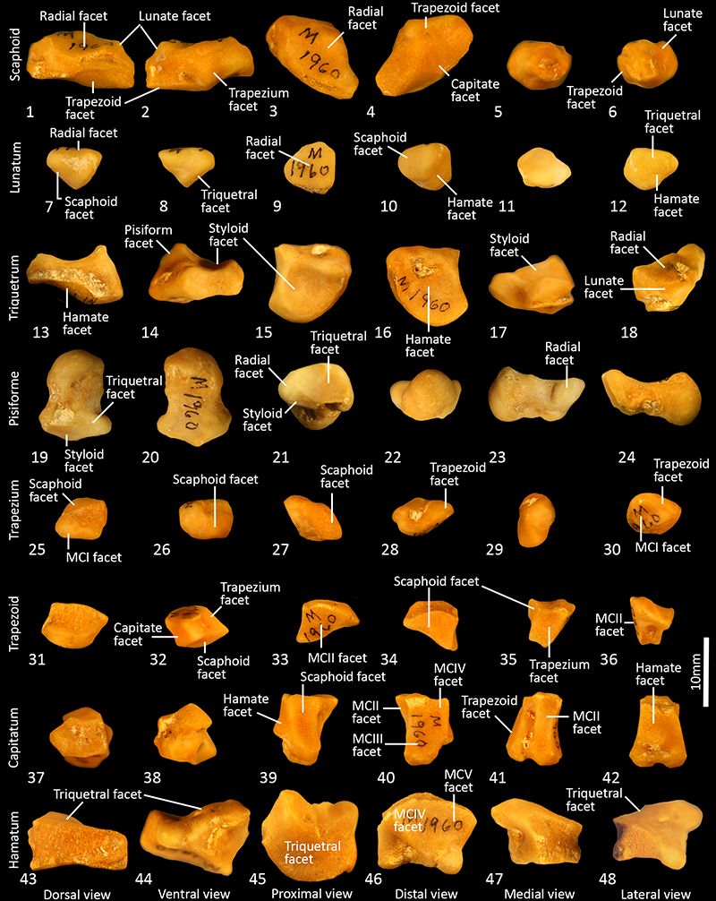
FIGURE 28. Metacarpals and phalanges (left) SAMA M1960. 1-4, Metacarpals proximal view (1); distal view (2); dorsal view (3); ventral (palmar) view (4); 5-7, phalanges dorsal view (5); ventral (palmar) view (6); lateral view (7). Scale bar equals 20 mm.
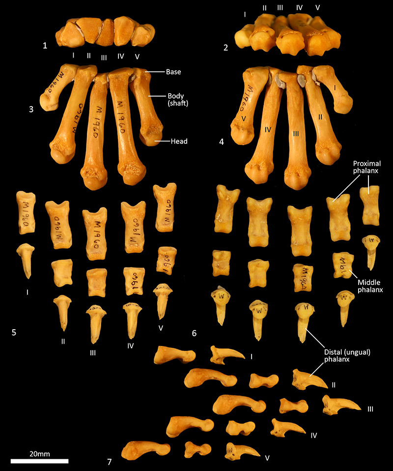
FIGURE 29. Pelvis SAMA M1960 (landscape). 1, ventral view; 2, dorsal view. Scale bar equals 20 mm.
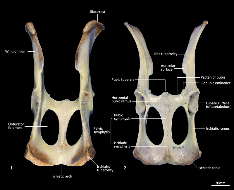
FIGURE 30. Pelvis SAMA M1960. 1, left lateral view; 2, cranial view; 3, caudal view. Scale bar equals 20 mm. 4, Epipubic bones. Scale bar equals 10 mm.
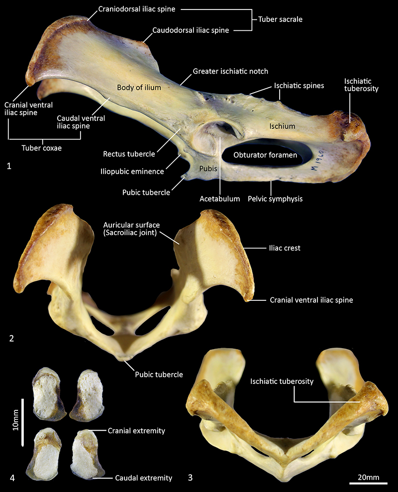
FIGURE 31. Pelvis WAM M195 (M3318) (landscape). 1, dorsal view; 2, ventral view; 3, right lateral view. Note missing epiphyses and unfused pelvic symphysis. Scale bar equals 20 mm.
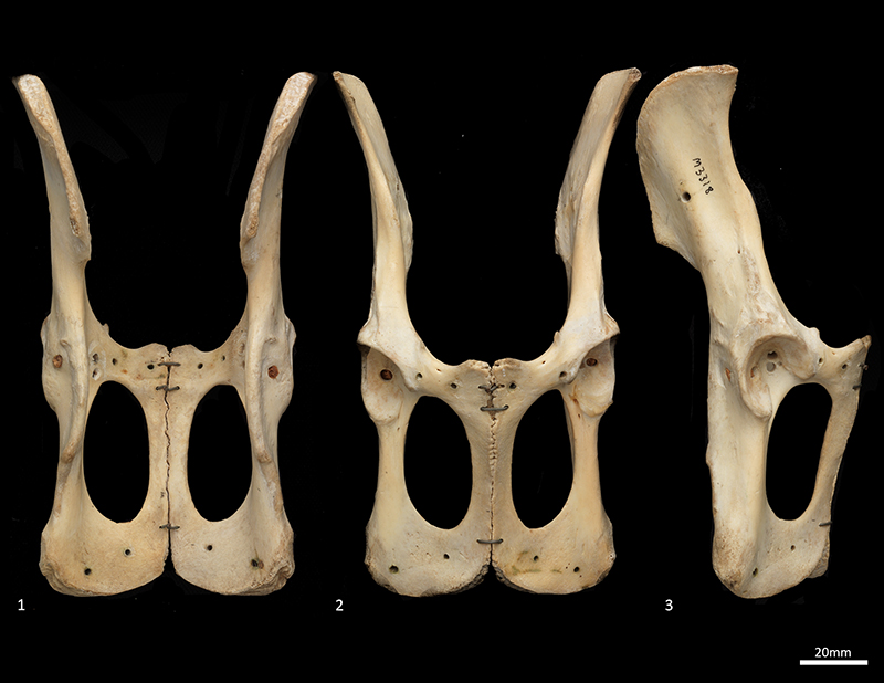
FIGURE 32. Femur SAMA M1960. 1, cranial view; 2, caudal view; 3, lateral view; 4, medial view; 5, proximal view; 6, distal view. Scale bar equals 20 mm.
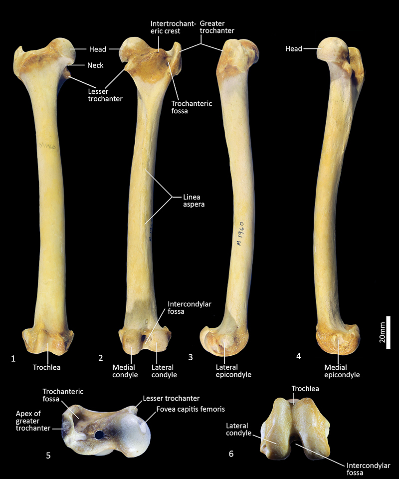
FIGURE 33. Femur WAM M195 (M3318). 1, cranial view; 2, caudal view; 3, lateral view; 4, medial view; 5, proximal view; 6, distal view. Scale bar equals 20 mm.
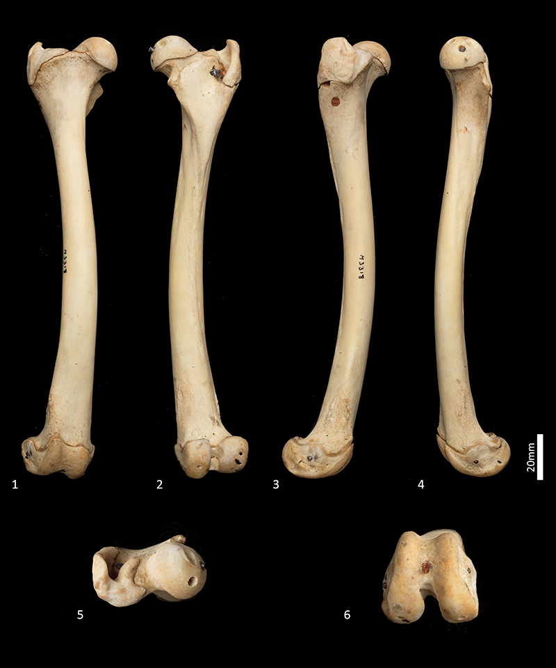
FIGURE 34. 1-2, Stifle (knee) (1) and ankle regions (2) of the left hindlimb of WAM M195 (M3318). Lateral view.
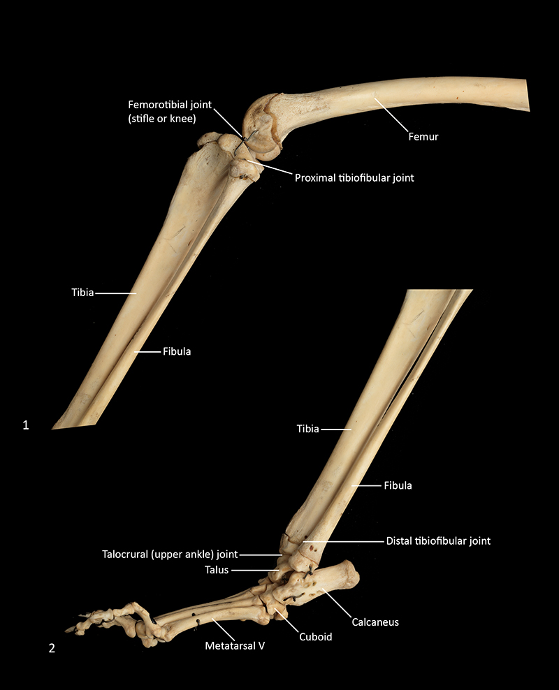
FIGURE 35. Tibia and Fibula (right) SAMA M1960. 1, fibula cranial view; 2, tibia cranial view; 3, tibia caudal view; 4, fibula caudal view; 5, fibula lateral view; 6, tibia lateral view; 7, fibula medial view; 8, tibia medial view. Scale bar equals 20 mm.
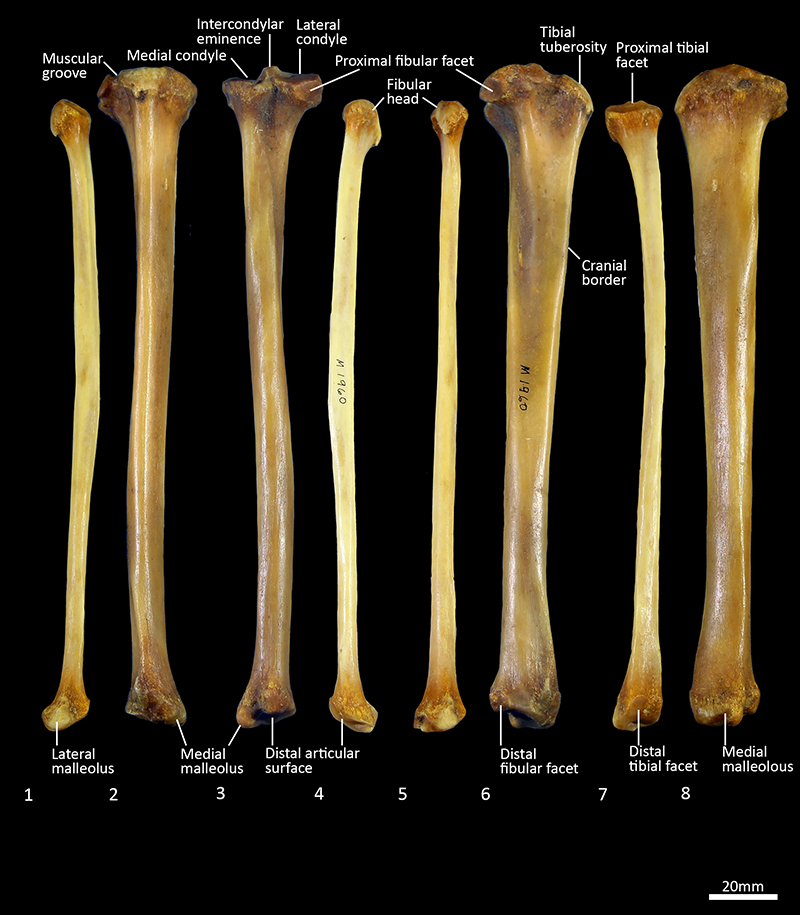
FIGURE 36. Tibia and fibula (right) WAM M195 (M3318). 1, fibula cranial view; 2, tibia cranial view; 3, tibia caudal view; 4, fibula caudal view; 5, tibia lateral view; 6, fibula medial view; 7, tibia medial view; 8, tibia proximal view; 9, tibia distal view. Scale bar equals 20 mm.
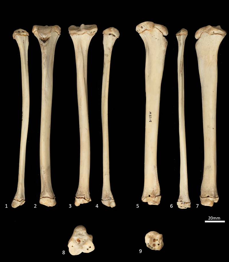
FIGURE 37. Right pes SAMA M1960. 1, plantar view (plantigrade posture); 2, dorsal view (plantigrade posture); 3, cranial view (digitigrade posture); 4, lateral view (digitigrade posture); 5, medial view (digitigrade posture). Scale bar equals 20 mm.
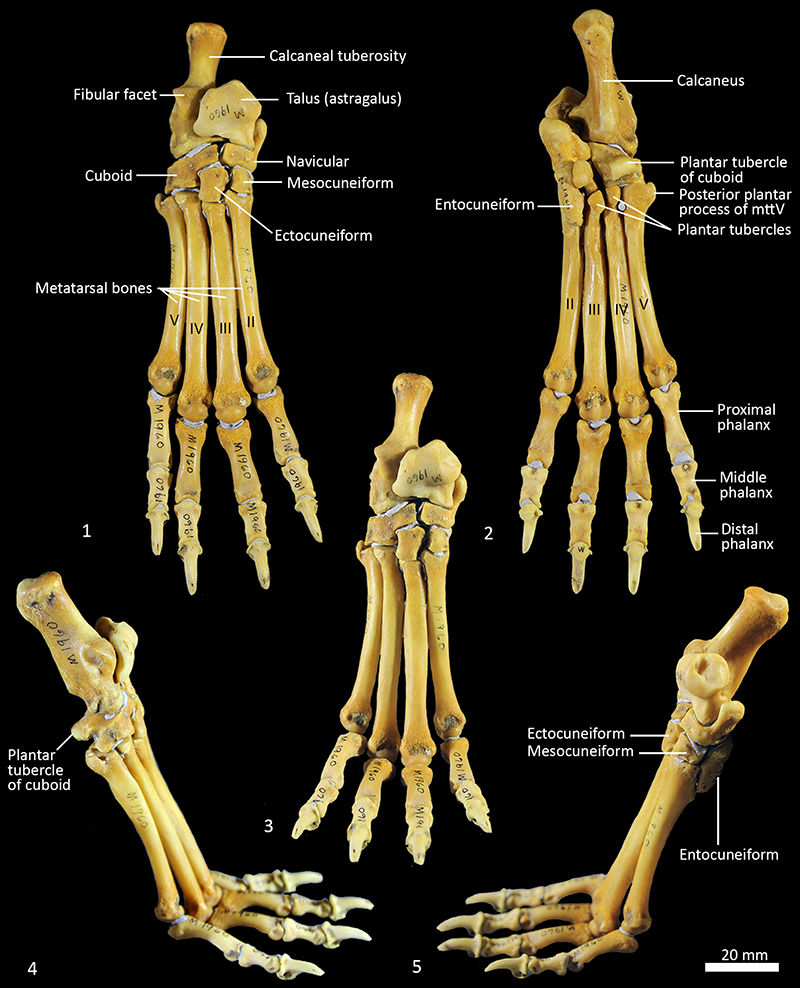
FIGURE 38. Proximal tarsal bones (right pes) SAMA M1960. 1-5, Calcaneus dorsal (1); ventral (2); medial (3); lateral (4); distal views (5); 6-10, talus (astragalus) dorsal (6); ventral (7); medial (8); lateral (9); proximal (10); 11-17, distal views; navicular, (central tarsal bone) dorsal (12); ventral (13); medial (14); lateral (15); proximal (16); distal (17) views. Scale bar equals 10 mm.
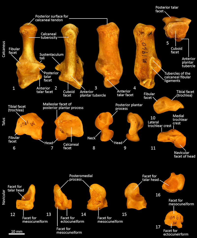
FIGURE 39. Distal tarsal bones (right pes) SAMA M1960. Dorsal, ventral, medial, lateral, proximal and distal views. 1-6, cuboid; 7-12, ectocuneiform; 13-18, mesocuneiform; 19-24, entocuneiform. Scale bar equals 10 mm.
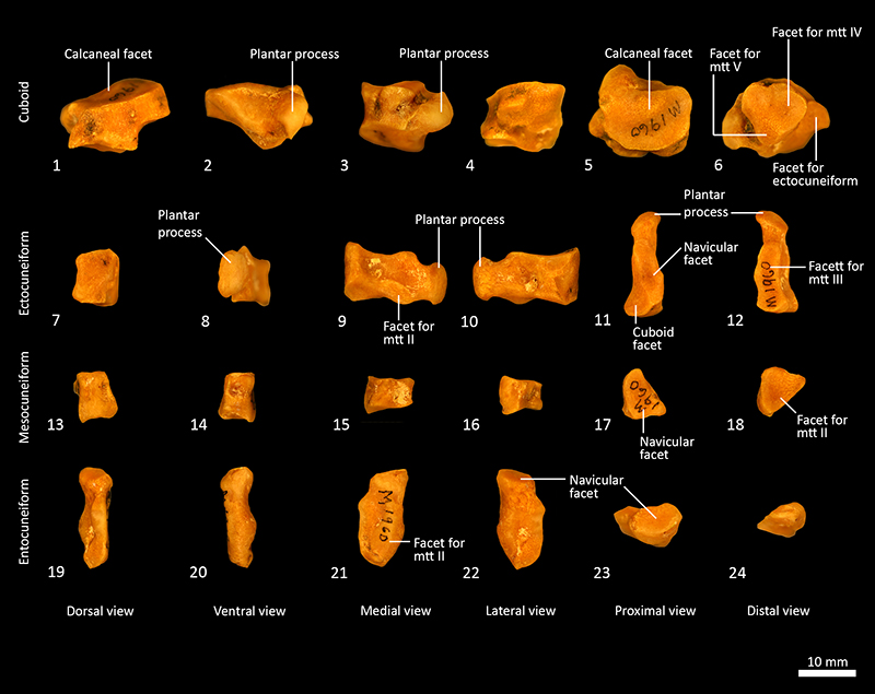
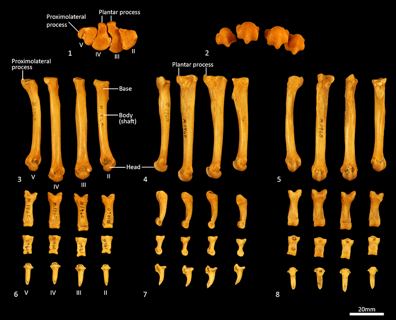
FIGURE 40. Metatarsals and phalanges (right pes) SAMA M1960. 1-5, metatarsals proximal view (1) (not to scale); distal view (2) (not to scale); dorsal view (3); lateral view (4); plantar view (5); 6-8, proximal middle and distal phalanges dorsal (6); lateral (7); plantar view (8). Scale bar equals 20 mm.