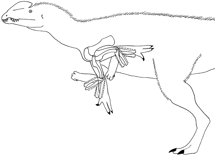
FIGURE 1. Humerus, radius, and ulna of the holotype of Dilophosaurus wetherilli. 1-4, left (on left) and right (on right) humerus in lateral (1), medial (2), anterior (3), and posterior (4) views, with views standardized according to the orientation of the distal condyles. 5-7, left radius and ulna in medial (5), lateral (6), and proximal (7) views. 8-10, right radius and ulna in proximal (8), lateral (9), and medial (10) views. Broken black lines in 1 indicate the midlines of the posterior (retractor) surface of the left humerus and the homologous surface of the right humerus, showing the strong degree of abnormal torsion of the diaphysis of the right humerus. Broken white line in 5 delineates the edge of a pathological cavitation. Abbreviations: ag, abnormal growth on ulna; bt, bony tumors; f, fracture of radius. Scale bar equals 50 mm.
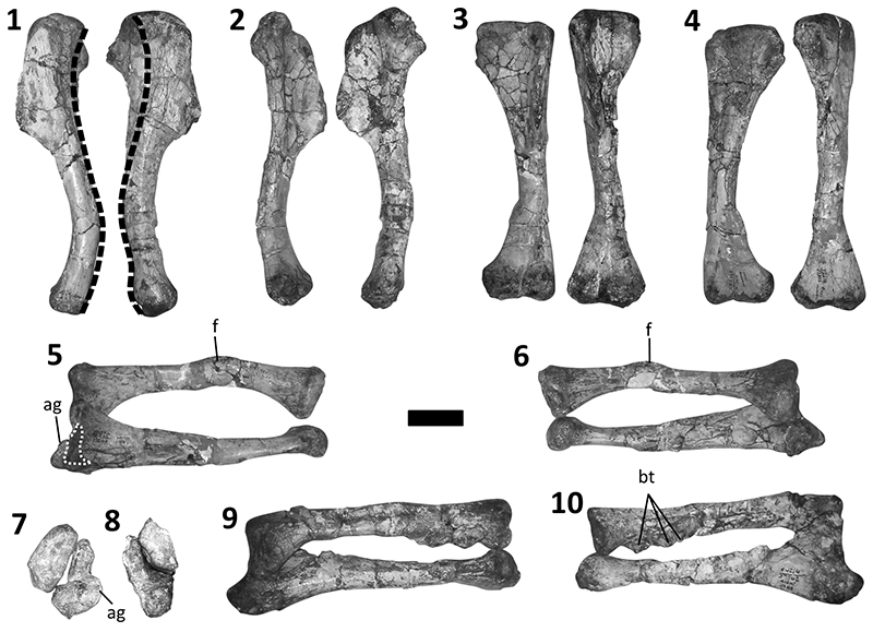
FIGURE 2. Asymmetry between bones of the left and right hands. In each panel of the figure, the bone from the left hand is on the left, and the bone from the right hand is on the right. 1-2, metacarpal III in posterior (1) and anterior (pollical) (2) views. 3-6, distal end of metacarpal III in posterior (3), anterior (4), flexor (palmar) (5) and extensor (lateral) (6) views. 7-10, manual phalanx I-1 in posterior (7), anterior (8), flexor (9) and extensor (10) views. 11-14, manual phalanx III-1 in posterior (11), anterior (12), flexor (13) and extensor (14) views. Broken white lines in 3-6 and 12-14 indicate edges of articular surfaces. Broken white lines in 7 and 10 indicate pathological pitting. Broken black lines in 13 and 14 indicate orientations of homologous surfaces of the phalanges of the two hands. Abbreviation: ef, extensor fossa. Scale bar equals 50 mm in 1-2 and 7-14. Scale bar equals 10 mm in 3-6.
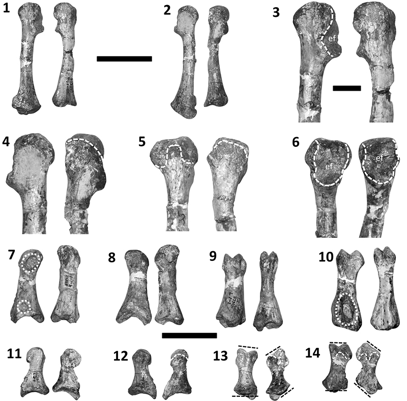
FIGURE 3. Distal carpal element (distal carpals 1 + 2) of right hand of holotype of Dilophosaurus wetherilli. 1, proximal view. 2, distal view. 3, extensor view. 4, flexor view. 5, posterior view. 6, anterior view. 7 - 8, distal carpal in articulation with the first and second metacarpals in extensor (7) and oblique proximo-extensor (8) views. Dotted white outlines indicate edges of extensor fossae. Scale bar equals 15 mm. 7 and 8 not to scale.
FIGURE 4. Range of motion in the forelimb of the holotype of Dilophosaurus wetherilli. 1–3. right shoulder in lateral (1), anterior (2), and dorsal (3) views. 4. right elbow in lateral view. 5–7. left shoulder in lateral (5), anterior (6), and dorsal (7) views. 8. left elbow in lateral view. 9. anterior view of right metacarpal I and phalanx I-1 of the holotype, with phalanx I-2 of UCMP 37303. 10. left metacarpal II and phalanx II-1 in anterior view. 11. left phalanges II-1 and II-2 in anterior view. 12. left digit III in anterior view. 13. left digit IV in posterior view. 14. right metacarpal I and phalanx I-1 in anterior view. 15. right metacarpal III and phalanx III-1 in anterior view. 16. left metacarpal II and phalanx II-1 in anterior (top left), posterior (top right), and extensor (bottom) views. 17. left metacarpal III and phalanx III-1 in anterior (top left), posterior (top right), and extensor (bottom) views. 18. right metacarpal III and phalanx III-1 in anterior (top left), posterior (top right), and extensor (bottom) views, with broken white outline indicating distal end of proximal phalanx, and solid white outline indicating proximal end of proximal phalanx. Note that the range of elevation measured in 2 and 6 is elevation by abduction, although the distal end of the humerus can be lifted higher during retraction (as shown by the retracted humerus in those panels of the figure). Note, too that the view of the elevated humerus obscures the view of the protracted humerus in 1 but not 5, which shows the influence of paleopathology in the misshapen right humerus, as does the long-axis rotation of the right humerus during retraction as shown in 1.
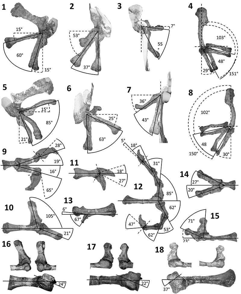
FIGURE 5. Bones of the left hand of the holotype of Dilophosaurus wetherilli in flexion (all bones except phalanx IV-1) and hyperextension (metacarpus and thumb only), and bones of the right hand (metacarpal I, metacarpal II, and thumb) in flexion and hyperextension. In 1-6, the bones of the left hand are on the left side, and the bones of the right hand are on the right side. 1-3, flexion in lateral (1), palmar (2), and anterior (3) views. 4-6, hyperextension in lateral (4), palmar (5), and anterior (6) views. 7-8, flexion of left hand in posterior view (7) and from the perspective of a distal view of the metacarpus (8). 9-10, hyperextension of left thumb in posterior view (9) and from the perspective of a distal view of the metacarpus (10). 11, flexion of phalanx I-1 of the right hand, from the perspective of a distal view of the metacarpus. Abbreviations: I - III, number of digit or metacarpal.
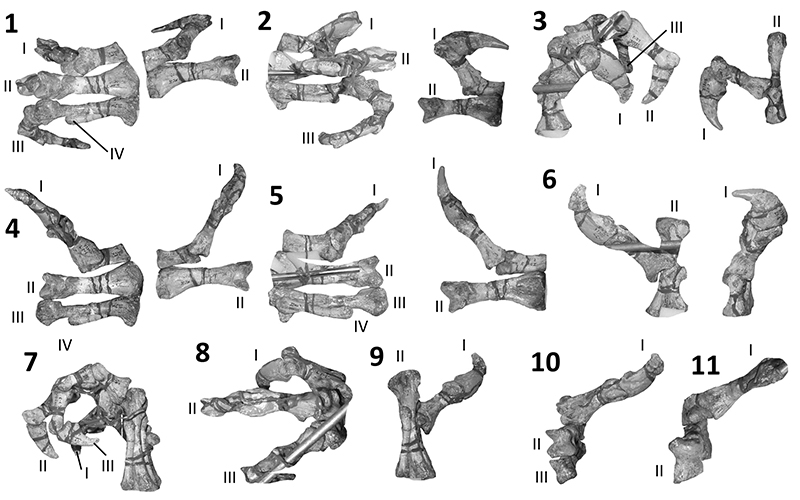
FIGURE 6. Range of motion at the elbow in specimens of Alligator mississippiensis. 1, a skeletal specimen: right forelimb of TMM 3004. 2, an intact specimen: left forelimb of Cadaver 2, showing that the humeral orientation relative to the viewer during elbow flexion matches that of the skeletal specimen.
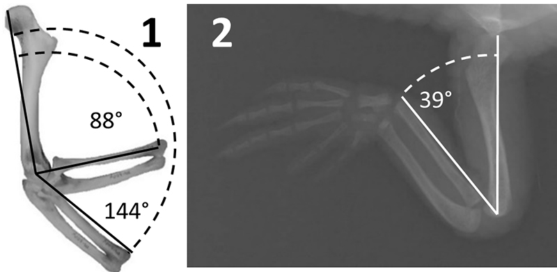
FIGURE 7. Reconstructed range of motion in the forelimb of Dilophosaurus wetherilli, with bare-bones range of elbow motion indicated by broken lines, showing that the influence of soft tissues allows for more elbow flexion and extension in the fully-fleshed animal than the bony articular surfaces at the elbow indicate.