FIGURE 1. Location of Seymour Island (Antarctica) and stratigraphy of its NE part. 1. Map of Antarctica showing the position of Seymour Island. 2. Map of Seymour Island showing the location of the Eocene La Meseta and Submeseta formations, and position of the locality DPV 13/84 with sampling site. 3. Simplified stratigraphic column of the Submeseta Formation with an arrow pointing at the sampling horizon, based on data from Montes et al. (2013). 4. View of the sampling site location from under the plateau of the Weddell Formation; photograph was taken in 2011 by TM. Abbreviations: cg, conglomerates and sandstones; t, Turritella (a gastropod).
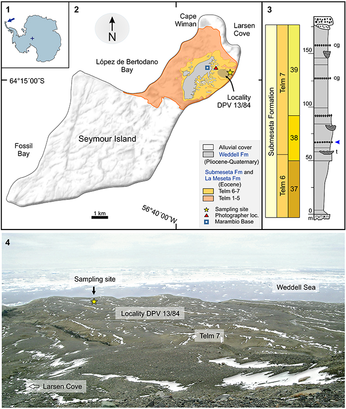
FIGURE 2. Partial foot skeleton of Eocene Delphinornis larseni, NRM-PZ A.994. 1. Right tarsometatarsus in dorsal view. 2. Left tarsometatarsus in dorsal view. 3. Same in medial view. 4. Same in lateral view. 5. Same in plantar view. 6. Same in proximal view. 7. Same in distal view. 8. Left tarsometatarsus of D. larseni, IB/P/B-0547 (Eocene, Seymour Island, Antarctica), in lateroplantar view. 9. Left tarsometatarsus of D. larseni, IB/P/B-0062 (Eocene, Seymour Island, Antarctica), in in dorsal view. 10. Same in medial view. 11. Same in lateral view. 12. Same in plantar view. 13. Same in proximal view. 14. Same in distal view. 15. Reconstruction of right-foot skeleton of D. larseni (NRM-PZ A.994) in dorsal view. 16. Outline of left-foot skeleton of extant Pygoscelis papua (unnumbered specimen from IB/P/B) in same view. 17. Reconstruction of left-foot skeleton of D. larseni (NRM-PZ A.994) in same view. 18. Schematic view of left-foot skeleton of D. larseni (NRM-PZ A.994) in dorsal view, as preserved in matrix, superimposed over X-ray image of same specimen. 19. X-ray-based heat map of portion of above fossil indicating location of tarsometatarsal dvf. 20. Left tarsometatarsus of Pygoscelis papua (unnumbered specimen from IB/P/B) in dorsal view. 21. Same in plantar view. Abbreviations: d2-4, digit 2-4; df, dermal flange; dvf, distal vascular foramen; ft, flexor tubercle; hs, hypotarsal sulcus; ice, intercotylar eminence; icf, infracotylar fossa; ier, impressions of extensor retinaculum; ihc, intermediate hypotarsal crests; in, intertrochlear notch; itc, insertion of tibial cranial muscle; lc, lateral cotyle; lhc, lateral hypotarsal crest; lis, lateral intermetatarsal sulcus; lm, lateral margin; lt, lateral (hypotarsal) tuberosity; ltm, left tarsometatarsus; lvf, lateral vascular foramen; m2-4, metatarsal 2-4; mc, medial cotyle; mhc, medial hypotarsal crest; mis, medial intermetatarsal sulcus; mm, medial margin; mvf, medial vascular foramen; ns, neurovascular sulcus; ota, origin surface for toe abductor; p2-4, phalanx 2-4; pu, ungual (terminal) phalanx; rtm, right tarsometatarsus; sd, supratrochlear depression; ta, tarsus; t2-4, trochlea 2-4. Asterisks denote comparative material. Scale bar equals 1 cm.
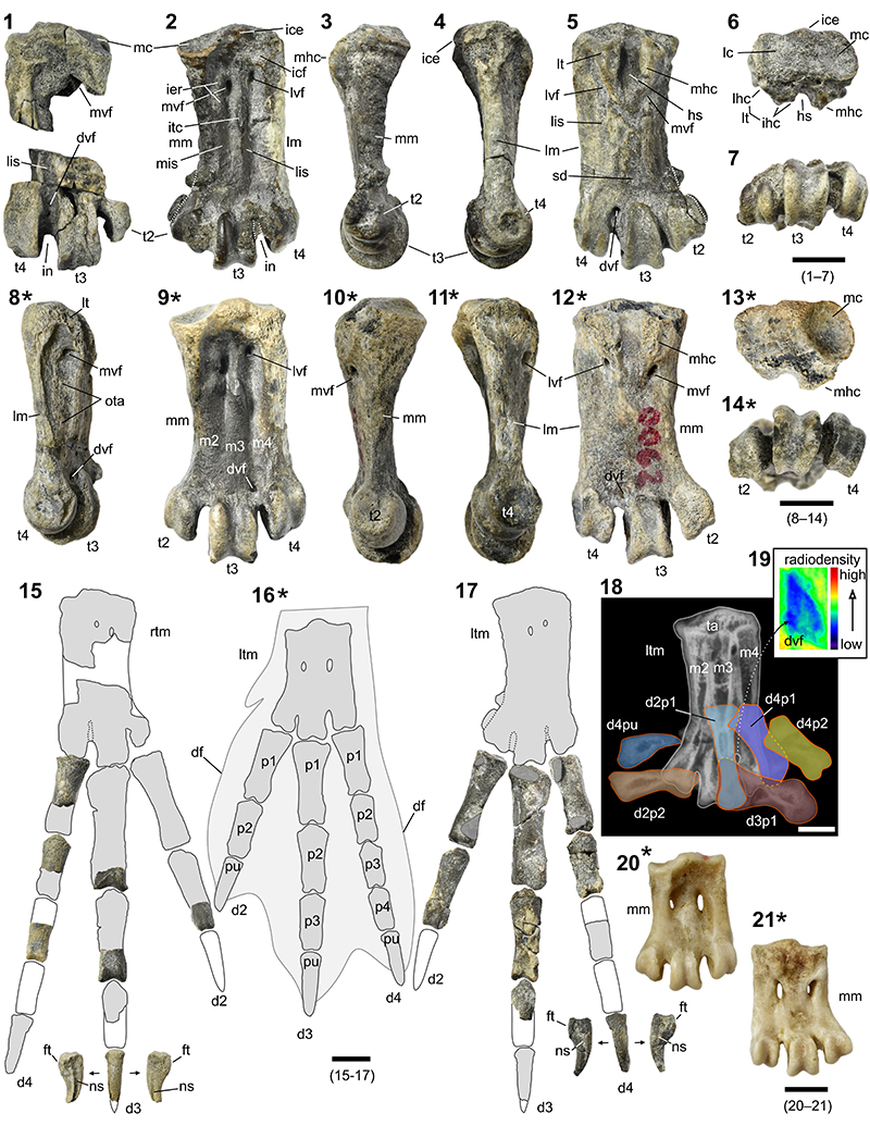
FIGURE 3. Comparison of tarsometatarsal measurements of selected present-day and Eocene penguins. The specimen maximum length and mediolateral midwidth are visualized as coordinates of a circle, whereas the dorsoplantar midwidth of the third trochlea - as its size. For specimen IDs, see Appendix 1.
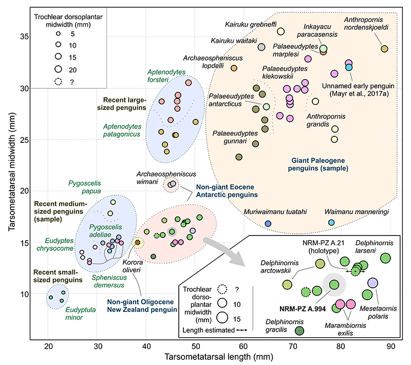
FIGURE 4. Minor fragments of pelvic skeleton and leg bones of Eocene Delphinornis larseni, NRM-PZ A.994. 1. Location of these skeletal elements within penguin body, in ventral view, is presented in red. Other bones included in NRM-PZ A.994, and shown in Figure 2.1-7, 2.15, 2.17-19, Figure 5.3-4, 5.6, 5.8-9, 5.13, 5.16, are in gray. 2. Heat map of left acetabular socket, based on X-ray. 3. Left acetabular socket in medial view with associated portion of synsacrum. 4. Same in lateral view with exposed cranial frame of obturator foramen. 5. Left ischium in medial view with associated portion of synsacrum. 6. Same in lateral view with exposed fragment of caudal frame of ilioischiadic foramen. 7. Right-side view of Eocene synsacrum IB/P/B-0149 (Eocene, Seymour Island, Antarctica). 8. Right femoral head in proximal view. 9. Same in caudal view. 10. Middle shaft of right femur (undetermined aspect of preserved wall). 11. Same in opposite view, showing pronounced stricture of medullary cavity. 12. Alleged fragment of proximal shaft of right tibiotarsus. 13. Middle shaft of left tibiotarsus in cranial view. 14. Distal shaft of same in cranial view. 15. Right femoral head in proximal view - close-up of ligamental pit. 16. Distal shaft of right tibiotarsus in cranial view. 17. Distal end of same in cranial view. 18. Distal end of left Eocene tibiotarsus IB/P/B-1033 (Eocene, Seymour Island, Antarctica), reversed. 19. Distal fragment of left (reversed) tibiotarsus of extant Pygoscelis papua (NHMUK/T 1900.8.17.1). Abbreviations: af, acetabular foramen; ec, extensor canal; es, extensor sulcus; fb, fibula; iif, ilioischiadic foramen; il, ilium; is, ischium; ivf, intervertebral foramen; lc, lateral condyle; lp, ligamental pit; of, obturator foramen; pb, pubis; s, synsacrum; rc, receptacle zone; rla, round ligament (one of its bundles) attachment point; sb, supratendinal bridge; sc, spinous crest of synsacrum. Asterisks denote comparative material. Scale bar equals 1 cm.
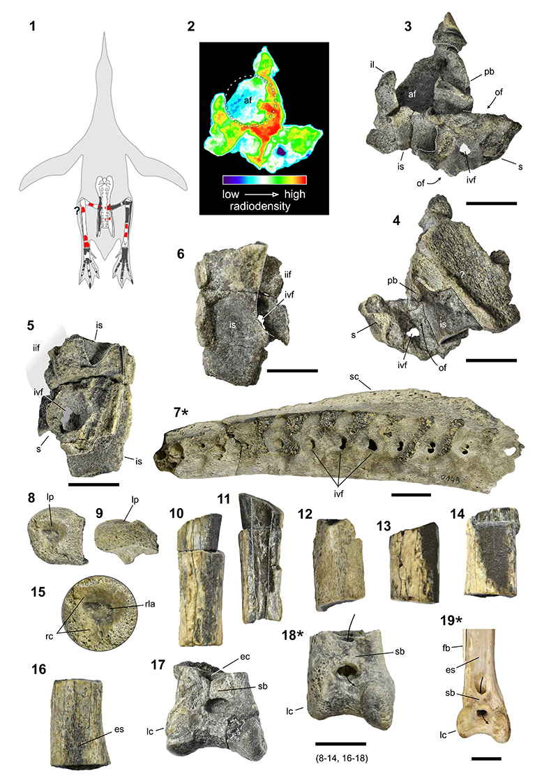
FIGURE 5. Incomplete pelvic skeleton and fragmentary leg bones of Eocene Delphinornis larseni, NRM-PZ A.994, major fragment (bones encased in a single lump of matrix). 1. Location of these skeletal elements within penguin body, in ventral view, is presented in red. Other bones included in NRM-PZ A.994, and shown in Figure 2.1-7, 2.15, 2.17-19, Figure 4.2-6, 4.8-17, are in gray. 2. X-ray-based schematic view of right hip bone and associated left leg bones, as preserved in matrix. 3. X-ray image of same. 4. Medial view of same. 5. Fragmentary right preacetabular ilium IB/P/B-0211 (Eocene, Seymour Island, Antarctica) in medial view. 6. Lateral view of right hip bone and associated left leg bones (NRM-PZ A.994). 7. Right (reversed left) hip bone of extant Spheniscus demersus (Linnaeus, 1758), NHMUK/T S/1998.23.2, in medial (left) and lateral view. 8. Dorsomedial view of two major hip-bone foramina of NRM-PZ A.994. 9. Ventromedial view of obturator foramen of same specimen. 10. Medial view of right hip bone, IB/P/B-0488 (Eocene, Seymour Island, Antarctica). 11. Lateral view of same. 12. Ventromedial view of three major hip-bone foramina of extant Pygoscelis papua (unnumbered specimen from IB/P/B). 13. Dorsomedial view of nutrition foramen of left tibiotarsus (NRM-PZ A.994). 14. Same view of tibiotarsus IB/P/B-1033 (Eocene, Seymour Island, Antarctica). 15. Caudal view of reversed right femur IB/P/B-0130 (Eocene, Seymour Island, Antarctica). 16. Same view of middle shaft of NRM-PZ A.994. Abbreviations: aa, articular area; af, acetabular foramen; ar, adductor ridge; at, antitrochanter; cil, caudal intermuscular line; dfe, distal end of femur; fc, fibular crest; feh, femoral head; fes, femoral shaft; iif, ilioischiadic foramen; iil, insertion site for ischiopubic ligament; iip, ilioischiadic pillar; il, ilium; im, insertion of iliofibular muscle; is, ischium; ipl, ischiopubic ligament; iss, ilioischiadic suture; lc, lateral condyle; lfb, left fibula; lfe, left femur; ltt, left tibiotarsus; lp, ligamental pit; mx, matrix; nf, nutrition foramen; of, obturator foramen; pa, pronounced angularity within ischium; pb, pubis; pdp, correlate of proximal dorsal process; ptt, proximal end of tibiotarsus; rf, renal fossa; rhb, right hip bone; s, synsacrum; syn, iliosynsacral synostosis; tb, tubercle; tts, tibiotarsal shaft. Asterisks denote comparative material. Scale bar equals 1 cm.
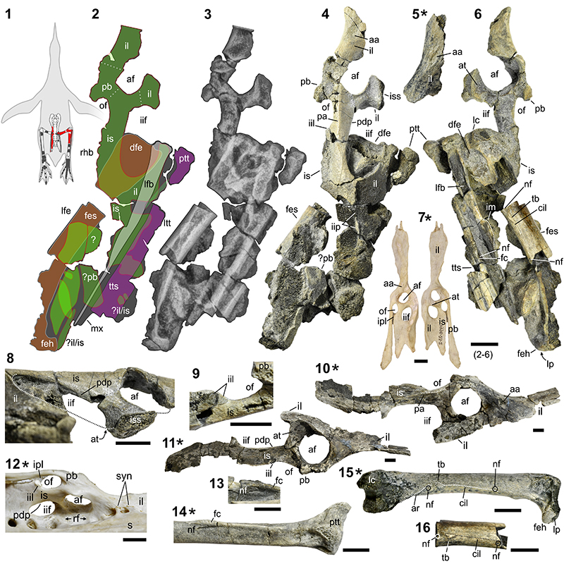
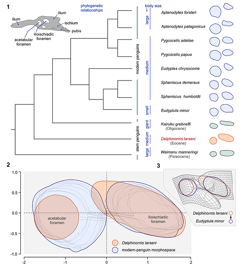
FIGURE 6. Shape analyses of outlines of major hip bone foramina. 1. Comparison of foramen configurations and shapes of eight extant and three stem-group penguins, including the Eocene specimen NRM-PZ A.994 (Delphinornis larseni); foramen pairs are not to scale, branching pattern for present-day species used in the cladogram follows Subramanian et al. (2013, figure 1). 2. Position of NRM-PZ A.994 relative to the extant-penguin morphospace, based on aligned, centered and scaled oulines. 3. Deformation grid between configurations representing NRM-PZ A.994 (D. larseni) and NHMUK/T S/2002.2.1 (extant Eudyptula minor). For other specimen IDs, see Appendix 1; for PCA plots, see Appendix 4.