FIGURE 1. 1. Original drawing of “Actinoporella sulcata Alth” (Pia, 1920: plate VII, figure 8). Outer surface of a rock sample from the Tithonian of Nyzhniv, Ukraine. 2. Original drawing of the type of “Clypeina marginiporella” [sic] (Michelin, 1845: plate 46, figure 27.a-b). Étampes, Essone, Paris Basin. 3. Clypeina marginoporella with two whorls joined together. Lutetian of Chambors, Oise, Paris Basin, E.P. Munier-Chalmas Collection (Génot, 1987: plate 26, figure 5). Note the absence of scars between these two fertile whorls (scale bar equals 500 µm).
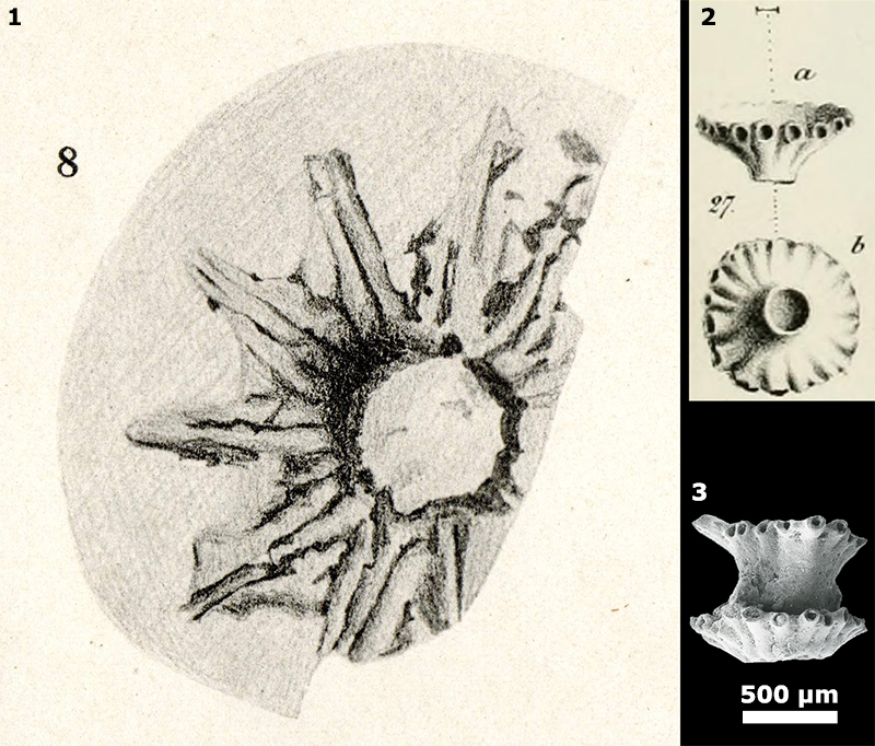
FIGURE 2. Weathered rock surfaces with Aloisalthella gen. nov. sulcata comb. nov., from the Upper Jurassic strata of Algeria, leg. R. Karpoff, J. Emberger Collection (1, 3: scale bar equals 1 mm, 2: scale bar equals 500 µm).
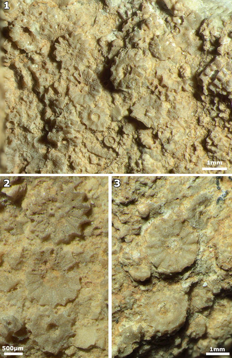
FIGURE 3. Aloisalthella gen. nov. sulcata comb. nov., from the Berriasian of Vuache, Savoie, E France, leg. P.O. Mojon, B. Granier Collection (scale bar equals 500 µm).
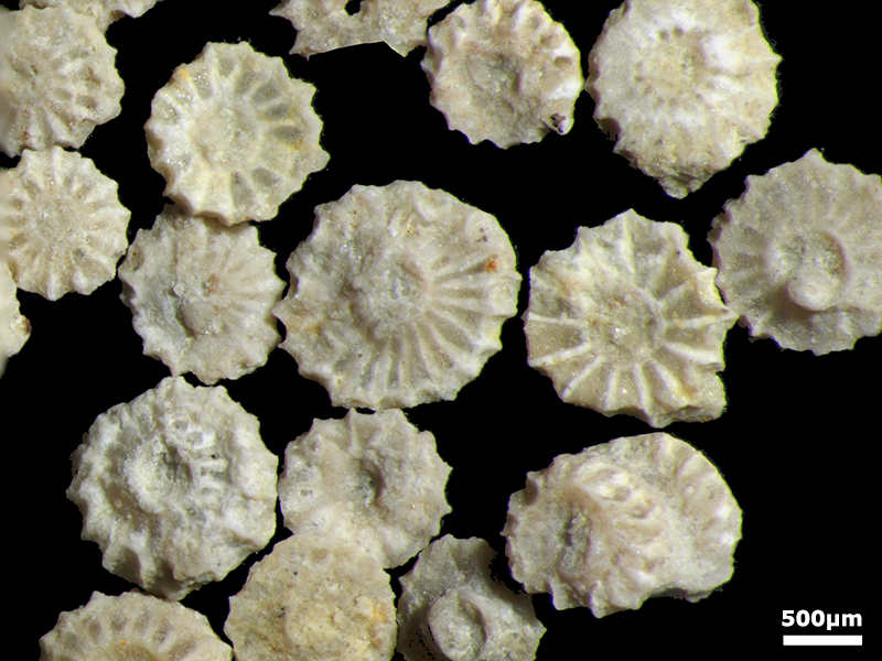
FIGURE 4. Aloisalthella gen. nov. sulcata comb. nov., from the Berriasian of Vuache, Savoie, E France, leg. P.O. Mojon, B. Granier Collection. 1-10. lower side views of single fertile whorls with a neck and upper side views without neck. 11. four (?) successive fertile whorls. 12-15. two (?) successive fertile whorls. All SEM photos same scale (scale bar equals 500 µm).
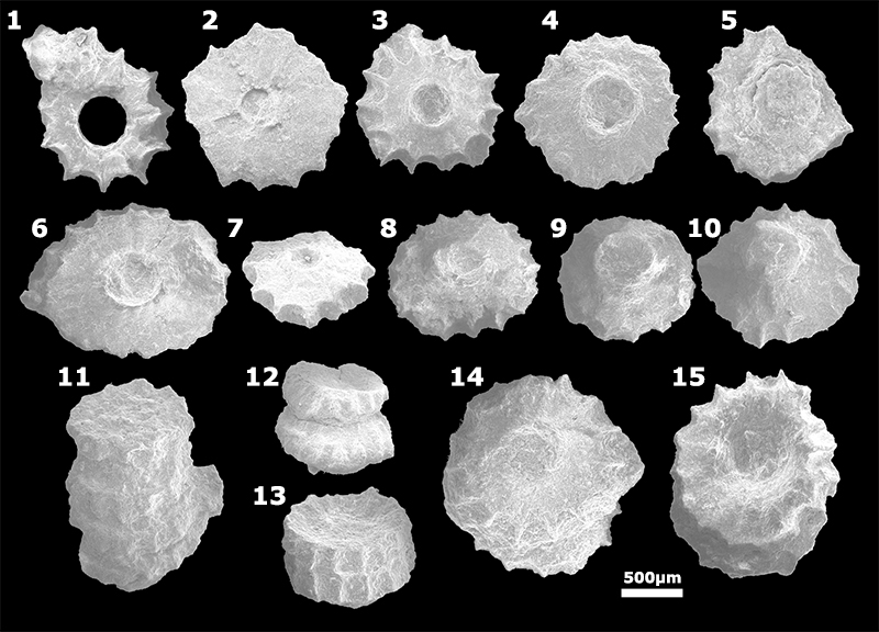
FIGURE 5. Original thin sections studied by Pfender (1927a). 1. no. 79, Nans, Provence, SE France - J. Pfender Collection. 2. no. 18, Sormiou, Provence, SE France - J. Pfender Collection. 3. no. 84, Méounes-lès-Montrieux, Provence, SE France - J. Pfender Collection. 4. without number, Tlemcen, Algeria - leg. Lemesle, E.P. Munier-Chalmas Collection. 5. no. 53, ? label illegible, Provence, SE France - J. Pfender Collection. 6. no. 7 bis, ? label illegible, Provence, SE France - J. Pfender Collection. 7. no. 8, Plan-d'Aups-Sainte-Baume, Provence, SE France - J. Pfender Collection. All photos same scale (scale bar equals 1 cm).
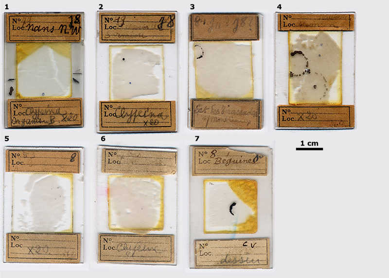
FIGURE 6. 1. Clypeina sp., Lutetian of Montjavoult, Oise, Paris Basin, E.P. Munier-Chalmas Collection (Génot, 1987: plate 3, figure 14). 2. Clypeina sp., Lutetian of Chambors, Oise, Paris Basin, E.P. Munier-Chalmas Collection (Génot, 1987: plate 3, figure 15). 3. Clypeina sp., Lutetian of Thiverval-Grignon, Seine-et-Oise, Paris Basin, P. Génot Collection. 4-10. Acetabularia caliculus Lamouroux in Quoy and Gaimard, 1824, Holocene of Abu Dhabi, UAE, B. Granier Collection: 4-5, 8: top views of the cap; 6-7: side views of the cap; 9: various views of main axes and caps, with or without coronas (note that one rare specimen has a bifurcated thallus); 10: detail of the calcareous coating of a thallus with scars corresponding to the emplacement of former sessile sterile laterals (arrows). Almost all photos same scale (scale bar equals 250 µm), except 9 (scale bar equals 1 mm).
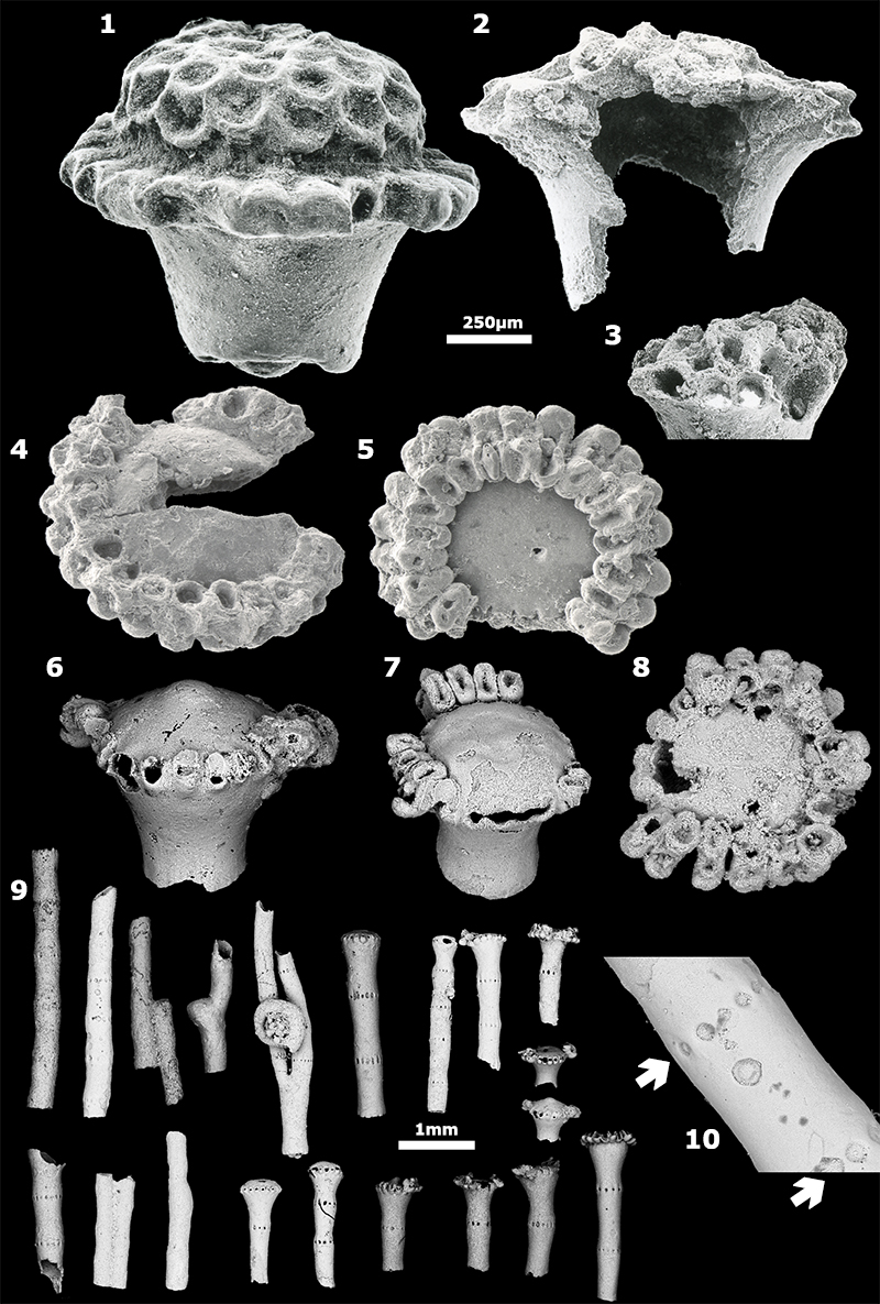
FIGURE 7. 3D reconstruction of the fossil genus Clypeina sensu L. and J. Morellet (1918) based on their 2D reconstruction (op. cit.: text-figure 1). Click on the image to play or download animation.
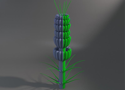
FIGURE 8. 1, 5, 10. Aloisalthella gen. nov. sulcata comb. nov., from the Berriasian of Provence, SE France, J. Pfender Collection. 1: thin section no. 79, Nans, Var: subtransverse section, high in a whorl (= plate V, figure 2 in Pfender, 1927a); 5, 10: thin section no. 49, road from Mazargues to Sormiou, now part of Marseilles’ urban area, Bouches-du-Rhône: 5: deep tangential section with two whorls (= plate V, figure 3 in Pfender, 1927a); 10: oblique section in a whorl (= plate V, figure 1 in Pfender, 1927a). 2-4, 6-7, 9, 11. Aloisalthella gen. nov. sulcata comb. nov., from the Kimmeridgian of Tlemcen, NW Algeria, leg. Lemesle, E.P. Munier-Chalmas Collection. 2: oblique section; 3: subtransverse section, low in a whorl; 4: oblique to subaxial section (arrows point to sterile scars along the main axis); 6: (= plate V, figure 5 in Pfender, 1927a); 7: transverse section, low in a whorl (= text-figure 3.B lower in Pfender, 1927a); 9: tangential oblique section; 11: oblique section of a whorl (= text-figure 3.B upper in Pfender, 1927a). 8. “verticille terminal de poils stériles” according to Pfender (1927a), i.e., uppermost verticil made of sterile hairs (= text-figure 2 in Pfender, 1927a), thin section no. 8, Plan-d’Aups-Sainte-Baume, Var, Berriasian of Provence, SE France, J. Pfender Collection. All photomicrographs same scale (scale bar equals 250 µm).
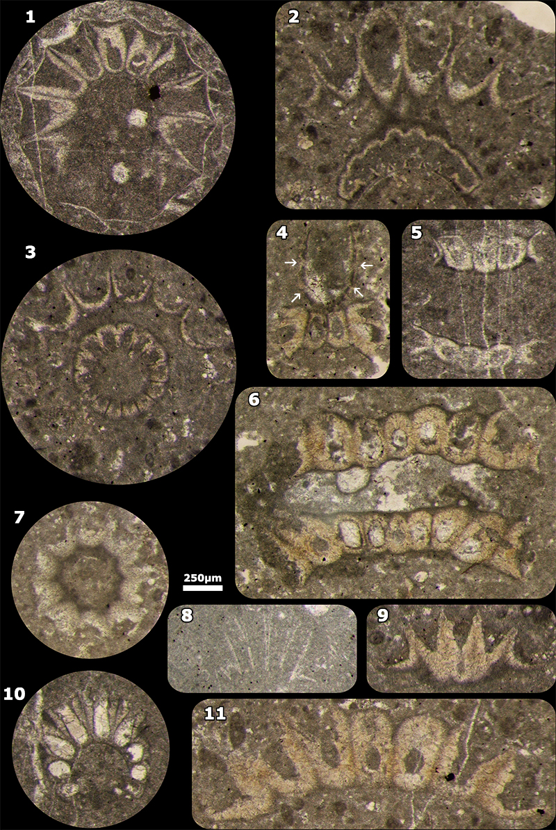
FIGURE 9. Aloisalthella gen. nov. sulcata comb. nov. 1, 4-5. Thin section no. 519 m, Upper Jurassic, PE-1, core 2, Sierra Perenchiza, Valencia, Spain, leg. R. Deloffre, B. Granier Collection. 1: subaxial section with 8 fertile verticils; d: subaxial section with 3 fertile verticils with clots (scars of sterile laterals) in between; e: longitudinal section of an isolated fertile verticil. 2-3. Thin section no. 8878035, Upper Kimmeridgian of Abu Dhabi, United Arab Emirates. 2: subtransverse sections of 2 main axes suggesting a branching of a single thallus; 3: subaxial section with 2 fertile verticils with clots (scars of sterile laterals) in between. 6-8. Berriasian of Provence, SE France, J. Pfender Collection. 6: longitudinal section of an isolated fertile verticil, thin section no. 7 bis, ? label illegible (= text-figure 3.A lower in Pfender, 1927a); 7: subtransverse section of an isolated fertile verticil, thin section no. 53, ? label illegible (= text-figure 3.A middle left in Pfender, 1927a); 8: subaxial section with 3 fertile verticils, thin section no. 84, Méounes-lès-Montrieux, Var (= text-figure 1 in Pfender, 1927a). All photomicrographs same scale (scale bar equals 250 µm).
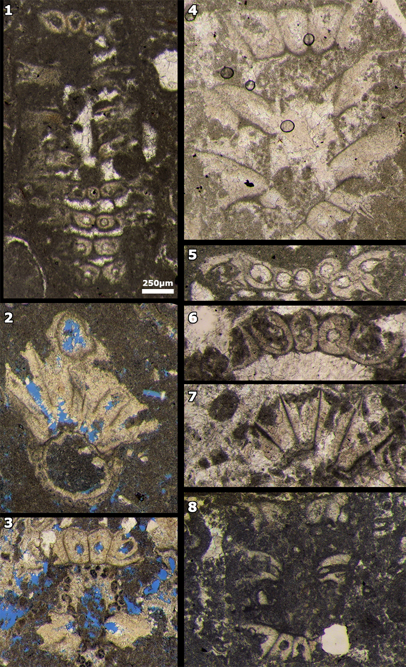
FIGURE 10. Aloisalthella gen. nov. sulcata comb. nov. 1-3, 5-6, 8-9. Upper Kimmeridgian of Abu Dhabi, UAE. 1: subtransverse section of a main axis with pores that correspond to the sterile laterals, thin section no. 8706034; 2-3: subtransverse section of a fertile verticil and detail of its fertile laterals with twisted walls, thin section no. 9855076; 5: subtransverse section of a fertile verticil with bitumen linings on the walls of its laterals, thin section no. 8822034; 6: subtransverse section of a fertile verticil with its fertile laterals with twisted walls, thin section no. 8957035; 8: oblique section of a fertile verticil with bitumen linings on the walls of its laterals, thin section no. 8902034; 9: subtransverse section of a fertile verticil with its fertile laterals with twisted walls, thin section no. 9998177. 4. Subtransverse sections of 2 main axes suggesting a branching of a single thallus, thin section no. 519 m, Upper Jurassic, PE-1, core 2, Sierra Perenchiza, Valencia, Spain, leg. R. Deloffre, B. Granier Collection. 7. random section of a fertile verticil, thin section no. 53, ? label illegible (= text-figure 3.A upper left in Pfender, 1927a), Berriasian of Provence, SE France, J. Pfender Collection. 1, 3, 5, 8 (scale bar equals 100 µm), and 2, 4, 6-7, 9 (scale bar equals 250 µm).
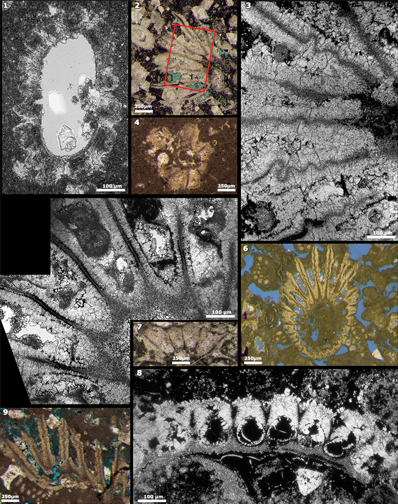
FIGURE 11. Aloisalthella gen. nov. sulcata comb. nov. Thin section no. 8730034, Upper Kimmeridgian of Abu Dhabi, UAE. 1. microfacies (scale bar equals 250 µm). 2. arrows pointing to some scars (clots) of sterile laterals (scale bar equals 100 µm).
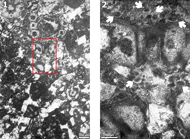
FIGURE 12. Aloisalthella gen. nov. sulcata comb. nov. 1-2, 4-5, 7-8, 11. Upper Jurassic, PE-1, core 2, Sierra Perenchiza, Valencia, Spain, leg. R. Deloffre, B. Granier Collection. 1, 11: thin section no. 519; 2, 8: thin section no. 519.25; 4: thin section no. 519.75; 5: thin section no. 518.50; 7: thin section, 518.75. 3, 6, 9-10. 8878035, Upper Kimmeridgian of Abu Dhabi, UAE, B. Granier Collection. Arrows in 7 and 11 points to places where the laterals are bent (the “elbow”). All photomicrographs same scale (scale bar equals 250 µm).
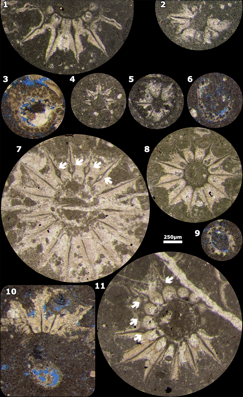
FIGURE 13. 3D reconstruction of an uncalcified fertile verticil of Aloisalthella gen. nov. sulcata comb. nov. It is assumed that on these fertile laterals bulges are located below the row (inferior). Click on image to play or download animation.
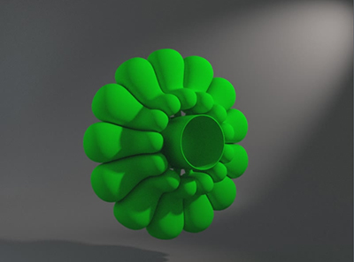
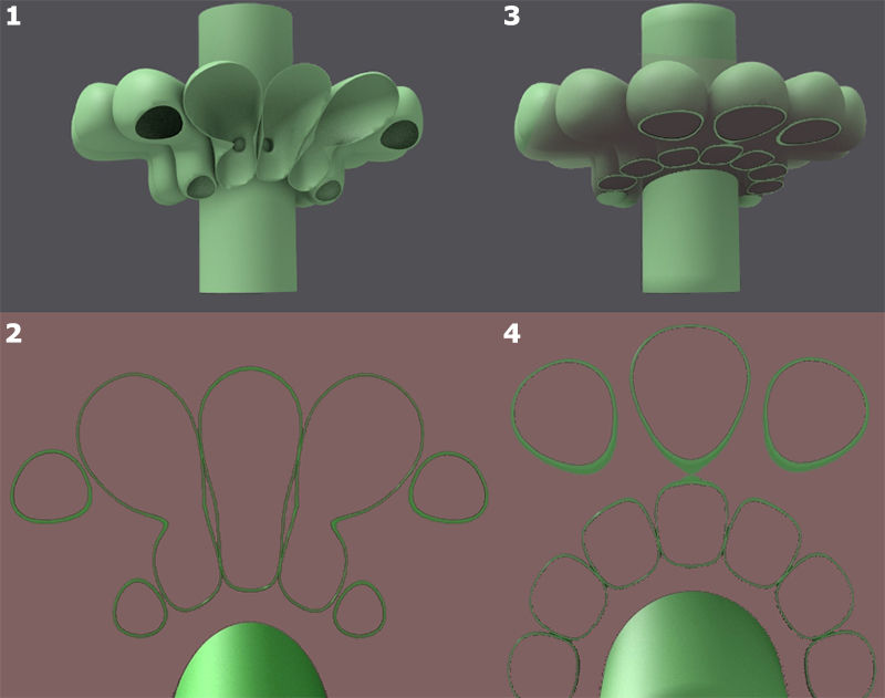
FIGURE 14. 3D reconstruction of an uncalcified living thallus of Aloisalthella gen. nov. sulcata comb. nov., with sets of sterile rows (of scars) in alternation with fertile rows. Click on image to play or download animation.
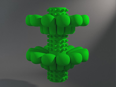
FIGURE 15. The laterals have the general shape of elbow tubes closed at both ends here (at the start of the reproductive stage). They communicate with the main axis through a small pore on the side of the elbow. 1-2. oblique sections 3D and 2D to compare with Figure 12.11. 3-4. oblique sections 3D and 2D to compare with Figure 8.3.