FIGURE 1. Partial skull of new referred specimen of Olsoniformes indet., OMNH 79340, in dorsal profile. 1, photograph; 2, line drawing. Scale bar equals 1 cm.
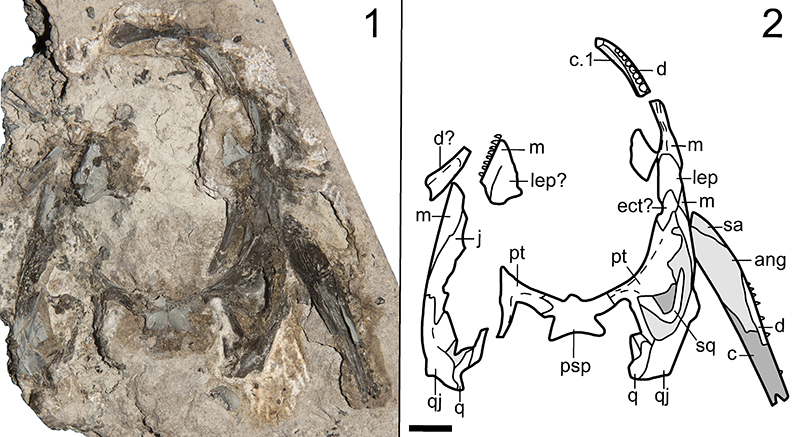
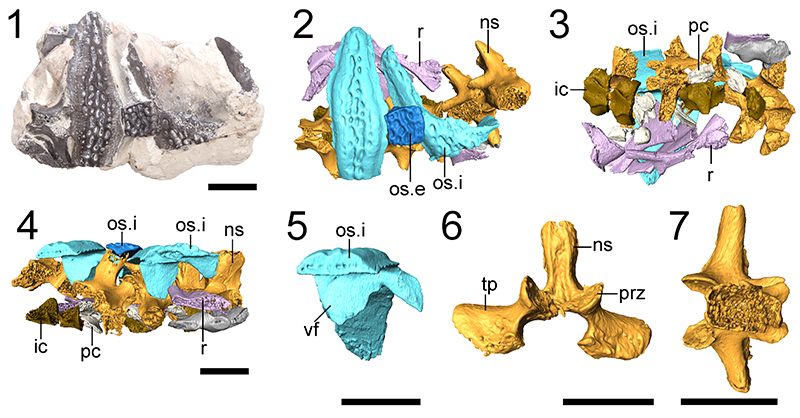
FIGURE 2. Isolated vertebral material of new referred specimens of Cacops sp. 1, photograph of ROMVP 80078 (neural spine with internal osteoderm) in anterior profile; 2, illustration of the same; 3, segmented rendering of the same; 4, photograph of ROMVP 80079 (neural spine) in anterior profile; 5, photograph of ROMVP 80078 in posterior profile; 6, illustration of the same; 7, segmented rendering of the same; 8, photograph of ROMVP 80079 in posterior profile; 9, segmented rendering of ROMVP 80078 in dorsal profile; 10, the same with the osteoderm removed to show the spinal groove (sg); 11, segmented rendering of the osteoderm of ROMVP 80078 in anterior profile. Scale bars equal 1 cm.
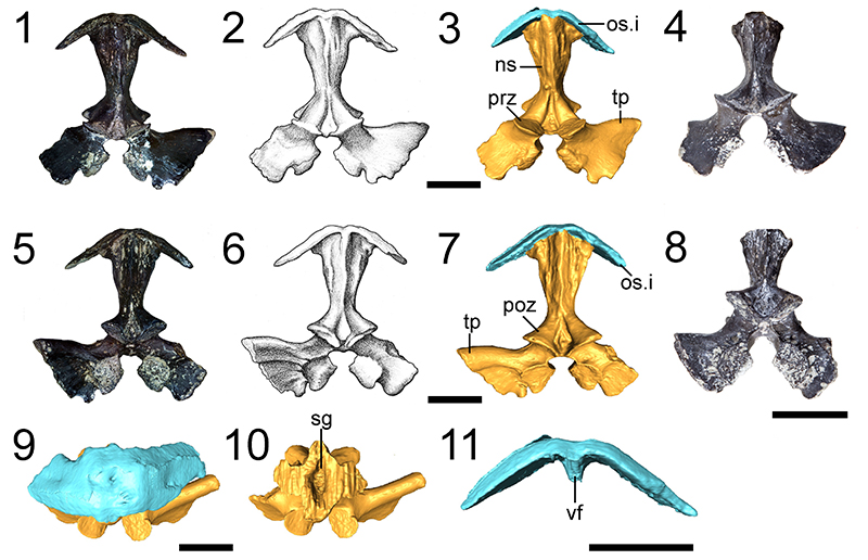
FIGURE 3. Photograph of right scapula of new referred specimen of Cacops sp., OMNH 79341, in lateral profile. Scale bar equals 1 cm.
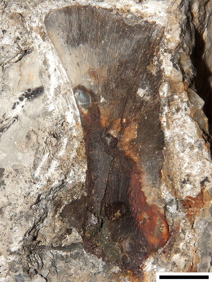
FIGURE 4. Skull and associated postcrania of new referred specimen of Cacops morrisi, OMNH 79339, in dorsal profile. 1, photograph; 2, line drawing. Only elements belonging to the dissorophid are illustrated in part 2. Scale bar equals 1 cm.
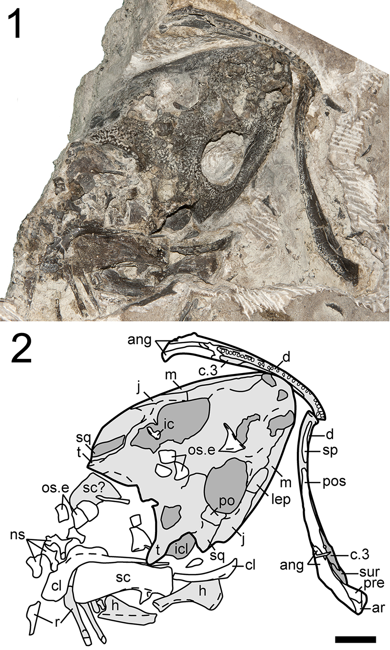
FIGURE 5. Right mandible of new referred specimen of Cacops morrisi, OMNH 79339, in lingual profile. 1, photograph; 2, line drawing. Scale bar equals 1 cm.
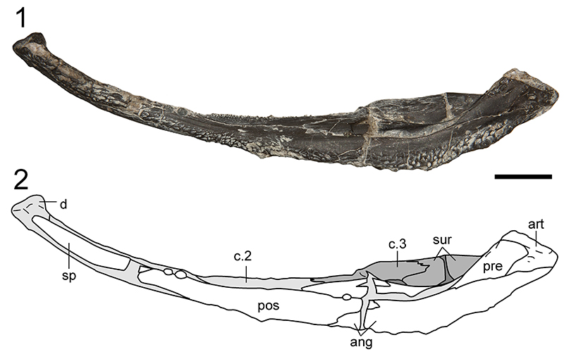
FIGURE 6. Skull of new referred specimen of Cacops woehri, OMNH 79338, in dorsal profile. 1, photograph; 2, line drawing. Scale bar equals 1 cm.
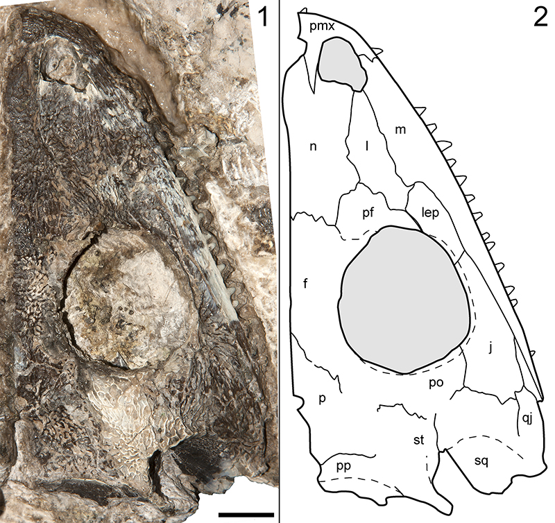
FIGURE 7. Skull of new referred specimen of Cacops woehri, OMNH 79338, in lateral profile. 1, photograph; 2, line drawing. Scale bar equals 1 cm.
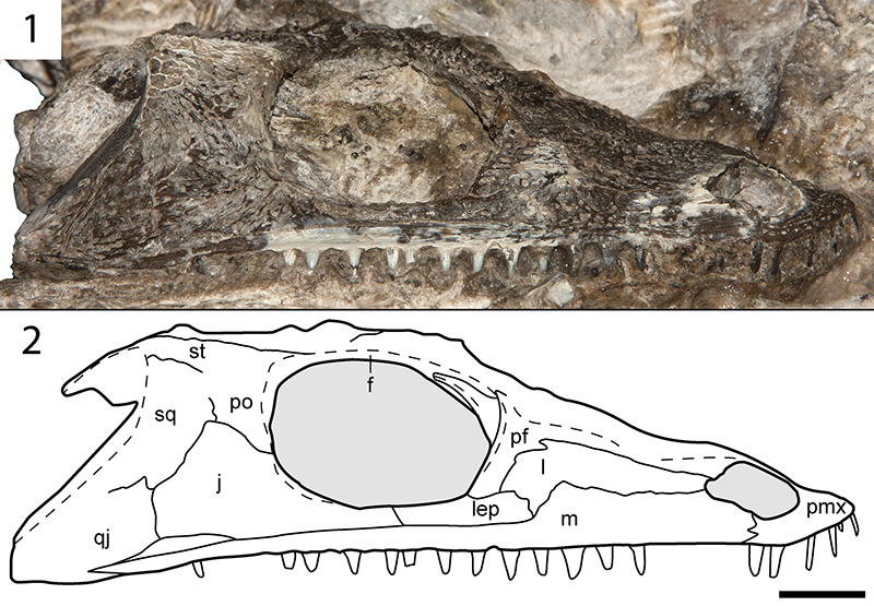
FIGURE 8. Partial snouts of new referred specimens of Cacops woehri, ROMVP 80800 and ROMVP 80081. 1, photograph of ROMVP 80800 in dorsal profile; 2, line drawing of the same; 3, photograph of ROMVP 80080 in ventral profile; 4, line drawing of the same; 5, photograph of ROMVP 80081 in dorsal profile; 6, line drawing of the same; 7, photograph of ROMVP 80081 in ventral profile; 8, line drawing of the same. Scale bars equal 1 cm.
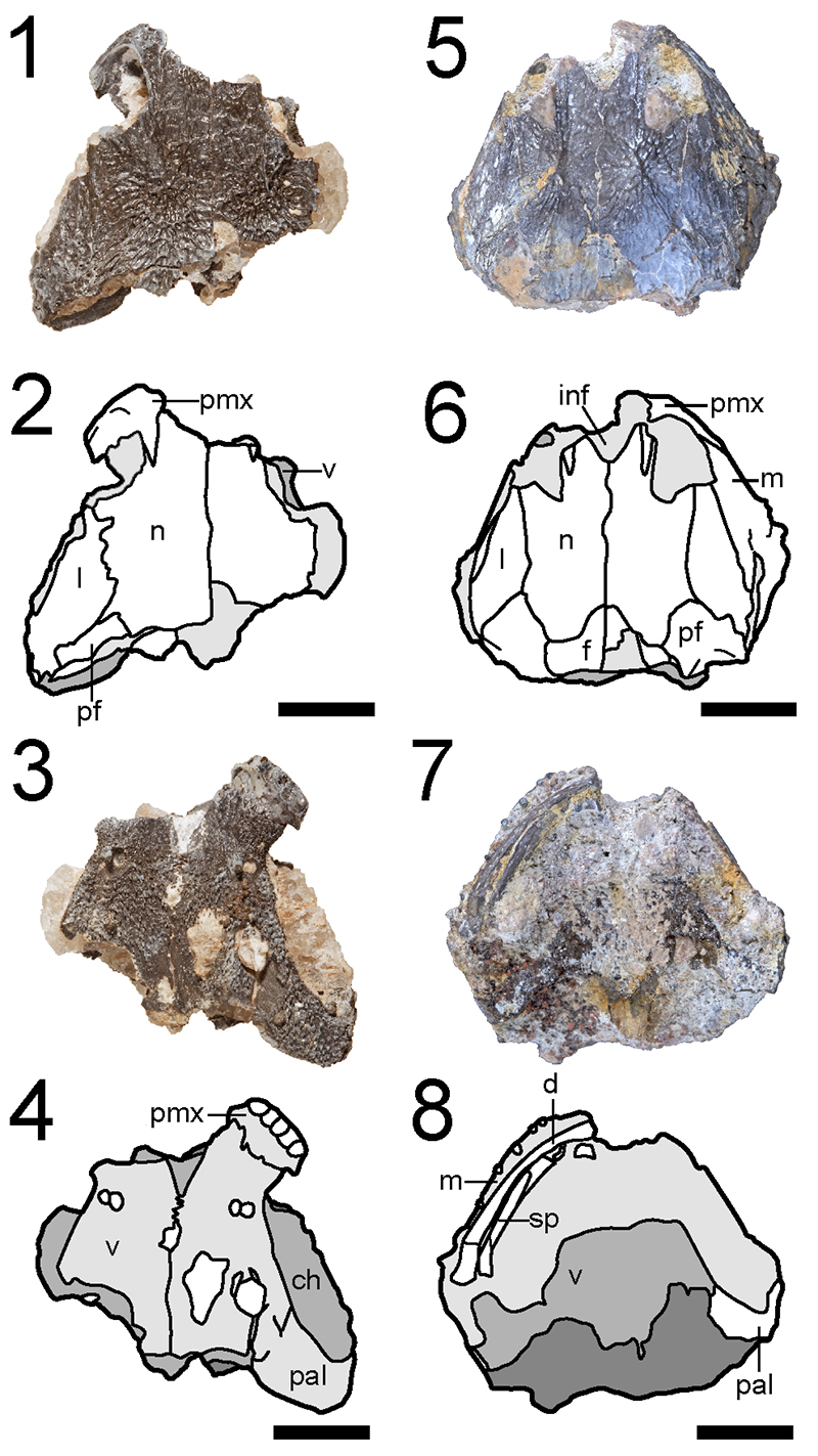
FIGURE 9. Cranial fragments of new referred specimens of Cacops woehri, ROMVP 80082 and ROMVP 80083. 1, photograph of ROM VP 80082 in anteroventral profile; 2, the same in dorsal profile; 3, the same in right lateral profile; 4, line drawing of ROMVP 80082 in anteroventral profile; 5, the same in dorsal profile; 6, the same in right lateral profile; 7, photograph of ROMVP 80083 in dorsal profile; 8, the same in posterior profile; 9, the same in left lateral profile; 10, the same in medial profile; 11, line drawing of ROMVP 80083 in dorsal profile; 12, the same in posterior profile; 13, the same in left lateral profile; 14, the same in medial profile. Scale bars equal to 1 cm.
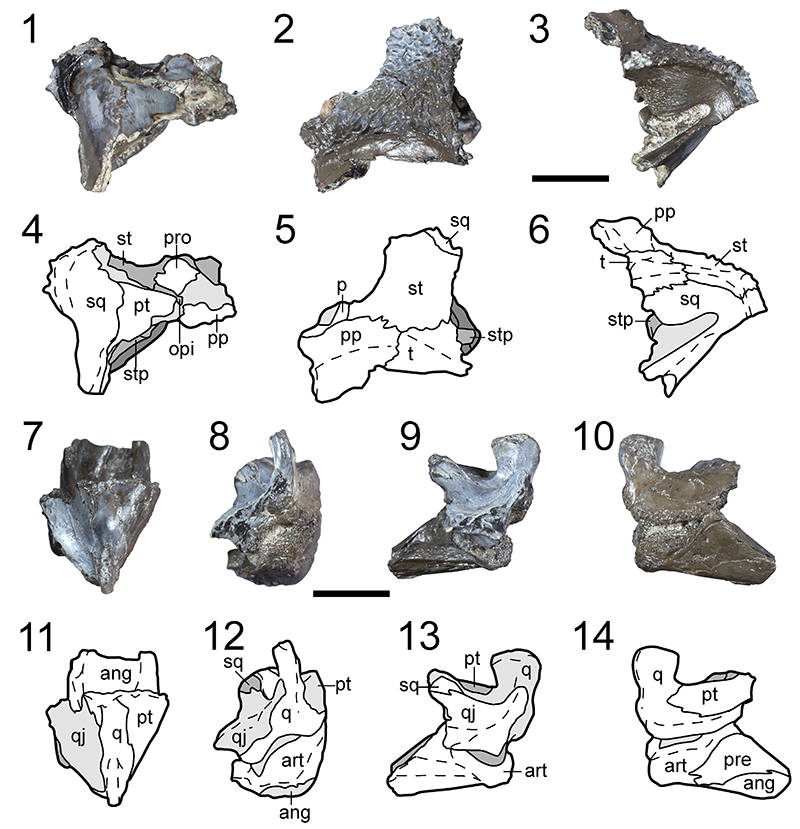
FIGURE 10. Osteoderms and neural spines of new referred specimen of Aspidosaurus sp., ROMVP 80069. 1, photograph in right lateral profile; 2, illustration of the same; 3, segmented rendering of the same; 4, photograph in anterior profile; 5, illustration of the same; 6, segmented rendering of the same; 7, photograph in dorsal profile; 8, line drawing of the same; 9, segmented rendering of the same; 10, photograph in ventral profile; 11, line drawing of the same; 12, segmented rendering of the same. Scale bars equal 1 cm.
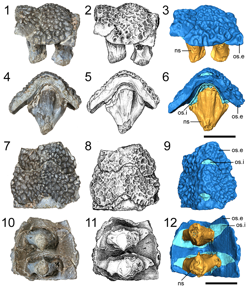
FIGURE 11. Comparison of osteoderms and neural spines of new referred specimen of Aspidosaurus sp., ROMVP 80069, and new referred specimen of Cacops sp., ROMVP 80078. 1, Segmented rendering in ventral profile showing planes of digital sections; 2, sagittal section; 3, transverse section; 4, segmented rendering of ROMVP 80078 in dorsal profile showing planes of digital sections; 5, sagittal section; 6, transverse section; note that a small portion of the transvere process was removed for spacing and because it does not contribute information regarding the osteoderm-spine contact.
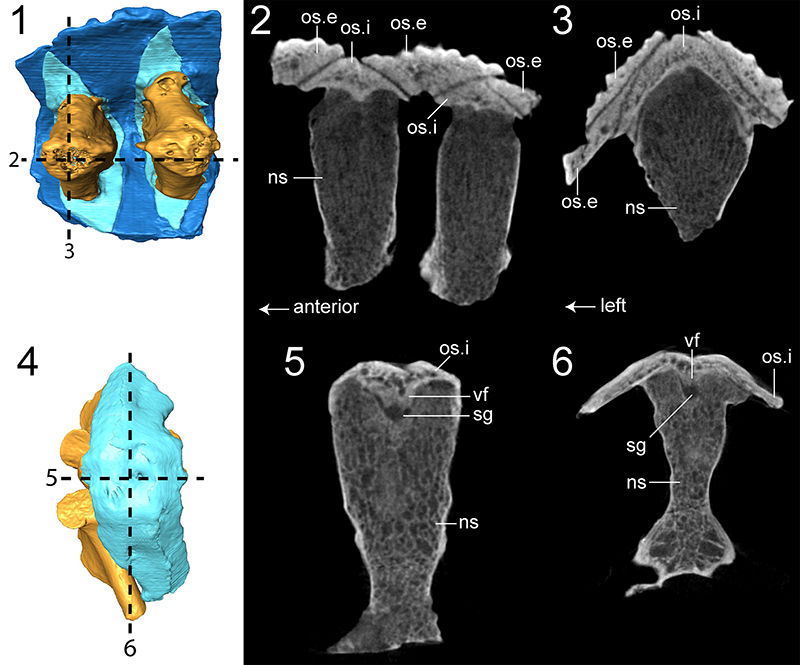
FIGURE 12. Postcrania of new referred specimens of Dissorophinae indet. 1, Photograph of ROMVP 80070 (partial humerus) in anterior profile; 2, the same in posterior profile; 3, photograph of partial ROMVP 80071 (partial scapulocoracoid) in posterolateral profile; 4, the same in anteromedial profile; 5, photograph of ROMVP 80103 (complete humerus) in flexor profile; 6, the same in anterior profile; 7, the same in extensor profile; 8, the same in posterior profile. Scale bars equal 1 cm.
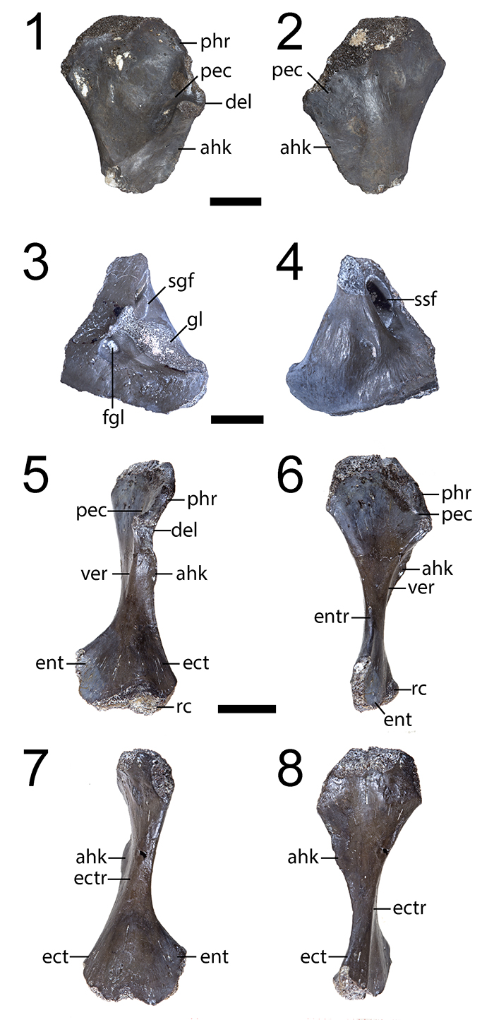
FIGURE 13. Partial skull and postcrania of new referred specimen of Dissorophus cf. multicinctus, ROMVP 80073. 1, photograph in dorsal profile; 2, line drawing of the same. Scale bar equals 1 cm.
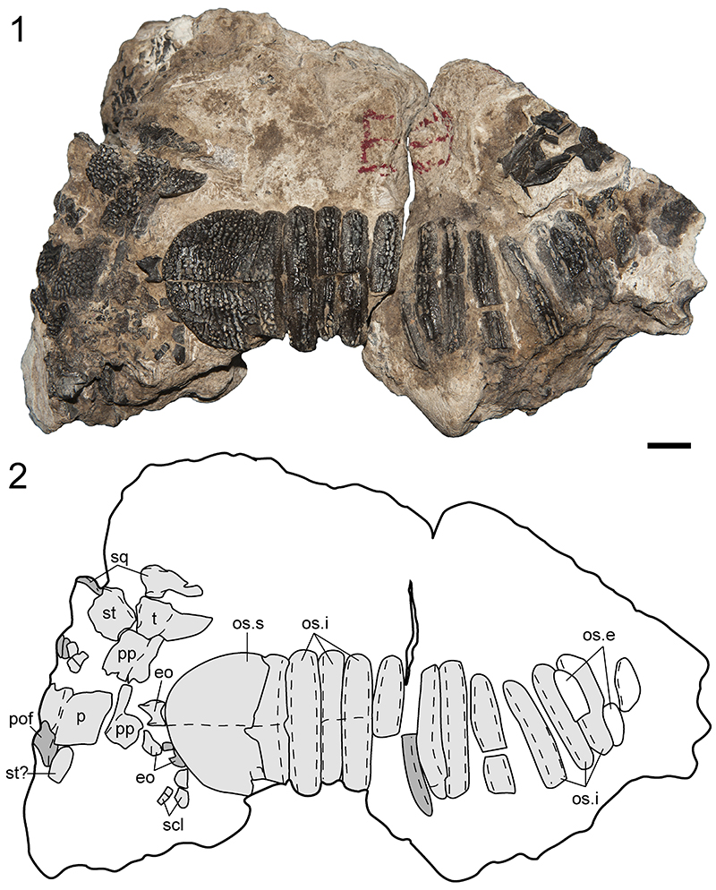
FIGURE 14. Partial skull and postcrania of new referred specimen of Dissorophus cf. multicinctus, ROMVP 80072. 1, photograph in dorsal profile; 2, line drawing of the same. Inset is a photograph of the stapes in posterior profile. Scale bar equals 1 cm for the main figure and 2 mm for the inset.
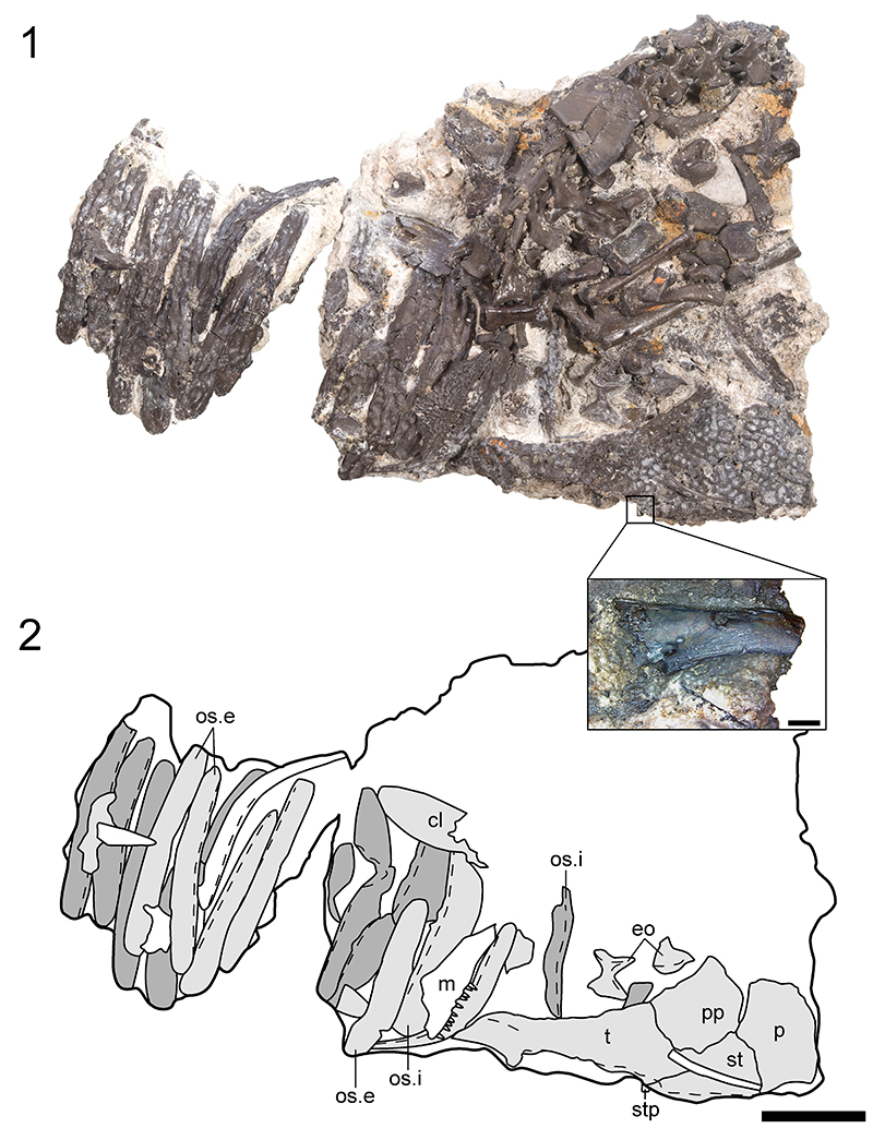
FIGURE 15. Photographs of new referred specimens of Dissorophus cf. multicinctus. 1, ROMVP 80074 in dorsal profile; 2, ROMVP 80075 in dorsal profile; 3, ROMVP 80077 in dorsal profile; 4, the same in ventral profile. Scale bars equal 1 cm.
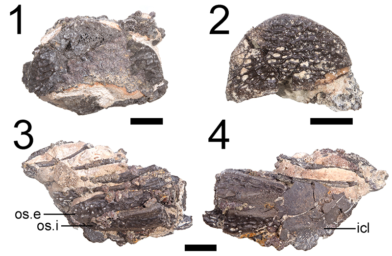
FIGURE 16. Postcrania of new referred specimen of Dissorophus cf. multicinctus, ROMVP 80076. 1, photograph in dorsal profile; 2, segmented rendering in dorsal profile; 3, segmented rendering in ventral profile; 4, segmented rendering in left lateral profile; 5, isolated external osteoderm in left ventrolateral profile showing bifurcated ventral flange. 6, isolated neural arch and spine in anterior profile; 7, the same in dorsal profile. Scale bars equal 1 cm..