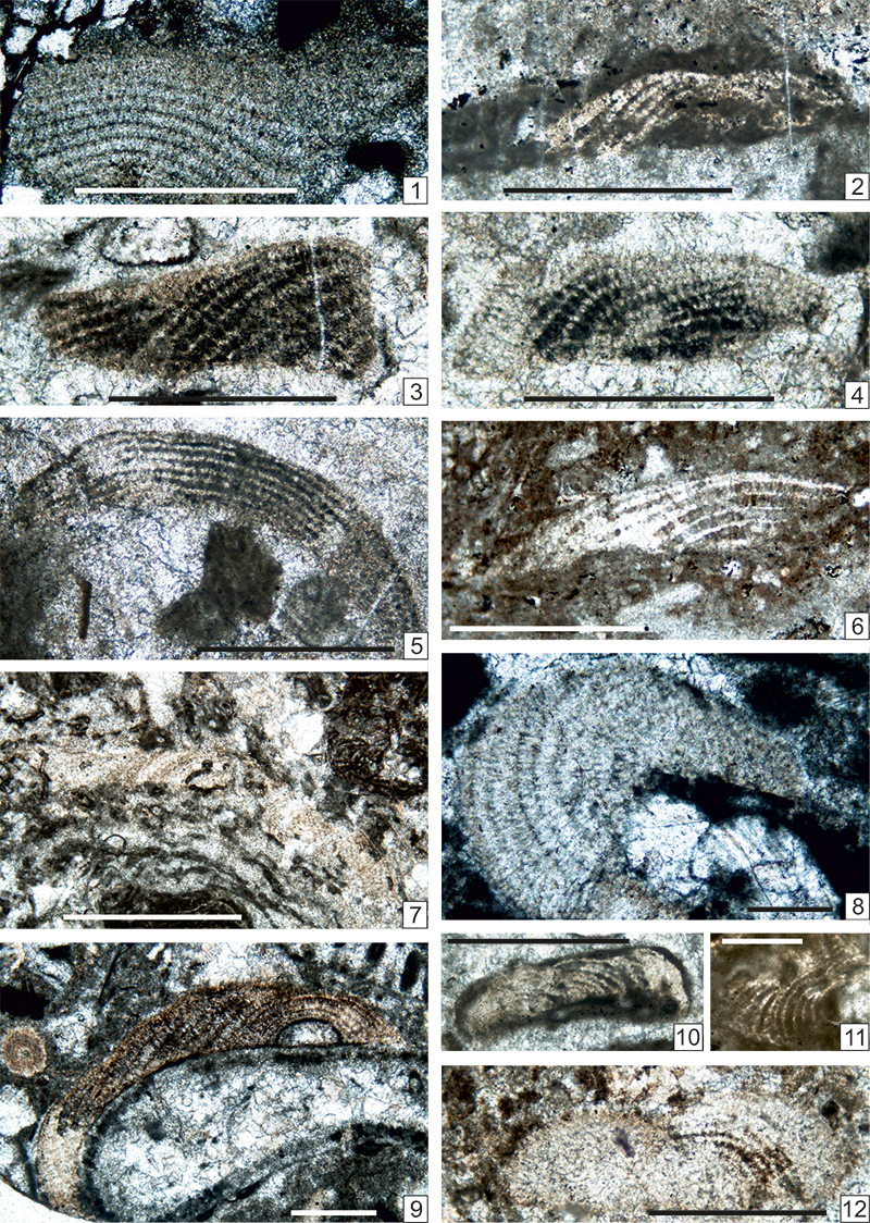
FIGURE 1. Map of the Trogkofel area in the Carnic Alps (southern Austria). GB: section Garnitzenbach of the uppermost part of the Grenzland Formation and Zweikofel Formation, GBT: Section Garnitzenbach of the Trogkofel Formation.
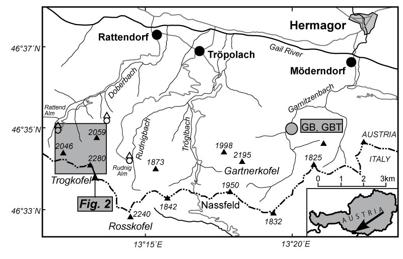
FIGURE 2. Detailed geologic map of the Trogkofel area. ZK: type section of the Zweikofel Fm, ZKO: section through the uppermost part of the Zweikofel Fm at Zweikofel East, TNA, TNB and TNC: sections of the Zottachkopf Fm on the northern side of the Trogkofel Massif, ZT: section through the upper part of the Zottachkopf Fm and basal part of the Trogkofel Fm, Z: section on the northern side of Zottachkopf through the upper part of the Zottachkopf Fm and basal Trogkofel Fm, TKW: section Trogkofel West through the uppermost part of the Zottachkopf Fm and basal part of the Trogkofel Fm, TKS: section Trogkofel South including the uppermost part of the Zottachkopf Fm and basal Trogkofel Fm; TK: type section of the Trogkofel Fm.
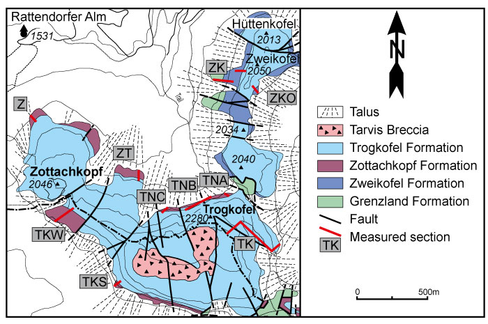
FIGURE 3. Correlation of Permian stages.
FIGURE 4. Correlation of Yakhtashian and Bolorian regional stages.
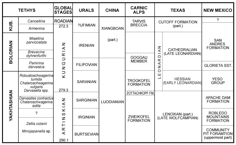
FIGURE 5. Pennsylvanian and Cisuralian series in the Carnic Alps.
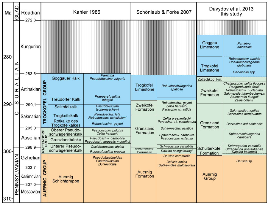
FIGURE 6. Algal classification adopted here.
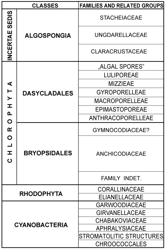
FIGURE 7. Hypothetical algal phylogeny proposed in this paper, mainly at the level of the classes.
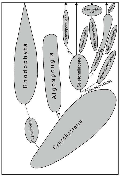
FIGURE 8. Hypothetical algal phylogeny, mainly at the level of orders and families.
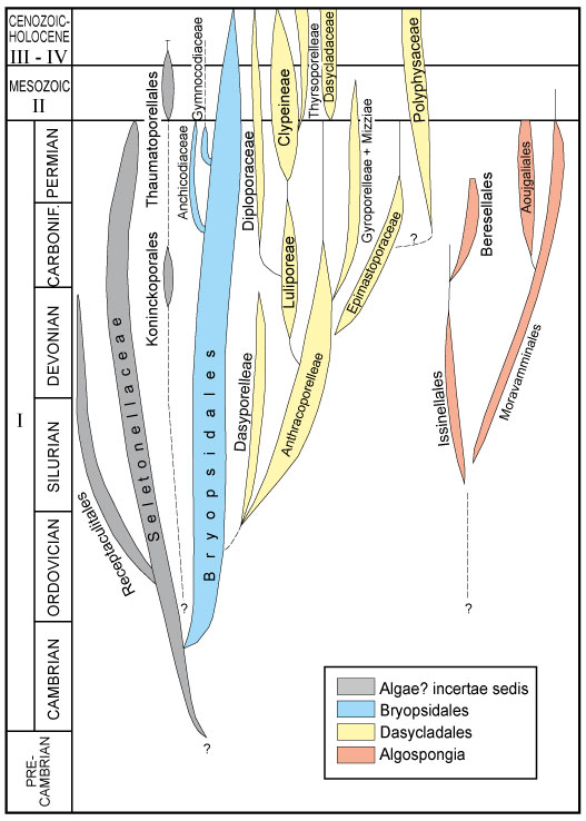
FIGURE 9. Possible phylogeny of cyanobacterial groups mentioned in this study.
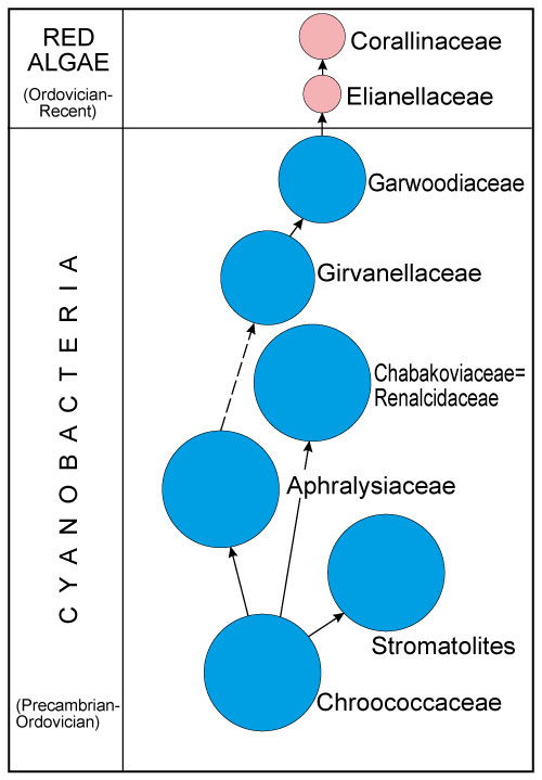
FIGURE 10. Another possible phylogeny of the Bryopsidales and Dasycladales mentioned in this study, mainly at the level of families and tribes.

FIGURE 11. 1, 2. Nostocites vesiculosa Maslov, 1929. 1. Random section. Sample TK26_3/00011. 2. Random section. Sample ZK203_3. 3. Gahkumella sp. Two transverse sections (bottom, left) and an axial section (top, right). Sample GB 157_13. 4. Girvanella sp. Longitudinal and transverse sections perhaps boring an Eugonophyllum magnum (Endo, 1951) emend. Konishi and Wray, 1961 encrusted by a tuberitinid (bottom) and palaeonubeculariid (top). Sample GB35_4. 5. Mitcheldeania sp. Longitudinal section showing the large filaments and their bifurcations. Sample GB17_2b. 6. Stromatolite. Axial sections. Sample TKW2_1. 7-10. Archaeolithoporella hidensis Endo, 1961a. 7. Longitudinal section attached on a tubiphytid. Sample GBT4_1. 8. Longitudinal section attached on tubiphytid and a bryozoa. Sample GBT11_2. 9. Large colony, in transverse section, subrounded by botryoidal cement. Sample TK68_1. 10. Thin colonies encrusting large botryoidal cement. Sample TK60_AP. Scale bars: 0.1 µm (Figures 11.1 - 11.3) and 1 µm (Figures 11.4 - 11.10).
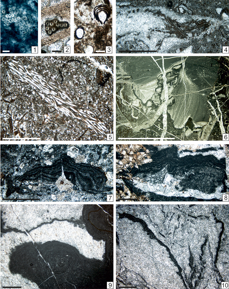
FIGURE 12. 1. Renalcis cf. granosus Vologdin, 1932. Longitudinal section. Sample TK20_1_1. 2, 12? Clinortonella cf. goggauensis (Flügel and Flügel-Kahler, 1980). 2. Axial section with typical filaments. Sample TK6_4. 12. Axial section with trichomes or sponge microscleres. Sample TNA_4_2. 3-4, 6-10. Koivaella ex gr. permiensis Chuvashov, 1974. 3. Bifurcated filament. Sample TK9_1. 4. Another type of bifurcated filament. Sample TNB.13_4. 6. A bifurcated filament preceeded by a basal part more or less similar to a tubiphytid. Sample TNB3_1_6. 7. Several filaments. Sample TNB8_1_7. 8. Bifurcated filament. Sample TK50_1_7. 9. Another typical filament. Sample TK50_1_5. 10. Another bifurcated filament. Sample TK50_1_4. 5. Garwoodia sp. Longitudinal section showing the typical bifurcation (right). Sample TK5_3. 11. Archaeolithoporella hidensis Endo, 1961a attached on a coral? Sample TK60_1. 13. Terebella sp. Longitudinal tube. Sample TNC5_1. Scale bars: 0.1 µm (Figures 12.1 - 12.4, 12.6-12.10, 12.12) and 1 µm (Figures 12.5, 12.11, 112.13).
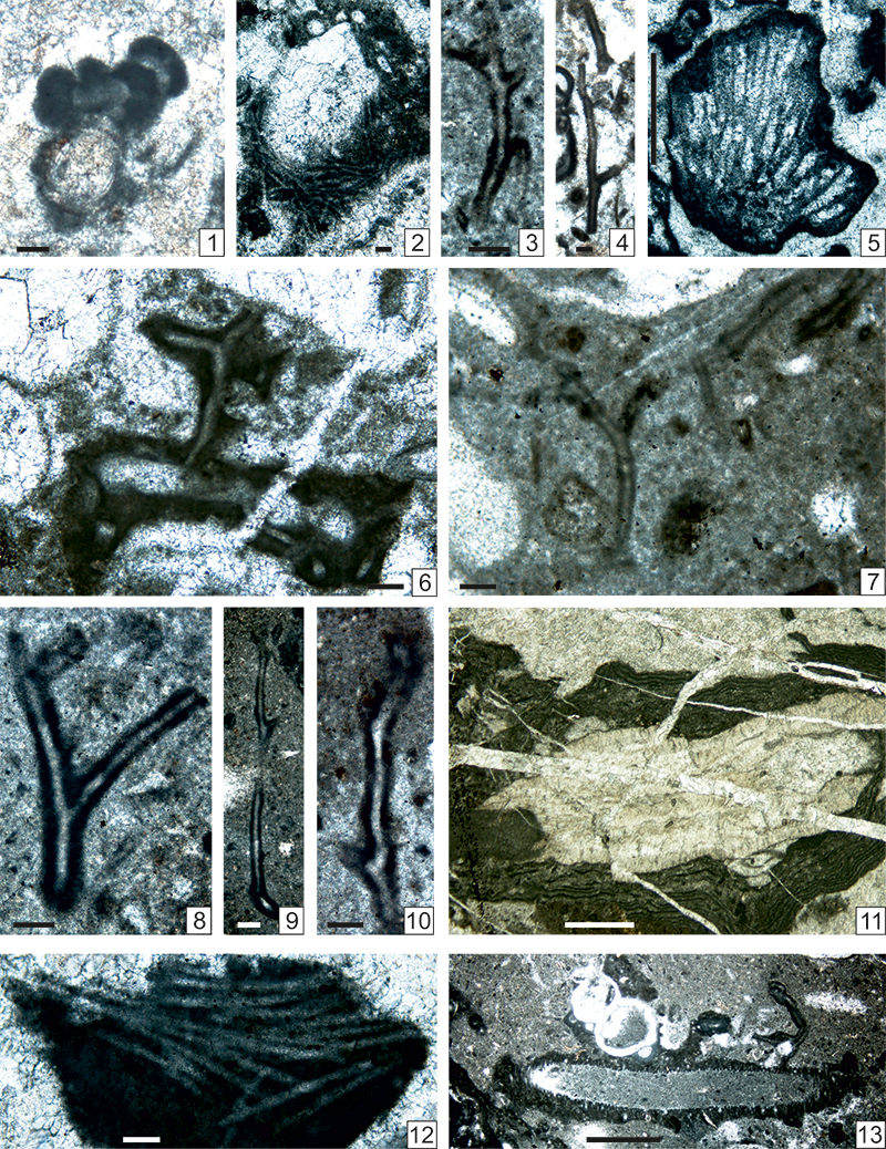
FIGURE 13. 1, 3, 4, 5. Parachaetetes ortonelloides (Endo, 1961c) n. comb. 1. Poorly preserved axial section with relatively broad recrystallized cellular files. Sample GB168_3. 3. Recrystallized axial section. Sample GB136_1. 4. Less recrystallized axial section. Sample Z9B_1. 5. Poorly preserved section resembling Ivanovia. Sample GB159_2. 2. Archaeolithophyllum lamellosum Wray, 1964. Several encrusting thalli, in axial section, attached on bryozoan, cyanobacterial crust and phylloid alga. Sample GB77_A2. 6-16. Homannisiphon morikawai (Endo, 1954). Several oblique sections. 6. Sample GB51_1 (previously published by Vachard and Krainer, 2001b, plate 13, figure 15). 7. Sample GB52_2. 8. Sample ZK99_5. 9. Sample Z1_4c. 10. Sample GB5_8_4. 11. Sample Z1_5. 12. Sample Z1_2. 13. Sample TNA2_1_2. 14. Sample TNA2_1_1. 15. Transverse oblique section. Sample Z1_3_4. 5. 16. Sample GB50_1 (previously published by Vachard and Krainer, 2001b, plate 13, figure 14). Scale bars: 0.2 µm (Figures 13.1, 13.2, 13.3); 0.5 µm (Figures 13.8, 13.11, 13.14); and 1 µm (Figures 13.4-13.9, 3.10, 12, 13.13, 13.15, 13.16).
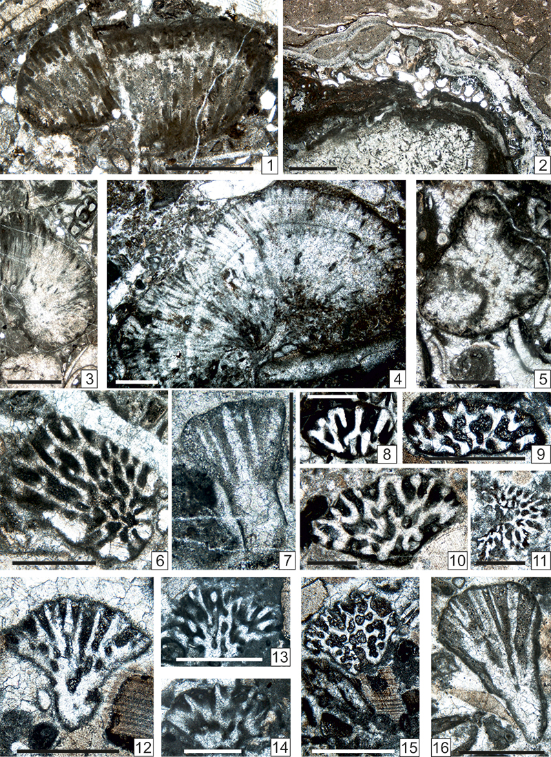
FIGURE 14. 1-5. Homannisiphon morikawai (Endo, 1954). Several oblique sections. 1. Sample TK18_4. 2. Sample TNB10_4. 3. Sample Z1_4a. 4. Sample Z1_4b. 5. Sample TNA3_3. 6-8. Ivanovia tenuissima Khorova, 1946. 6. Longitudinal section. Sample GB_76_3. 7. Longitudinal section. Sample TNA2_1_4c. 8. Tangential section. Sample TNC5_2. 9, 10. Anchicodium japonicum Endo, 1953a. 9. Longitudinal section. Sample TKS3_1. 10. Longitudinal section. Sample ZK67_A. 11, 12. Eugonophyllum magnum (Endo, 1951) emend. Konishi and Wray, 1961. 11. Longitudinal section. Sample Z2_1. 12. Longitudinal section. Sample Z7_1_width 9.5 µm. Scale bars: 0.1 µm (Figure 14.6), 0.5 µm (Figures 14.1 - 14.5), and 1 µm (Figures 14.7 - 14.11).
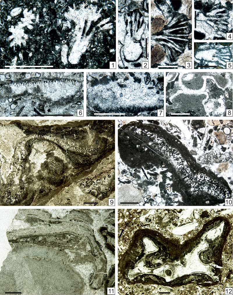
FIGURE 15. 1-12. Eugonophyllum magnum (Endo, 1951) emend. Konishi and Wray, 1961. 1. Two transverse sections as nuclei of oncoids. Sample TKS1_1b. 2. Transverse section as nucleus of an oncoid. Sample TKS3_2. 3. Two oblique sections with a Tetrataxis sp. Sample Z12B_3. 4. Longitudinal section showing a conceptacle. Sample TKS6_1. 5. Longitudinal section with a loop. Sample TKS13_2. 6. Longitudinal section as nucleus of an oncoid. Sample TKS13_3. 7. Longitudinal section. Sample TKS14_1a. 8. Longitudinal section. Sample TSK14_1b. 9. Two longitudinal sections (one with a conceptacle). Sample Z6B_1. 10. Two longitudinal sections (one with a bifurcation of the thallus). Sample TKW6_1b. 11. Typical longitudinal section (with two loops). Sample Z6B_3a. 12. One typical section (center) with four other ones. Sample Z6B_3b. Scale bars: 0.5 µm (Figure 15.3); all others = 1 µm.
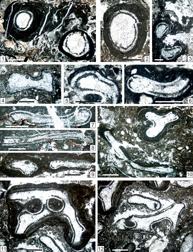
FIGURE 16. 1-3, 10-13. Eugonophyllum magnum (Endo, 1951) emend. Konishi and Wray, 1961. 1. Typical, ramified axial section. Sample. Z9B_2a. 2. Longitudinal section. Sample TK51A_2. 3. Transverse section. Sample GBT3_1. 10. Longitudinal section. Sample Z9_2. 11. Longitudinal section. Sample Z9_2. 12. Longitudinal section encrusted with girvanellaceans and a tuberitinid (center, right). Sample TKW10_4. 13. Transverse section with Globuliferoporella sp. (bottom, right). Sample TKS4_3. 4-9. Neoanchicodium catenoides Endo in Endo and Kanuma, 1954. 4. Longitudinal section. Sample TKW9_2. 5. Neomicrosparitized transverse section (left) and more typical transverse section (right). Sample TK_64_2. 6. Transverse section. Sample GBT4_3. 7. Longitudinal section. Sample TKW9_5. 8. Longitudinal section. Sample TKW9B_2. 9. Transverse section (top, center) and longitudinal section (bottom). Sample TNC2_4. Scale bars: 0.5 µm (Figures 16.4, 16.6); all others = 1 µm.
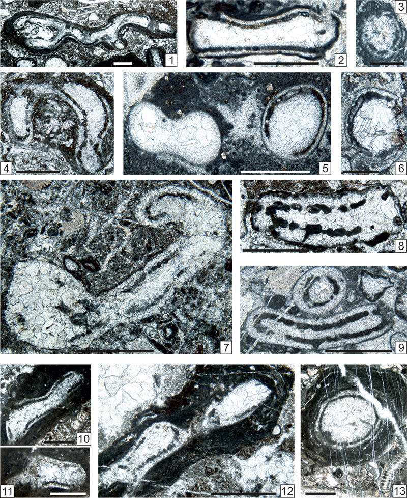
FIGURE 17. 1, 2. Eugonophyllum? konishi Kulik, 1978. Two fertile longitudinal sections. 1. Sample TM6A_1. 2. Sample TK49_2. 3, 4. Eugonophyllum magnum (Endo, 1951) emend. Konishi and Wray, 1961. Two longitudinal sections. 3. Sample. TK47_1. 4. Sample TK52_1. 5, 6, 8-11. Calcipatera schoenlaubi n. sp. Six subaxial sections. 8. Holotype. Sample TNA16_1_1. 6. Paratype. Sample ZK85A. 9. Paratype. Sample TK16_2_2. 10. As nucleus of an girvanellacean oncoid. Paratype. Sample TNA18_2_3. 11. As nucleus of an oncoid with girvanellacean and claracrustacean. Paratype. Sample TNA16_2_4. 7. Nanjinophycus? sp. Broken longitudinal section. Sample TNA16_2_1. Scale bar 1 µm.
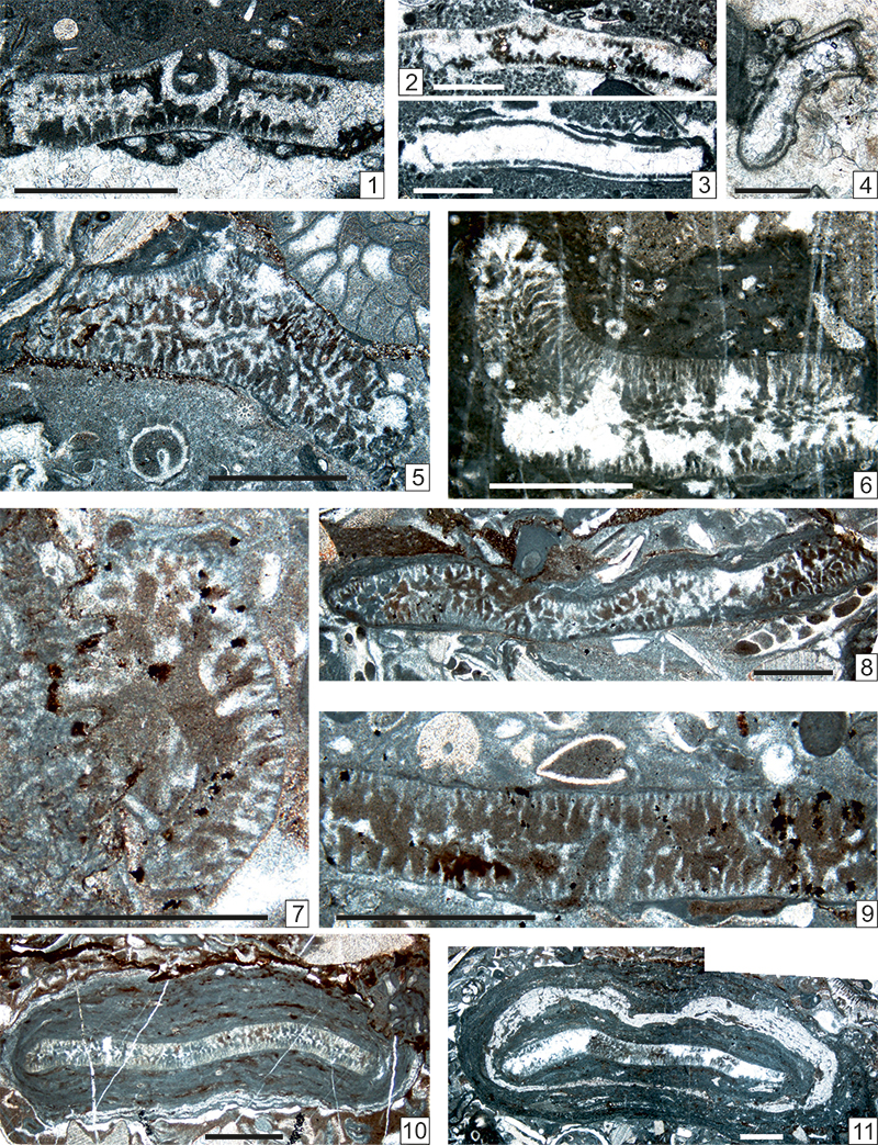
FIGURE 18. 1. Anthracoporella spectabilis Pia, 1920. Deformed, longitudinal section. Sample TNA2_1_4a. 2, 3. Anthracoporella vicina Kochansky and Herak, 1960. 2. Several sections in different planes. Sample TNA2_1_5. 3. Fertile specimen with endospores. Sample TNA2_2_1. 4, 5? Paraepimastopora kanumai (Endo in Endo and Kanuma, 1954). 4. Fragment of thallus in longitudinal section. Sample GB58_6. 5. Fragment of thallus in tangential section. Sample TK46_2. 6, 7, 9. Epimastopora japonica Endo, 1951 emend. Mamet, Roux and Nassichuk, 1987. Different longitudinal sections. 6. Sample TKS2_1. 7. Sample TNA18_2_1a. 9. Sample TNA18_2. 8, 10, 11. Epimastopora likana Kochansky and Herak, 1960 emend. herein. Two longitudinal sections. 8. Sample TKW9B_3. 10. Sample TNC5_4. 11. Sample ZK199_A. Scale bar = 1 µm.
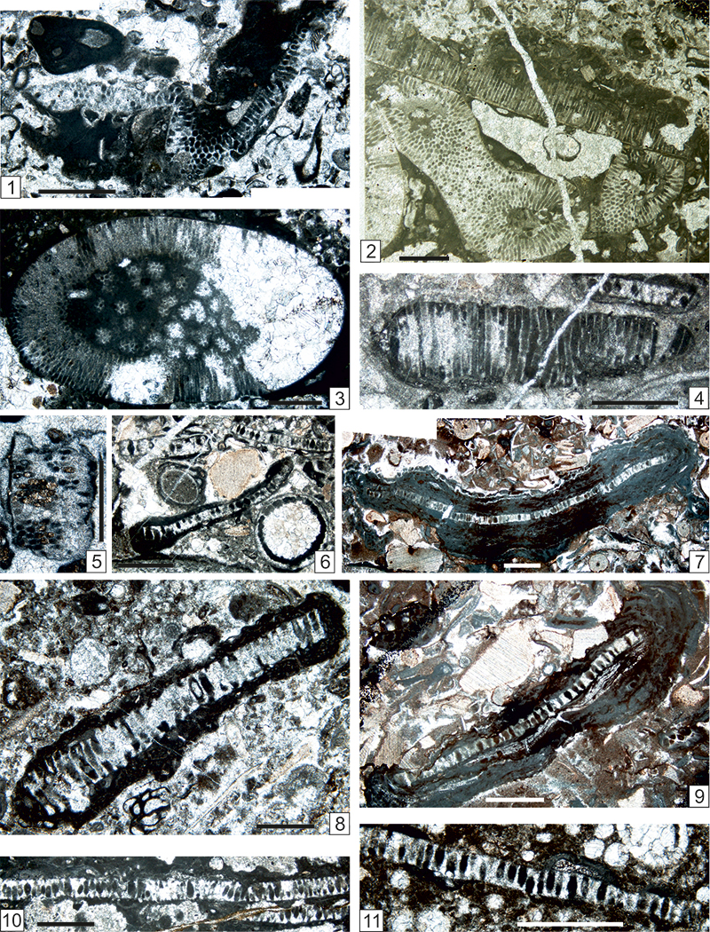
FIGURE 19. 1, 2, 6-9, 12 (left). Epimastopora japonica Endo, 1951 emend. Mamet, Roux and Nassichuk, 1987. Several longitudinal and oblique sections. 1. Sample TNC7_1. 2. Sample Z6_3. 3. Sample ZK98_18. 4. Sample ZT1_3. 6. Sample TKW5B_4b. 7. Sample TKW12_2a. 8. Sample TKW5B_3. 9. Sample Z5_1. 12. Sample ZK188_1_A. 3, 4. Epimastopora likana Kochansky and Herak, 1960 emend. herein. Several longitudinal sections. 3. Sample ZK98_18. 4. Sample ZT1_3. 5, 10. Epiastopora alpina (Kochansky and Herak, 1960) n. gen. n. comb. 5. Longitudinal section. Sample GB60_8. 10. Transverse section. Sample TNA1_2_2. 11. Epiastopora fluegeli (Kulik, 1978) n. comb. Longitudinal section. Sample TKW13B_8. 12, 13, 15. Globuliferoporella piai (Kordé, 1951). Several longitudinal sections. 12. (right: bottom and center) with Epimastopora japonica Endo, 1951 emend. Mamet, Roux and Nassichuk, 1987 (left), E. likana Kochansky and Herak, 1960 emend. herein (bottom center) and Eugonophyllum magnum (Endo, 1951) emend. Konishi and Wray, 1961 (center). Sample ZK188_1_A. 13. Sample ZK184_1. 15. Sample TKW10_2B. 14. Epimastopora cf. izawaikensis Endo, 1953a. Sample TK55_1. Scale bars: 0.5 µm (Figures 19.3, 19.5); all others = 1 µm.
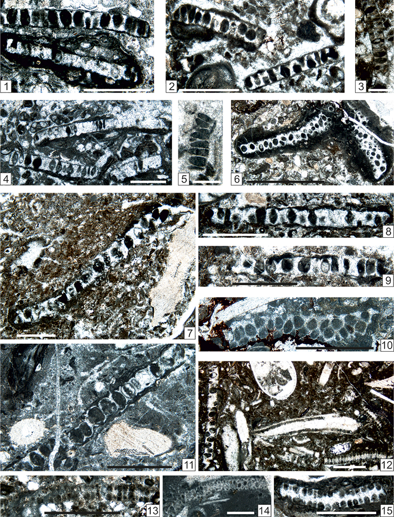
FIGURE 20. 1. Calcipatera schoenlaubi n. sp. Paratype encrusted by Claracrusta catenoides (Homann, 1972) emend. Vachard in Vachard and Montenat, 1981. Sample TM8_1. 2. Anthracoporella spectabilis Pia, 1920. Sample TM7_4. 3. Epimastopora likana Kochansky and Herak, 1960 emend. herein. Sample TM7_1a. 4. Eugonophyllum magnum (Endo, 1951) emend. Konishi and Wray, 1961. Sample ZK201_A. 5. Epiastopora alpina (Kochansky and Herak, 1960) n. gen. n. comb. Fragment of longitudinal section. Sample GB54_7. 6. Epimastopora cf. iwaizensis Endo, 1953a. Sample TM7_1b. Scale bars: 0.5 µm (Figure 20.5); all others = 1 µm.
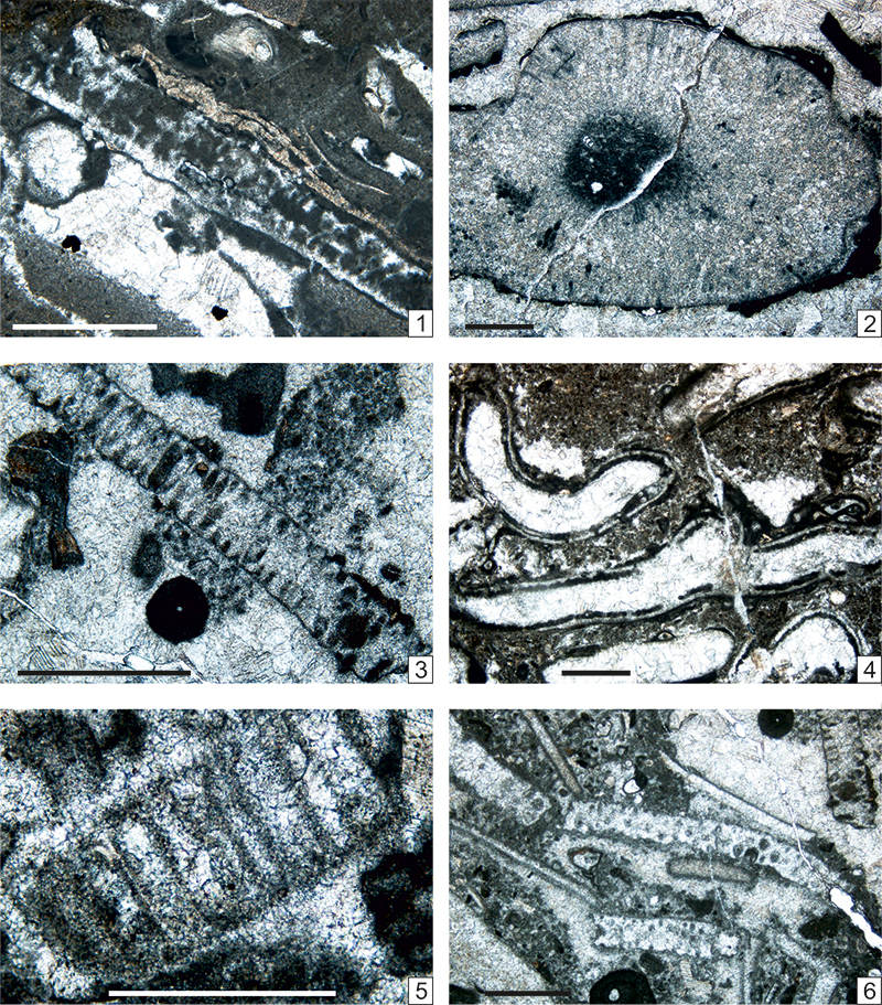
FIGURE 21. 1, 7?, 8-11. Globuliferoporella piai (Kordé, 1951). 1. Longitudinal section. Sample GB67_7a. 7? Tangential section. Sample Z10B_2. 8. Longitudinal section with Epiastopora alpina (Kochansky and Herak, 1960) n. gen. n. comb. (top right). Sample Z6B_5. 9. Oblique section. Sample TKW12B_2. 10. Longitudinal section. Sample TKW9_1. 11. Longitudinal section. Sample Z10_1. 2-5, 6? Globuliferoporella angulata Chuvashov, 1974. Three oblique longitudinal sections. 2. Sample GB153_A. 3. Sample ZK96_3. 4. Sample TKW9B_1. 5. Sample ZK187_2. 6. Sample TKW12_2b. Scale bars: 0.5 µm.
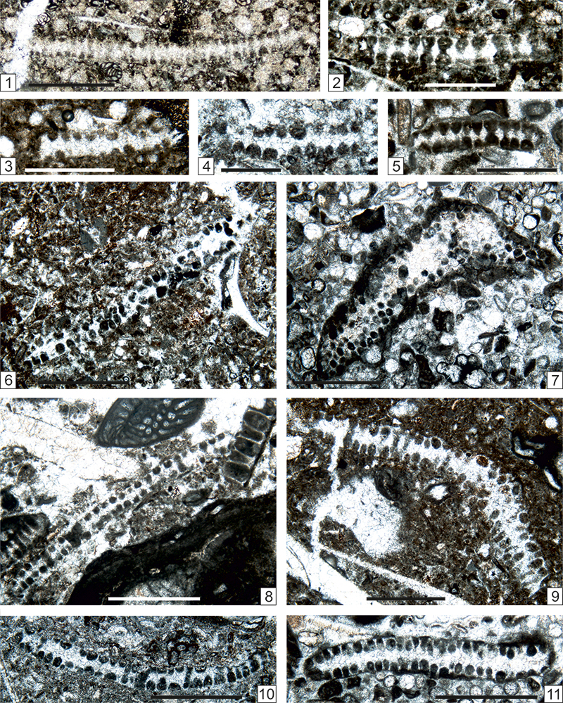
FIGURE 22. 1. Anthracoporella spectabilis Pia, 1920 emend. De Castro, 2002. Deformed longitudinal section. Sample Z3_2. 2, 5. Epiastopora alpina (Kochansky and Herak, 1960). 2. Several sections. Sample GB38_6. 5. Transverse section with Pseudovidalina sp. Sample TKW13_2b. 3, 6? Epimastopora likana Kochansky and Herak, 1960 emend. herein. 3. Broken longitudinal section. Sample ZK97_19. 6? Tangential section. Sample TNC5_3. 4. Nanjinophycus? sp. Transverse section with numerous, bifurcated, acrophore to phloiophore laterals. Sample TNA16_1_2. 7, 8. Mizzia cornuta Kochansky and Herak, 1960. Two recrystallized transverse sections. 7. Sample GB130_1. 8. Sample GB130_3. 9-11. Mizzia velebitana Schubert, 1908. Three transverse sections. 9. Sample GB129_3. 10. Sample GB67_10. 11. Encrusted by Palaeonubecularia sp. Sample ZK207_7. 12?, 13? Mizzia yabei (Karpinsky, 1909) emend. Pia, 1920. Two oblique sections. 12. Sample ZK96_11. 13. Sample GB163_5. Scale bars: 0.5 µm.
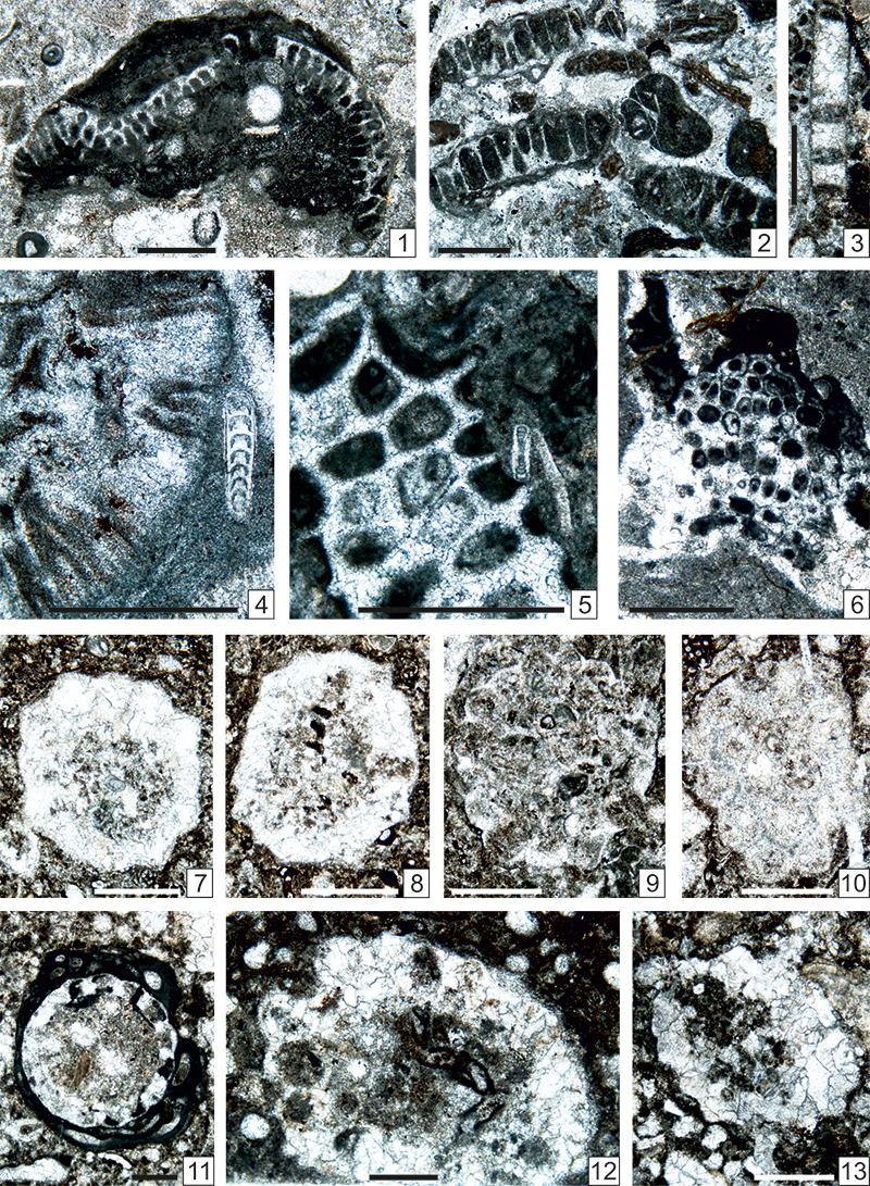
FIGURE 23. 1, 2. Macroporella cf. siamensis Endo, 1969. Longitudinal section. Sample GBT3_3. 2. Transverse section. Sample GBT3_4. 3, 5, 6, 8, 13, 18. Mizzia cornuta Kochansky and Herak, 1960. Several oblique transverse sections. 3. Sample GB40_5. 5. Sample GB40_6. 6. Sample GB70s_3. 7. Sample GB72_3. 8. Sample GB105_5. 13. Sample GB106_A. 18. Sample GBT1_4. 7, 9-12, 14? Mizzia velebitana Schubert, 1908. Several oblique transverse sections. 9. Sample GB109_2. 10. Sample GB111_4. 11. Sample GB129_6. 12. Sample GB66_11. 14? Sample GB156_9. 4, 15-17. Mizzia yabei (Karpinsky, 1909) emend. Pia, 1920. Several oblique sections. 4. Sample GB43_5. 15. Sample TK12_4. 16. Sample ZK202_6. 17. Sample Z14_1_2. Scale bars: Figures 23.1 - 23.3 = 1 µm; all others = 0.5 µm.
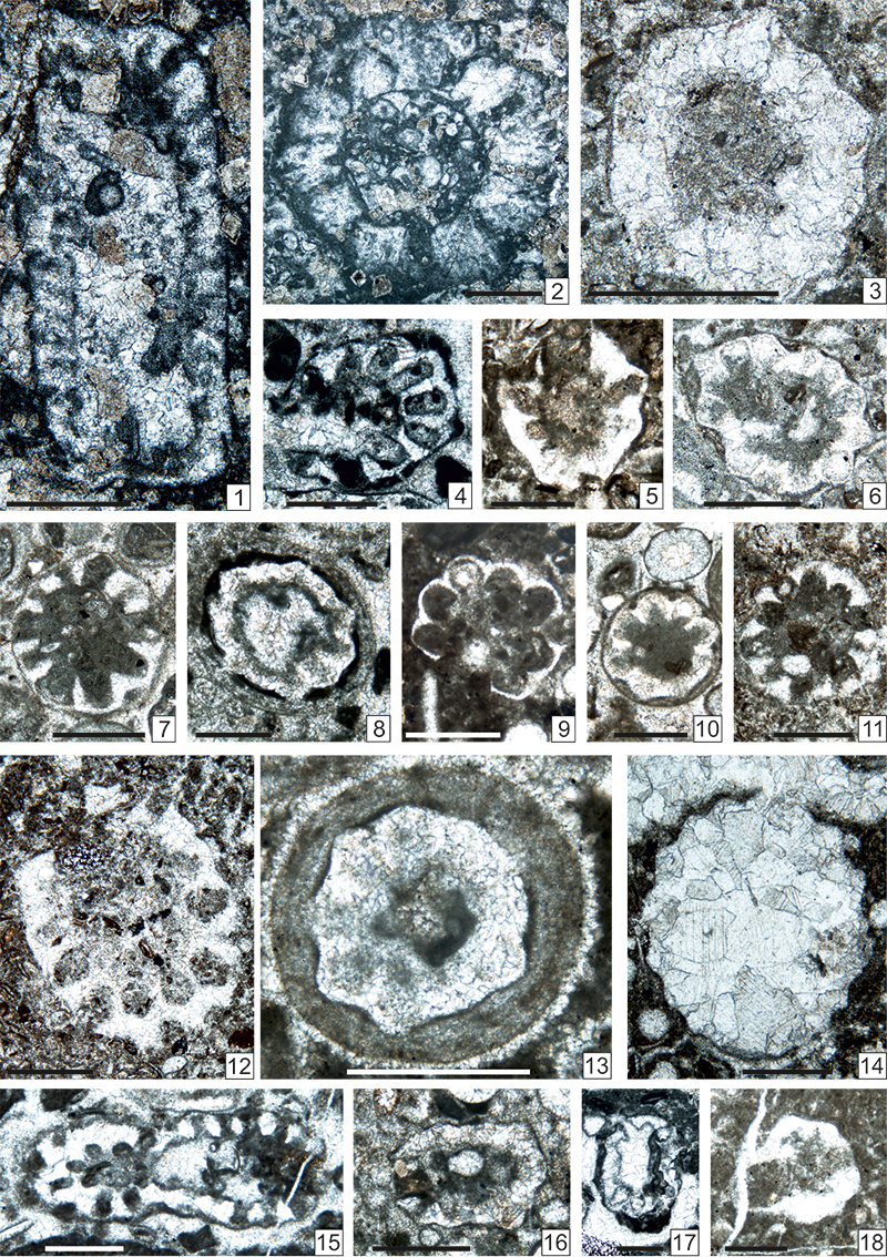
FIGURE 24. 1, 2, 5, 7, 8, 9 (bottom), 10. Mizzia yabei (Karpinsky, 1909) emend. Pia, 1920. 1. Oblique section. Sample GB43_5. 2. Oblique section. Sample GB71_1_1. 5. Oblique section. Sample Z13B_4. 7. Transverse section. Sample TSK12_2. 8. Transverse section. Sample TKS 12_3a. 9. Subaxial section (bottom, left). Sample ZK204_A. 10. Abraded tangential section. Sample ZK203_2. 3, 4, 6, 9 (top), 11-13, 14? Mizzia velebitana Schubert, 1908. 3. Transverse section. Sample GB146_2. 4. Oblique section. Sample GB163_10. 6. Transverse section superficially abraded. Sample TKS13_5. 9. Transverse section (top, right). Sample ZK204_A. 11. Transverse section. Sample GB175_11. 12. Tangential section showing the aspect of surface. Sample ZK94_5_4. 13. Transverse section. Sample ZK200_5. 14. Transverse section showing different stages of diagenesis. Sample ZK200_6. Scale bars: 0.5 µm.
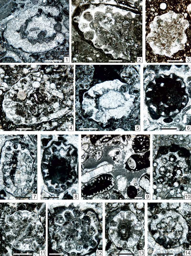
FIGURE 25. 1-12. Pseudoepimastopora carnica (Flügel, 1966) emend. herein, n. comb. 1. Transverse section. Sample GB36_4. 2. Oblique transverse section. Sample GB60_2_2. 3. Oblique section. Sample GB61_4. 4. Transverse section. Sample GB72_1b. 5. Transverse section. Sample Z5_2. 6. Oblique section. Sample Z5_3. 7. Transverse section. Sample Z7_2. 8. Longitudinal section. Sample Z7_3. 9. Transverse section. Sample Z9_1b. 10. Transverse section. Sample ZK99c_13. 11. Transverse section. Sample ZK94_7_4. 12. Transverse section. Sample ZK99a_10. 13. Globuliferoporella? sp. Transverse section. Sample Z19_3. Scale bars: 0.5 µm.
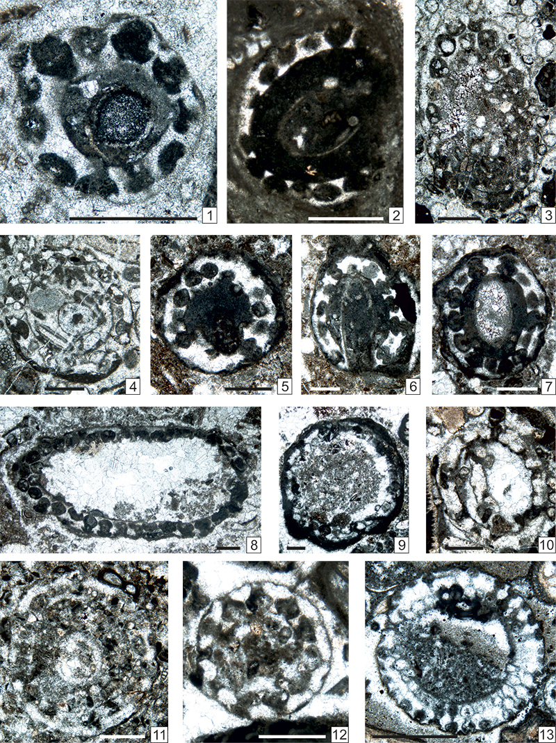
FIGURE 26. 1, 2, 4. Connexia slovenica Kochansky-Devidé, 1979. 1. Oblique section of two laterals. Sample GB40_2. 2. Fragment of longitudinal section. Sample GBT4_2. 4. Two laterals. Sample Z9B_7. 3. Connexia? sp. Sample GBT3_2. 5-8. Claracrusta catenoides (Homann, 1972) emend. Vachard in Vachard and Montenat, 1981. Longitudinal section with a tangential section in Figure 26.5. 5. Sample GB36_5. 6. Sample TNA18_1_2. 7. Sample TKW6B_3. 8. Sample TKW4_1. Scale bars: 1 µm.
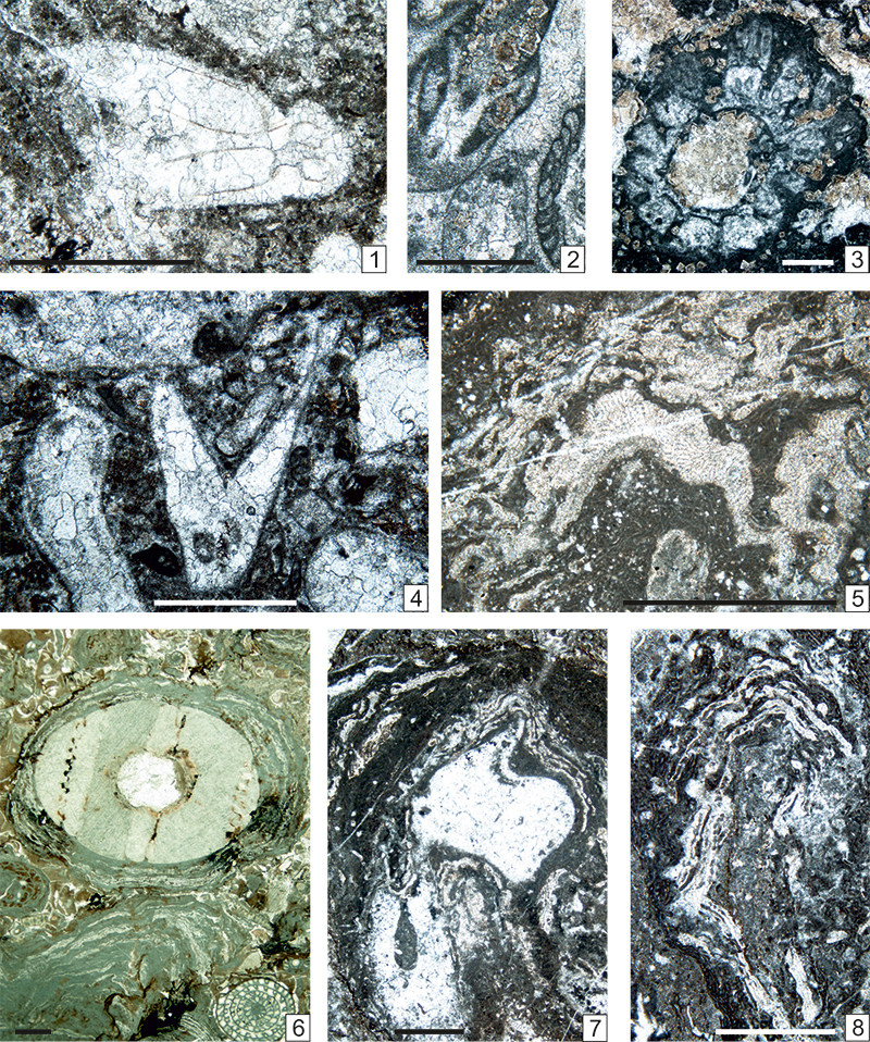
FIGURE 27. 1. Gyroporella sp. Transverse section. Sample ZT1_6. 2. Epimastopora likana Kochansky and Herak, 1960 emend. herein. Longitudinal section. Sample GB156_8. 3, 6. Pseudoepimastopora carnica n. comb., emend. herein. 3. Transverse section with Connexia slovenica Kochansky-Devidé, 1979 (top, right) in axial section. Sample ZT1_5. 6. Oblique transverse section. Sample Z5B_3. 4. Renalcis cf. granosus Vologdin, 1932. A colony associated with Tubiphytes carinthiacus (Flügel). Sample GBT11_2. 5. Epimastopora cf. izawaikensis Endo, 1953a. Longitudinal section (left) with Salopekiella? sp. (top right) in axial section. Sample TM5_2. 7, 8. Neoanchicodium catenoides Endo in Endo and Kanuma, 1954. Numerous sections. 7. Sample Z9B_3. 8. Sample TKS12_1_2. 9. Connexia slovenica Kochansky-Devidé, 1979. Small fragment with two laterals, in transverse and oblique sections. Sample TK32. 10-12. Efluegelia ex gr. johnsoni (Flügel, 1966). Three longitudinal sections more or less recrystallized. 10. Sample GB157_10. 11. Sample GB162_1. 12. Sample Z17B_1. 13-15. Ungdarella uralica Maslov, 1956. Several sections with “Algen Sporen”. 13. Sample ZK198_9. 14. Sample ZK200_2. 15. Sample ZK200_9. Scale bars: Figure 27.8 = 1 µm, Figure 27.11 = 0.25 µm, all others = 0.5 µm.
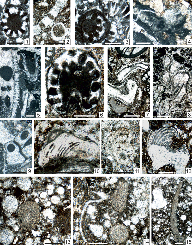
FIGURE 28. 1-9. Ungdarella uralica Maslov, 1956. Several oblique sections more or less recrystallized and one transverse section relatively well-preserved (Figure28.8). 1. Central parts of both branchs are chertified. Sample GB123_2a. 2. Sample GB125_2. 3. Sample GB 123_26. 4. Sample GB126_1. 5. Sample TNB15_1_1. 6. With Mendipsia conili (Nguyen Duc Tien, 1980). Sample GB130_5a. 7. Sample GBT13. 8. Sample ZK199_1. 9. Sample ZK200_10. Scale bars: Figure 28.7 = 1 µm; all others = 0.5 µm.
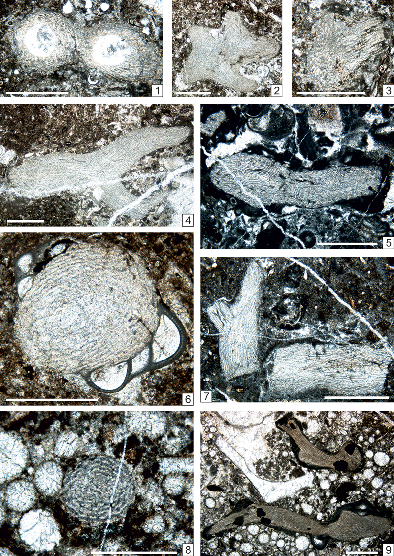
FIGURE 29. 1-9, 12. Efluegelia ex gr. johnsoni (Flügel, 1966). Various longitudinal and tangential sections. 1. Sample GB03_8. 2. Sample GB49_3. 3. Sample GB 56_2. 4. Sample GB_60_1_4. 5. Sample GB60_7a. 6. Sample GB60_4. 7. Sample GB68_6. 8. Sample TNB15_2_6. 9. Sample TK52_5. 12. Sample GB126_4. 10, 11. Efluegelia johnsoni (Flügel, 1966). 10. Sample GB109_4. 11. Sample GB117_1. Scale bars: Figures 29.6, 29.8 = 0.5 µm, all others = 1 µm.
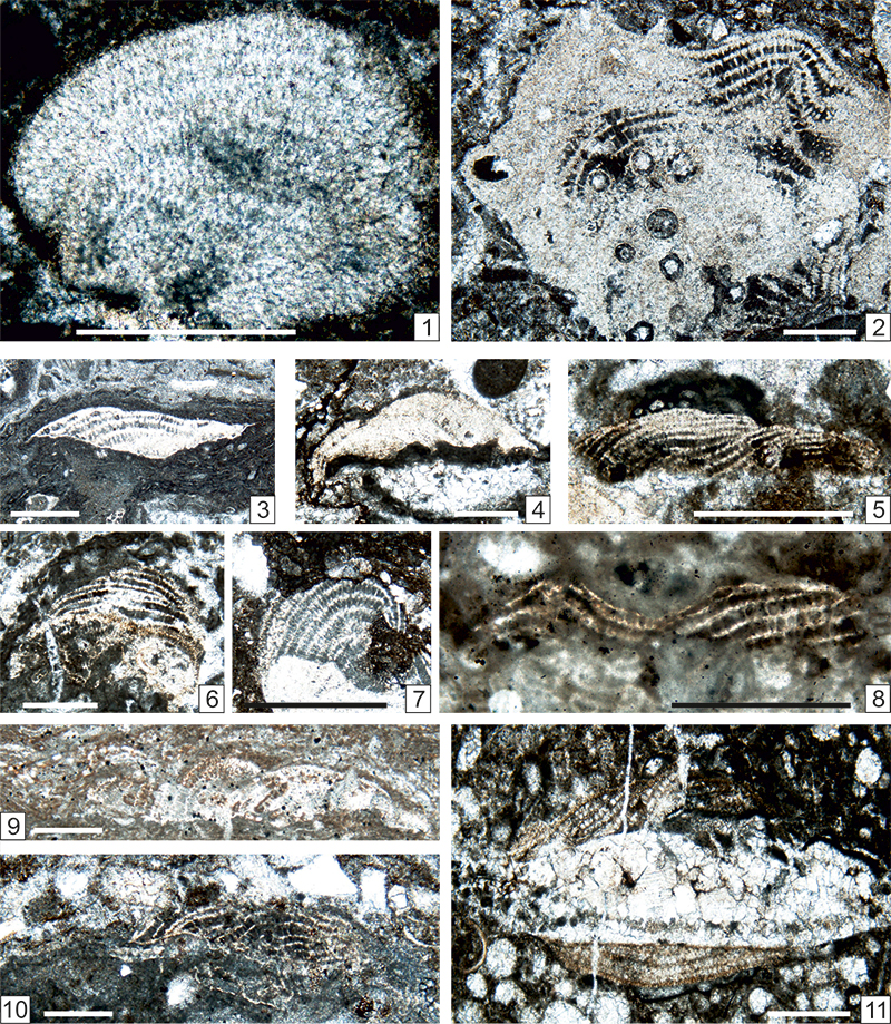
FIGURE 30. 1, 2, 5, 8-11. Efluegelia ex gr. johnsoni (Flügel, 1966). Various longitudinal and tangential sections 1. Sample GBT1_2. 2. Sample TKW9_6. 5. Sample ZK187_5. 8. Sample ZK95_gross_3. 9. Sample ZKa_5. 10. Sample ZK199. 11. Sample ZK200_1. 3, 4, 6, 7. Efluegelia johnsoni (Flügel, 1966). 3. Sample TKW138_4. 4. Sample Z6_1. 6. Sample Z15_3_4. 7. Sample GB174_5. Scale bars: Figures 30.8, 30.9, 30.10 = 0.2 µm, all others = 0.5 µm.