FIGURE 1. A locality map showing the counties in Texas, USA where the three new Belonostomus fossils were collected.
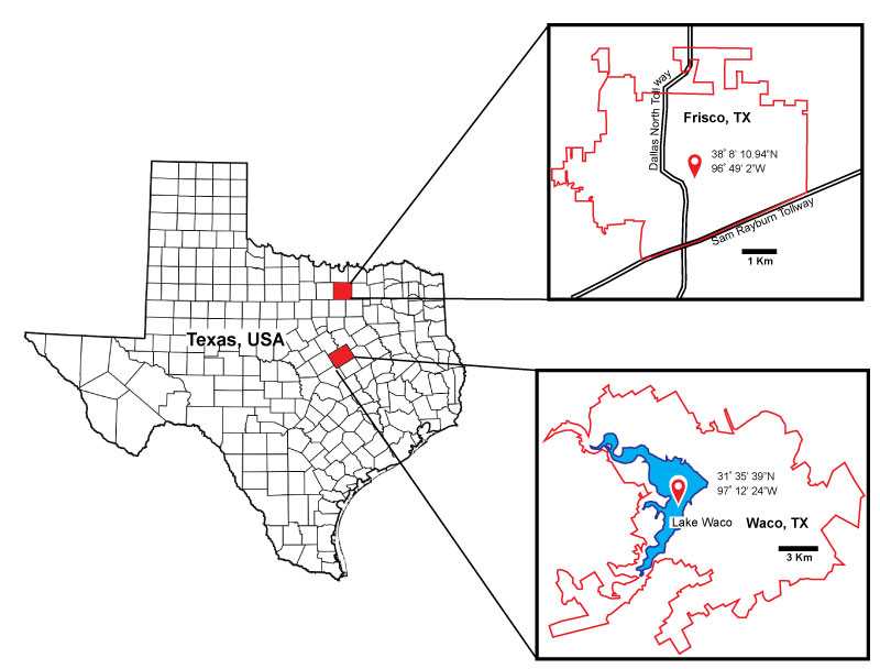
FIGURE 2. An expanded map showing all known localities in North America that have produced Belonostomus fossils. 1. Ashville Formation, Saskatchewan, Canada. 2. Frenchman Formation, Saskatchewan, Canada. 3. Kaskapau Formation, Alberta, Canada. 4. Foremost and Oldman formations, Alberta, Canada. 5. Dinosaur Park Formation, Alberta, Canada. 6. Horseshoe Canyon, Alberta, Canada 7. St. Mary River Formation, Alberta, Canada. 8. Tongue River Formation, North Dakota, USA. 9. Judith River Formation, Montana, USA. 10. Hell Creek Formation, Montana, USA 11. Judith River Formation, North Dakota, USA 12. Hell Creek Formation, North Dakota, USA. 13. Lance Formation, Wyoming, USA. 14. Mesa Verde Formation, Wyoming, USA, 15. Prairie Bluff Chalk, Mississippi, USA. 16. Tombigbee Sand Member of the Eutaw Formation, Alabama, USA 17. Paluxy Formation, Texas, USA 18. Austin Chalk Formation, Texas, USA 19. Del Rio Formation, Texas, USA, 20. Eagle Ford Formation, Texas, USA. 21. Agua Nueva Formation, Nuevo Leon, México. 22. Tlayua Formation, Puebla, México. 23. Cerro de la Virgen Formation, Oaxaca, México.
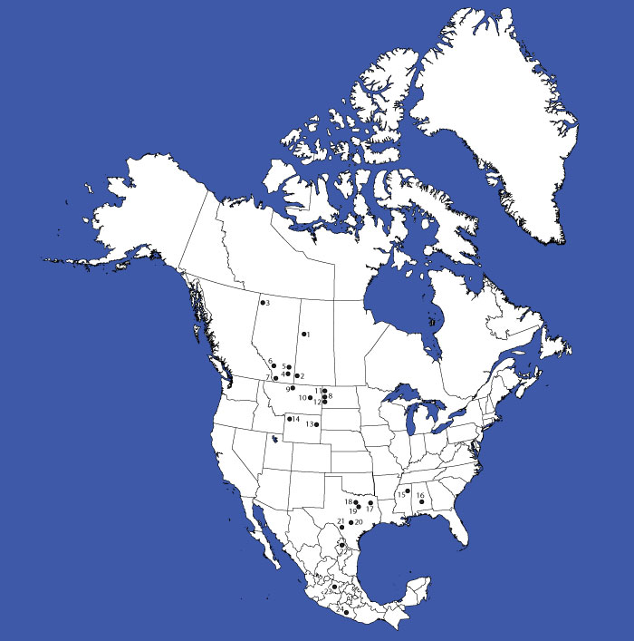
FIGURE 3. A correlated stratigraphic column representing formations in the North American Gulf Coastal Plain where Belonostomus fossils can be found. The stars represent where in the section Belonostomus fossils have been documented. The line indicates how deep into section they have been observed (modified from Scott et al., 2003).
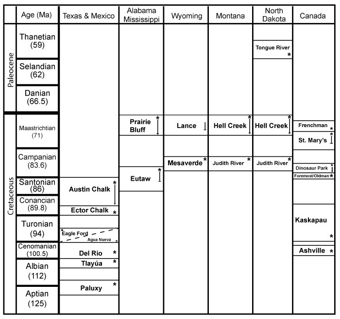
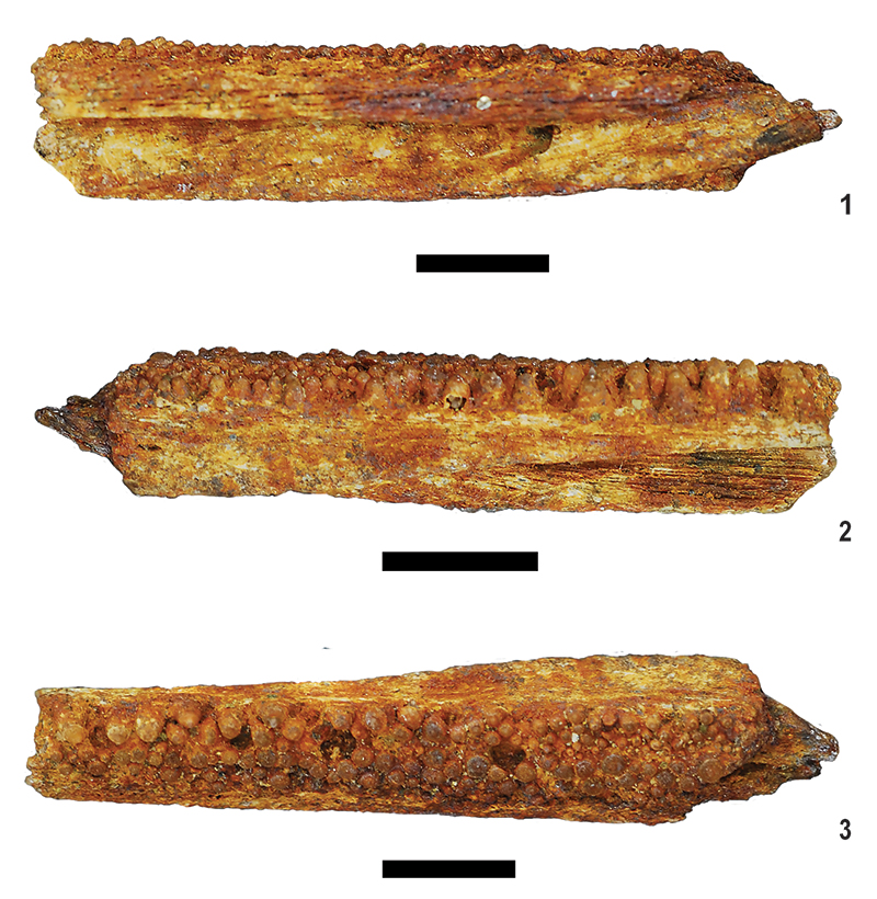
FIGURE 4. Belonostomus sp. SMU 77675. Fused left and right premaxillae. 1, dorsal view; 2, ventral view; 3, left lateral view; 4, close up of the teeth in lateral view. Scale bars equal 10 mm.
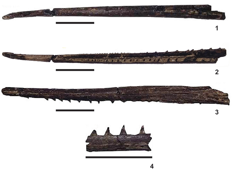
FIGURE 5. Belonostomus sp. SMU 77676. Partial predentary. 1, dorsal view; 2, ventral view. Abbreviation: pds: predentary symphysis. Scale bars equal 10 mm.
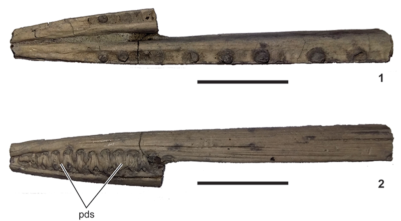
FIGURE 6. Belonostomus sp. SMU 77677. 1, ventral view; 2, Lateral view. Scale bars equal 10 mm.
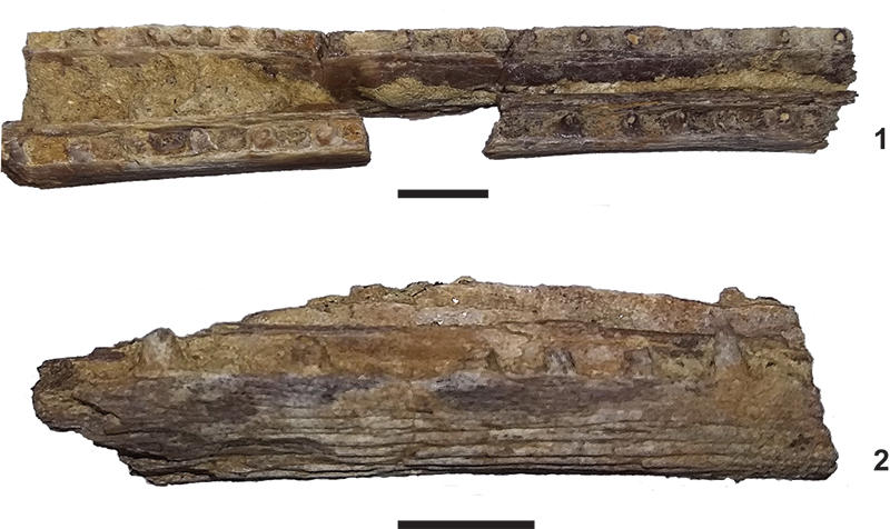
FIGURE 7. Belonostomus sp. ALMNH 1994.24.15 1, dorsal view; 2, right lateral view; and 3, ventral view. Scale bars equal 5 mm.
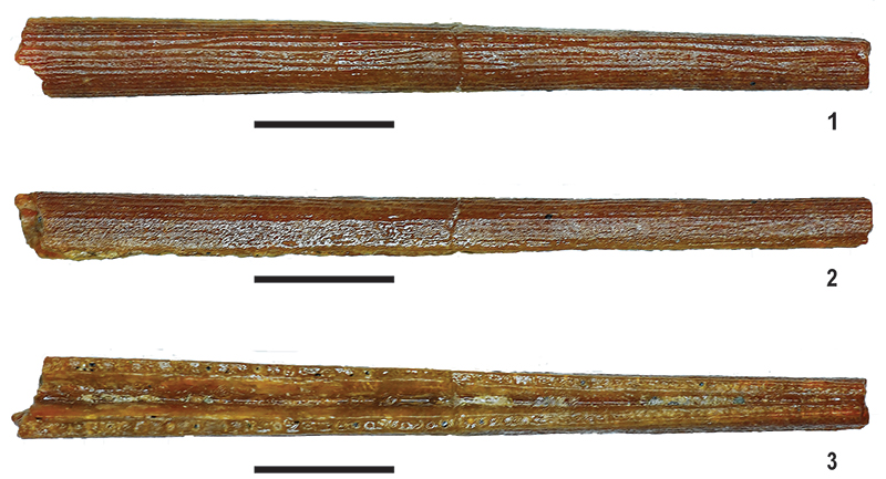
FIGURE 8. Belonostomus sp. MMNS 3152. Rostrum and premaxillae. 1, left lateral view. Anterior to the left. 2, ventral view. Scale bar equal 10 mm.

FIGURE 9. Belonostomus sp. ALMNH 1994.31.112. 1, lateral view; 2, medial view; 3, dorsal view. Scale bars equal 5 mm.