FIGURE 1. Map of Greece, with star indicating the latest Miocene/earliest Pliocene (MN 13/14) locality of Maramena.
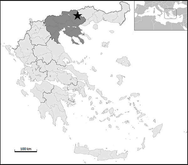
FIGURE 2. Cryptobranchidae indet. from Maramena: atlas (UU MAA 7441) in anterior (1), left lateral (2), posterior (3), ventral (4), and dorsal (5) views. Abbreviations: ct, cotyle; ffsn, foramen for the first spinal nerve; op, odontoid process.
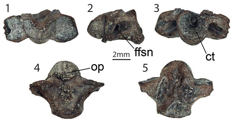
FIGURE 3. Various salamandrids from Maramena. Salamandrina sp. (1-5): trunk vertebra (UU MAA 7483) in anterior (1), left lateral (2), posterior (3), ventral (4), and dorsal (5) views. Lissotriton sp. (Lissotriton vulgaris group) (6-15): trunk vertebra (UU MAA 7518) in anterior (6), right lateral (7), posterior (8), ventral (9), and dorsal (10) views; trunk vertebra (UU MAA 7517) in anterior (11), right lateral (12), posterior (13), ventral (14), and dorsal (15) views. Lissotriton sp.(16-20): trunk vertebra (UU MAA 7486) in anterior (16), right lateral (17), posterior (18), ventral (19), and dorsal (20) views. Ommatotriton sp. (21-28): trunk vertebra (UU MAA 7489) in anterior (21), right lateral (22), posterior (23), ventral (24), and dorsal (25) views; right femur (UU MAA 7248) in anterior (26), posterior (27), and medial (28) views. Abbreviations: cd, condyle; ct, cotyle; ns, neural spine; pc, pericondylar constriction; pt, pterygapophysis; sf, subcentral foramen; t, trochanter; vl, ventral lamina; zy, zygosphene.
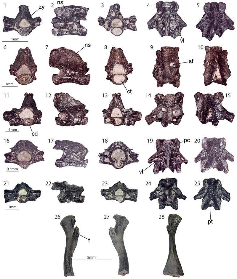
FIGURE 4. Salamandrids from Maramena: Salamandra sp. (1-5): trunk vertebra (UU MAA 7495) in anterior (1), right lateral (2), posterior (3), dorsal (4), and ventral (5) views. Salamandridae indet. (6-9): atlas (UU MAA 7490) in dorsal (6) and ventral (7) views; rib (UU MAA 7508) in anterior view (8); rib (UU MAA 7519) in posterior view (9). Abbreviations: cd, condyle; ct, cotyle; na, neural arch; neural spine; op, odontoid process; sf, subcentral foramen.
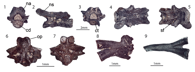
FIGURE 5. Latonia sp. from Maramena: left maxilla (UU MAA 7227) in labial (1) and lingual (2) views; right maxilla (UU MAA 7223) in labial (3) and lingual (4) views; right angular (UU MAA 7154) in dorsal view (5); left scapula (UU MAA 7520) in externalview (6); right humerus (UU MAA 7225) in ventral view (7); right ilium (UU MAA 7528) in lateral (8) and medial (9) views; costa (UU MAA 7523) in lateral (10) and ventral (11) views. Abbreviations: a, acetabulum; ch, caput humeri; cl, crista lateralis; cm, crista medialis; cp, coronoideus portion of the coronoid process; dec, dental crest; doc, dorsal crest; dt, dorsal tubercle; fcv, fossa cubitalis ventralis; fo, supra-acetabular fossa; gf, glenoid fossa; ig, interiliac groove; lh, lamina horizontalis; mg, Meckelian groove; mo, margo orbitalis; pa, pars ascendens; pd, pars descendens; pdp, posterior depression; pp, paracoronoideus portion of the coronoid process; ps, pars suprascapularis.
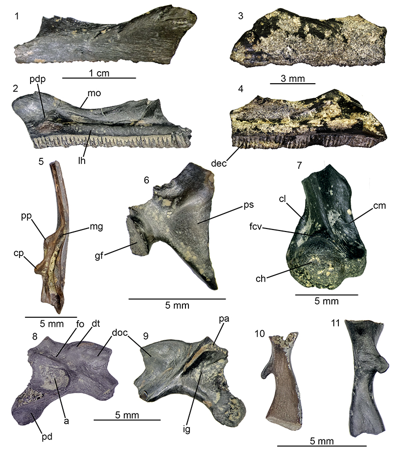
FIGURE 6. Various frogs from Maramena. Hyla sp. (1-2): left ilium (UU MAA 7118) in lateral view (1); left humerus (UU MAA 7474) in ventral view (2). Pelophylax sp. (3-6): right coracoid (UU MAA 7233) in internal view (3); left ilium (UU MAA 7232) in lateral (4) and medial (5) views; right angular (UU MAA 7194) in dorsal view (6). Ranidae indet. (7): right humerus (UU MAA 7231) in ventral view (7). Anura indet., different morphotypes (8-14). Anura indet. (morphotype A) (8-9): phalanx (UU MAA 7527) in ventral view (8); phalanx (UU MAA 7514) in ventral view (9). Anura indet. (morphotype B) (10-12): phalanx (UU MAA 7515) in ventral view (10); two phalanges (UU MAA 7524) in ventral view (11-12). Anura indet. (morphotype C) (13-14): phalanx (UU MAA 7516a) in ventral view (13); phalanx (UU MAA 7516b) in ventral view (14). Abbreviations: a, acetabulum; ch, caput humeri; cp, coronoid process; pa, pars ascendens; pd, pars descendens; pz, preacetabular zone; s, shaft; ts, tuber superior.
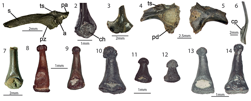
FIGURE 7. Mauremys aristotelica from Maramena: nuchal (UU MAA 7283) in dorsal (1) and visceral (2) views; neural (UU MAA 7146) in dorsal (3) and visceral (4) views; neural (UU MAA 7253) in visceralview (5); left costal VI (UU MAA 7284) in dorsal (6) and visceral (7) views; costal (UU MAA 7286) in dorsal (8) and visceral (9) views; costal (UU MAA 7288) in dorsal view (10); left peripheral III (UU MAA 7289) in dorsal (11) and visceral (12) views; bridge peripheral (UU MAA 7297) in dorsal (13) and visceral (14) views; peripheral (UU MAA 7293) in dorsal (15) and visceral (16) views; peripheral (UU MAA 7295) in dorsal (17) and visceral (18) views; right hyoplastron (UU MAA 7296) in visceral (19) and dorsal (20) views. Abbreviation: por, musk pore.
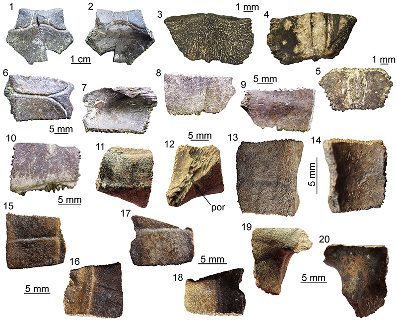
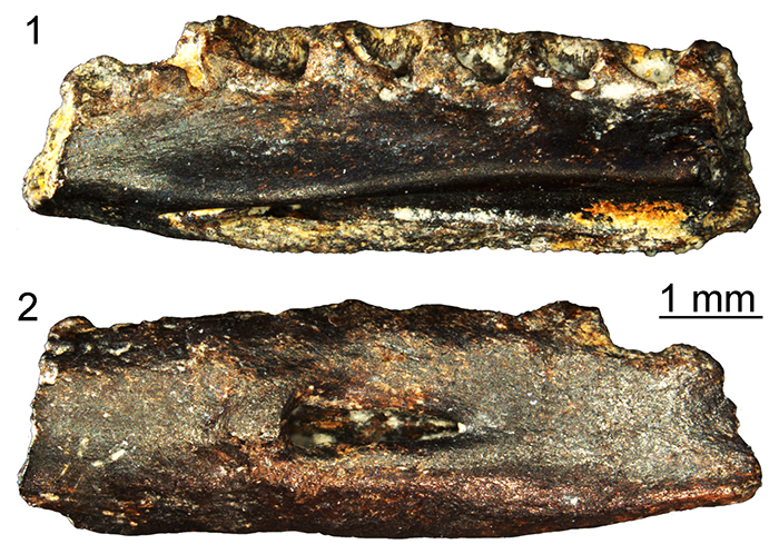
FIGURE 8. ?Testudinidae indet. from Maramena: ?osteoderm (UU MAA 7135) in dorsal (1), ventral (2), and lateral (3) views.
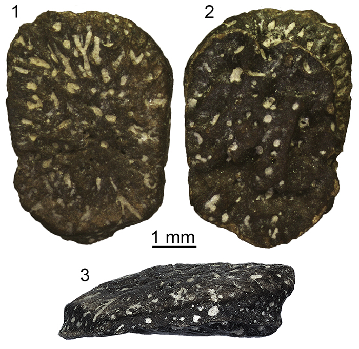
FIGURE 9. Agaminae indet. from Maramena: frontal (UU MAA 7191) in dorsal (1) and ventral (2) views; premaxilla (UU MAA 7076) in anterior (3) and posterior (4) views; premaxilla (UU MAA 7341) in anterior (5) and posterior (6) views; right maxilla (UU MAA 7043) in medial (7) and lateral (8) views; right dentary (UU MAA 7041) in medial (9) and lateral (10) views; left dentary (UU MAA 7042) in medial view (11); left dentary (UU MAA 7207) in medial view (12); right dentary (UU MAA 7305) in medial view (13); caudal vertebra (UU MAA 7062) in left lateral (14) and dorsal (15) views. Abbreviations: asn, articular surface with the nasal; cc, crista cranii; ig, interdental groove; lp, lateral process; mf, Meckelian groove; ss, subdental shelf.
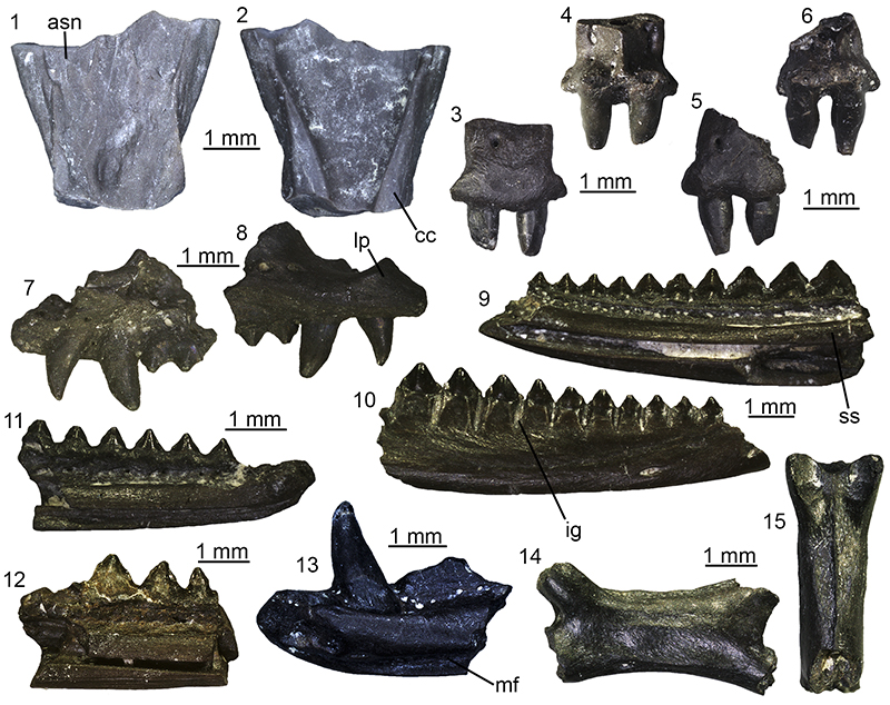
FIGURE 10. Lacertidae indet. from Maramena: right maxilla (UU MAA 7032) in lateral (1) and medial (2) views; right quadrate (UU MAA 7096) in anterior (3), posterior (4) and medial (5) views; left pterygoid (UU MAA 7099) in ventral (6) and dorsal (7) views; right pterygoid (UU MAA 7097) in dorsal (8) and ventral (9) views; right dentary (UU MAA 7048) in medial view (10); right dentary (UU MAA 7049) in medial view (11); right dentary (UU MAA 7050) in medial (12) and lateral (13) views. Abbreviations: bp, basipterygoid fossa;c, conch; fc, fossa columellae; ml, medial lamina; pf, pterygoid flange; pp, palatine process; qp, quadrate process; ss, subdental shelf.
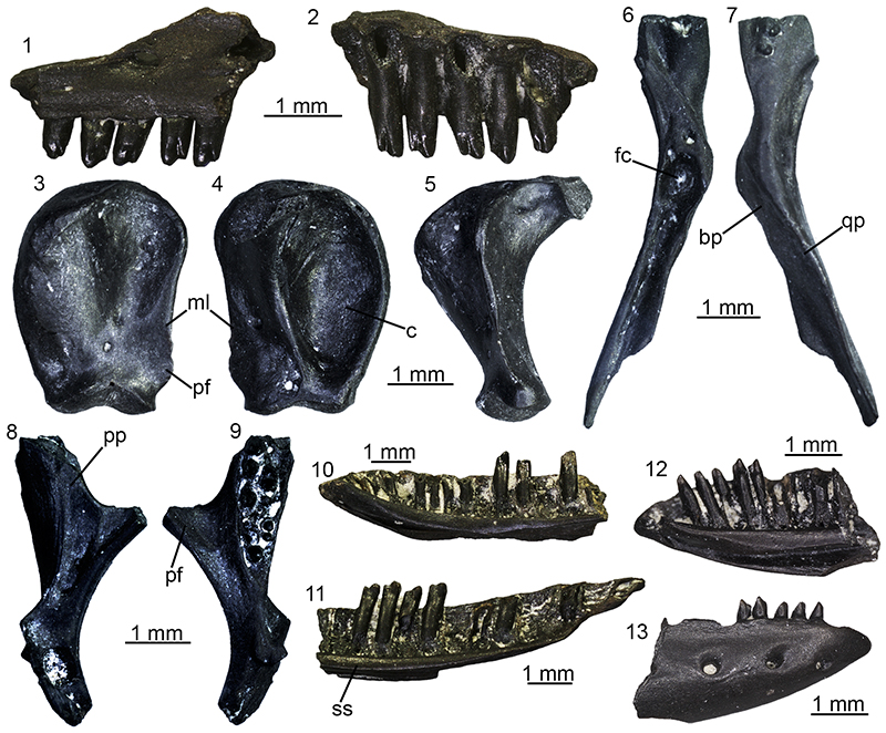
FIGURE 11. ?Lacertidae indet. from Maramena: fragment of a tooth bearing bone (UU MAA 7033) in dorsal (1), medial (2), and lateral (3) views.
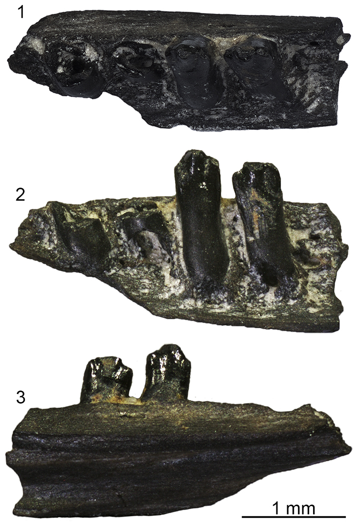
FIGURE 12. aff. Palaeocordylus sp. from Maramena: right maxilla (UU MAA 7046) in lateral (1) and medial (2) views; right maxilla (UU MAA 7047) in medial view (3); right maxilla (UU MAA 7077) in medial view (4); right maxilla (UU MAA 7078) in lateral view (5); left dentary (UU MAA 7003) in medial view (6); right dentary (UU MAA 7004) in lateral (7) and medial (8) views; right dentary (UU MAA 7006) in lateral (9) and medial (10) views; right dentary (UU MAA 7036) in medial view (11); left dentary (UU MAA 7199) in lateral (12) and medial (13) views. Abbreviations: pp, posterior process; ss, subdental shelf.
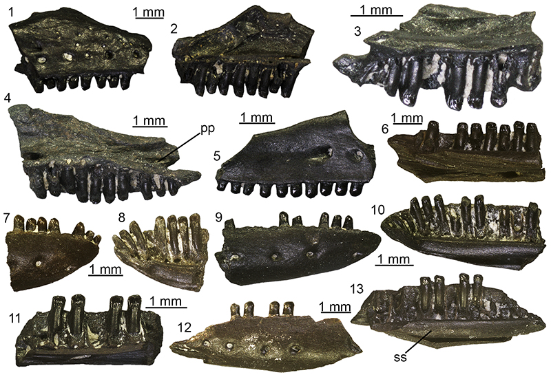
FIGURE 13. ?Scincidae indet. from Maramena: left dentary (UU MAA 7027) in lateral (1) and medial (2) views; right dentary (UU MAA 7107) in medial view (3). Abbreviation: ss, subdental shelf.
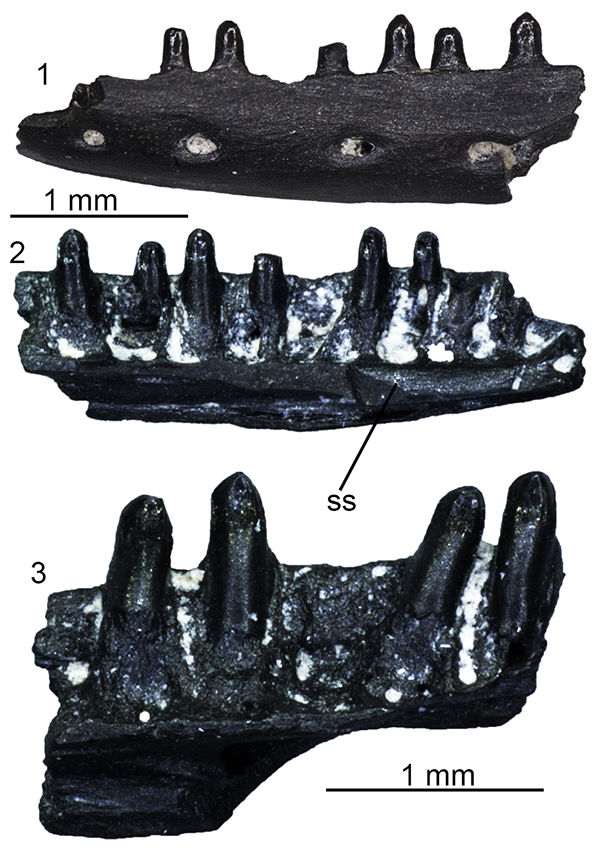
FIGURE 14. Anguis sp. from Maramena: trunk vertebra (UU MAA 7181) in dorsal (1), ventral (2), anterior (3), posterior (4), and left lateral (5) views; trunk vertebra (UU MAA 7263) in dorsal (6), ventral (7), anterior (8), posterior (9), and left lateral (10) views. Abbreviations: c, vertebral centrum; nc, neural canal.
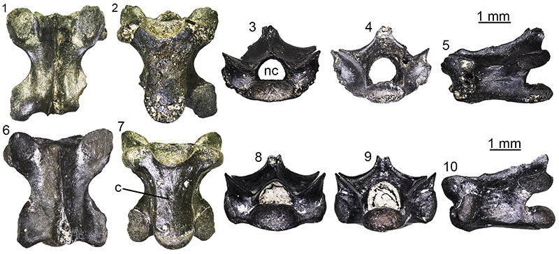
FIGURE 15. Ophisaurus morphotypes from Maramena. Ophisaurus sp. (morphotype 1) (1-3): right maxilla (UU MAA 7324) in medial view (1); right dentary (UU MAA 7176) in medial view (2); right dentary (UU MAA 7177) in medial view (3). Ophisaurus sp. (morphotype 2) (4-7): left maxilla (UU MAA 7180) in medial view (4); left dentary (UU MAA 7100) in medial view (5); right dentary (UU MAA 7186) in medial (6) and lateral (7) views. Ophisaurus sp. (morphotype 3) (8): left dentary (UU MAA 7313) in medial view (8). Ophisaurus sp. (morphotype 4) (9-10): left dentary (UU MAA 7175) in medial (9) and lateral (10) views. Ophisaurus sp. (morphotype 5) (11): left maxilla (UU MAA 7178) in medial view (11). Abbreviations: af, alveolar foramen; is, intramandibular septum; sp, surangular spine; ss, subdental shelf.
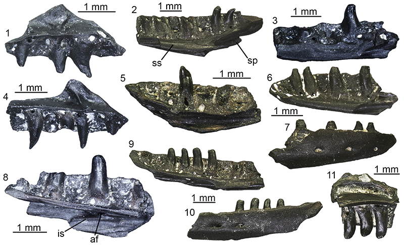
FIGURE 16. Ophisaurus from Maramena (indeterminate morphotype), vertebrae:trunk vertebra (UU MAA 7173) in anterior (1) and posterior (2) views; trunk vertebra (UU MAA 7215) in ventral view (3); trunk vertebra (UU MAA 7216) in dorsal view (4).
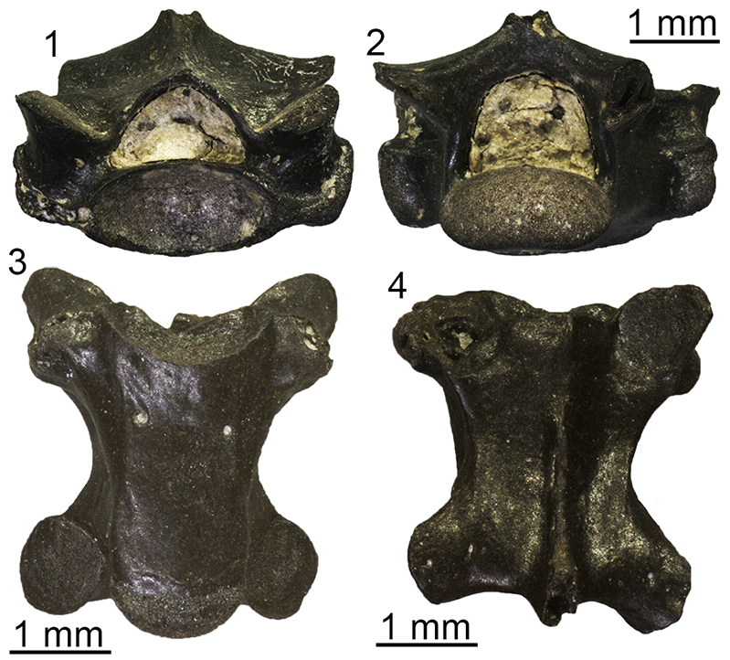
FIGURE 17. Pseudopus sp. from Maramena, cranial elements: right maxilla (UU MAA 7171) in lateral (1), medial (2), dorsal (3), and ventral (4) views; right dentary (UU MAA 7407) in lateral (5), medial (6), dorsal (7), and posterior (8) views; fragment of tooth bearing bone (UU MAA 7307) in lateral (9) and medial (10) views. Abbreviation: ss, subdental shelf.
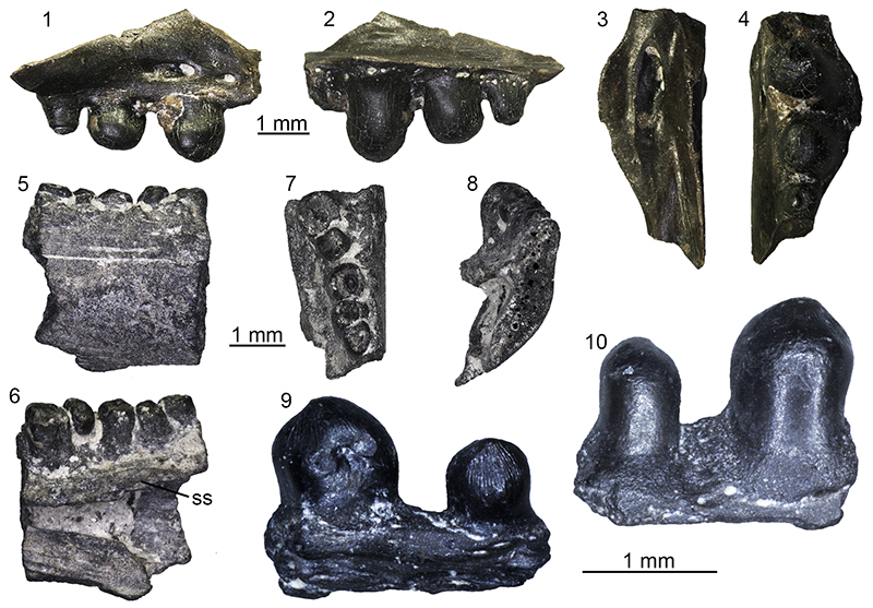
FIGURE 18. Pseudopus sp. from Maramena,vertebrae: trunk vertebra (UU MAA 7172) in left lateral (1), anterior (2), posterior (3), dorsal (4), and ventral (5) views; trunk vertebra (UU MAA 7328) in ventral view (6); cloacal vertebra (UU MAA 7329) in dorsal (7) and ventral (8) views. Abbreviation: c, vertebral centrum.
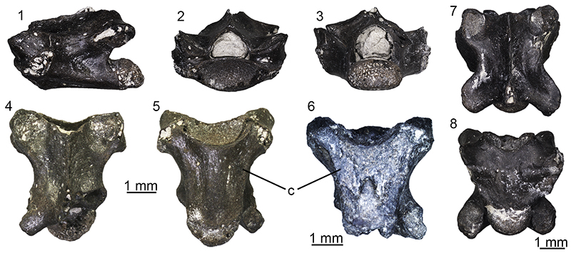
FIGURE 19. Non-Anguis Anguinae indet. from Maramena (1-12): osteoderm (UU MAA 7161) in external view (1); osteoderm (UU MAA 7164) in external view (2); osteoderm (UU MAA 7165) in external view (3); osteoderm (UU MAA 7167) in external view (4); osteoderm (UU MAA 7168) in external view (5); osteoderm (UU MAA 7169) in external view (6); osteoderm (UU MAA 7187) in external view (7); osteoderm (UU MAA 7262) in external view (8); osteoderm (UU MAA 7265) in external view (9); osteoderm (UU MAA 7271) in external view (10); osteoderm (UU MAA 7275) in external view (11); osteoderm (UU MAA 7315) in external view (12). Anguidae indet. (13-17): left quadrate (UU MAA 7408) in anterior (13), posterior (14), and medial (15) views; caudal vertebra (UU MAA 7182) in dorsal (16) and ventral (17) views. Abbreviations: ap, autotomy plane; lk, longitudinal keel; ll, lateral lamina; ml, medial lamina.
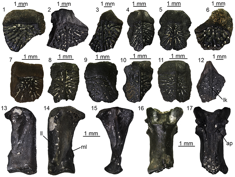
FIGURE 20. Varanus sp. from Maramena: tooth (UU MAA 7029) in labial (1) and lingual (2) views; tooth (UU MAA 7030) in lingual (3) and anterior (4) views; tooth (UU MAA 7148) in labial (5) and lingual (6) views; tooth (UU MAA 7149) in lingual view (7); tooth (UU MAA 7150) in lingual view (8); tooth (UU MAA 7151) in labial (9) and lingual (10) views; caudal vertebra (UU MAA 7028) in posterior (11) and dorsal (12) views; caudal vertebra (UU MAA 7198) in left lateral (13), dorsal (14), and ventral (15) views; caudal vertebra (UU MAA 7278) in anterior (16) and posterior (17) views. Abbreviations: c, cotyle; pe, pedestal; s, striae.
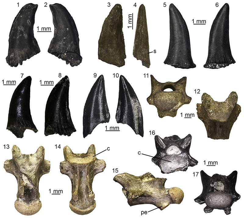
FIGURE 21. Scolecophidia indet. from Maramena: trunk vertebra (UU MAA 7373) in anterior (1), posterior (2), right lateral (3), left lateral (4), ventral (5), and dorsal (6) views; trunk vertebra (UU MAA 7374) in anterior (7), posterior (8), right lateral (9), left lateral (10), ventral (11), and dorsal (12) views; trunk vertrebra (UU MAA 7375) in anterior (13), posterior (14), right lateral (15), left lateral (16), ventral (17), and dorsal (18) views. Abbreviations: cd, condyle; ct, cotyle; nac, neural arch; po, postzygapophysis; prp, prezygapophyseal accessory process; scf, subcentral foramen; zy, zygosphene.
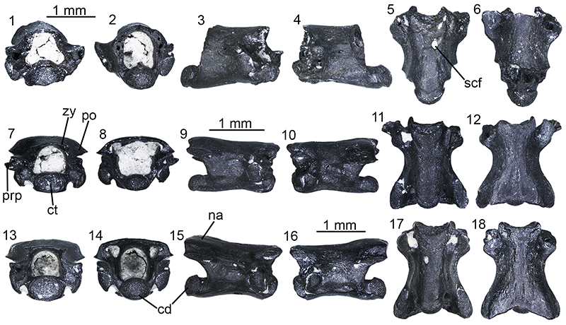
FIGURE 22. Periergophis micros gen. et sp. nov. from Maramena: holotype posterior trunk vertebra (UU MAA 7615) in dorsal (1), right lateral (2), ventral (3), anterior (4), and posterior (5) views. Abbreviations: cd, condyle; hkt, haemal keel tubercles; ns, neural spine; p, parapophysis; pctf, paracotylar foramina; prf, prezygapophyseal articular facet.
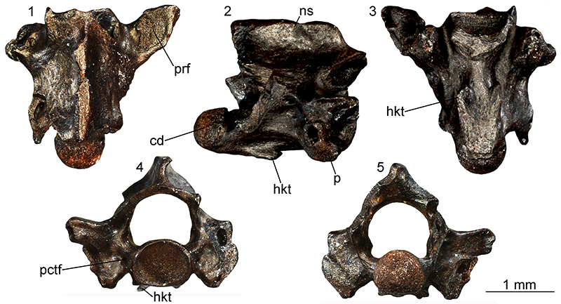
FIGURE 23. Periergophis micros gen. et sp. nov. from Maramena, paratypes: cervical vertebra (UU MAA 7551) in right lateral (1) and ventral (2) views; paratype mid-trunk vertebra (UU MAA 7614) in right lateral (3), dorsal (4), ventral (5), left lateral (6), anterior (7), and posterior (8) views; paratype cloacal vertebra (UU MAA 7618) in left lateral (9), dorsal (10), ventral (11), and anterior (12) views; paratype caudal vertebra (UU MAA 7636) in right lateral (13), dorsal (14), ventral (15), and anterior (16) views. Abbreviations: cd, condyle; ct, cotyle; d, diapophysis; hae, haemapophysis; haet, haemapophyseal tubercles; hy, hypapophysis; lf, lateral foramen; ly,lymphapophysis; na, neural arch; nc, neural canal; ns, neural spine; p, parapophysis; pctf, paracotylar foramen; pl, pleurapophysis; pof, postzygapophyseal articular facet; pr, prezygapophysis; prf, prezygapophyseal articular facet; prp, prezygapophyseal accessory process; scf, subcentral foramen; scg, subcentral groove; scr, subcentral ridge; zg, zygantrum; zy, zygosphene.
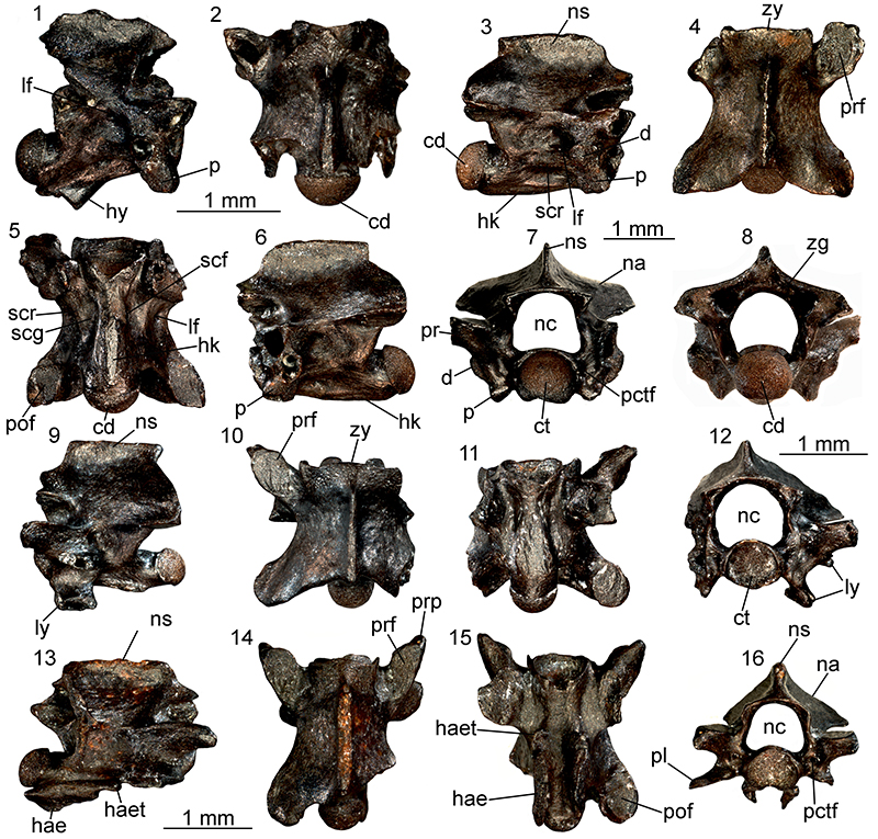
FIGURE 24. Explanatory drawings of the haemal keel tubercles and the haemapophyseal keel tubercles in posterior trunk and caudal vertebrae of Periergophis micros gen. et sp. nov. Holotype posterior trunk vertebra UU MAA 7615 in right lateral (1) and ventral (2) views; paratype caudal vertebra UU MAA 7636 in right lateral view (3).
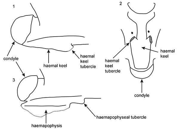
FIGURE 25. Periergophis micros gen. et sp. nov. from Maramena, referred specimens: cervical vertebra (UU MAA 7552) in right lateral (1), dorsal (2), ventral (3), and anterior (4) views;mid-trunk vertebra (UU MAA 7740) in dorsal (5) and ventral (6) views; posterior trunk vertebra (UU MAA 7616) in anterior (7), dorsal (8), and ventral (9) views. Abbreviations: cd, condyle; ct, cotyle; hk, haemal keel; hkt, haemal keel tubercle; hy, hypapophysis; lf, lateral foramen; nc, neural canal; ns, neural spine; p, parapophysis; pctf, paracotylar foramen; po, postzygapophysis; pp, parapophyseal process; pr, prezygapophysis; prf, prezygapophyseal articular facet; scf, subcentral foramen; scr, subcentral ridge; zy, zygosphene.
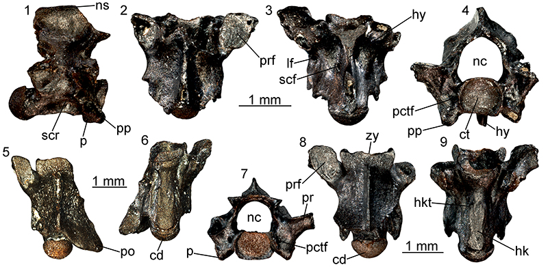
FIGURE 26. Periergophis micros gen. et sp. nov. from Maramena, referred specimens: mid-trunk vertebra (UU MAA 7820) in ventral (1) and anterior (2) views; mid-trunk vertebra (UU MAA 7848) in anterior (3) view; mid-trunk vertebra (UU MAA 7903) in left lateral view (4); mid-trunk vertebra (UU MAA 7890) in ventral view (5); mid-trunk vertebra (UU MAA 7922) in dorsal (6) and ventral (7) views; posterior trunk vertebra (UU MAA 7790) in right lateral (8), anterior (9), and ventral (10) views; posterior trunk vertebra (UU MAA 7780) in anterior (11), right lateral (12), dorsal (13), and ventral (14) views; posterior trunk vertebra (UU MAA 7839) in ventral view (15); posterior trunk vertebra (UU MAA 7894) in ventral view (16); cloacal vertebra (UU MAA 7797) in anterior (17) and ventral (18) views; caudal vertebra (UU MAA 7907) in dorsal view (19); caudal vertebra (UU MAA 7779) in ventral view (20); caudal vertebra (UU MAA 7827) in ventral view (21); caudal vertebra (UU MAA 7921) in left lateral view (22). Abbreviations: cd, condyle; hae, haemapophysis; hk, haemal keel; hkt, haemal keel tubercle; ly, lymphapophysis; ns, neural spine; prf, prezygapophyseal articular facet; prp, prezygapophyseal accessory process.
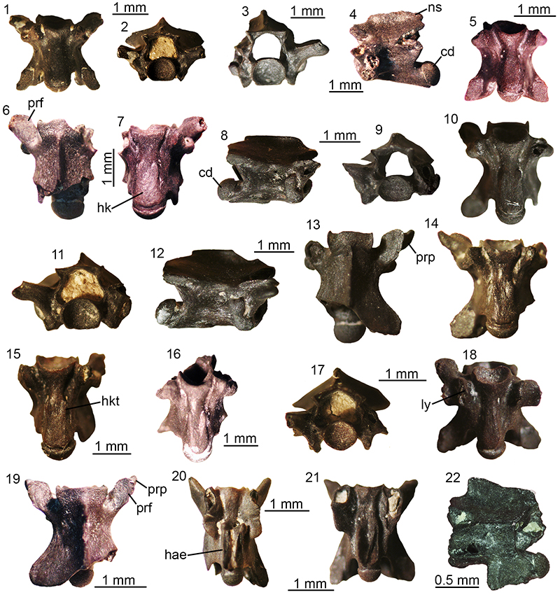
FIGURE 27. Paraxenophis spanios gen. et sp. nov. from Maramena: holotype mid-trunk vertebra (UU MAA 7645) in left lateral (1), dorsal (2), ventral (3), posterior (4), and anterior (5) views. Abbreviations: cd, condyle; ct, cotyle; hk, haemal keel; ir, interzygapophyseal ridge; lf, lateral foramen; lnhk, lateral notch of the haemal keel; na, neural arch; nc, neural canal; pof, postzygapophyseal articular facet; prf, prezygapophyseal articular facet; prp, prezygapophyseal accessory process; scf, subcentral foramen; scg, subcentral groove; scr, subcentral ridge; zg, zygantrum; zy, zygosphene.
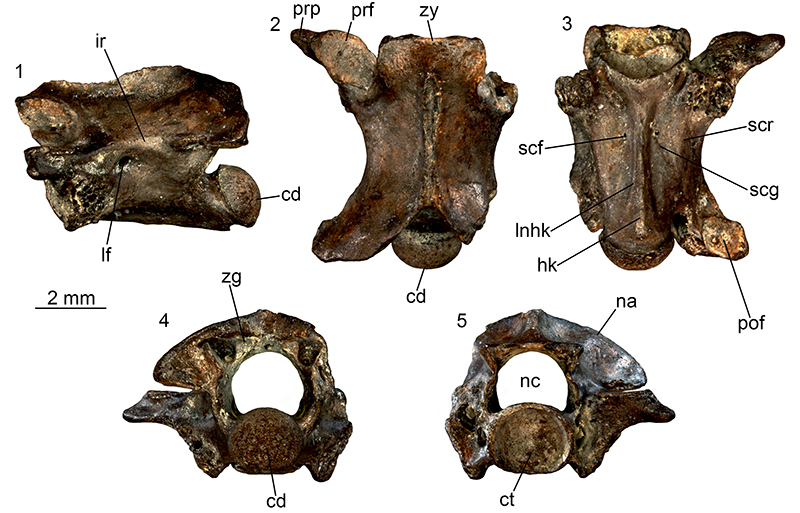
FIGURE 28. Explanatory drawing of a vertical cross section of a trunk vertebra of Paraxenophis spanios gen. et sp. nov., showing the distinctive lateral notches of the haemal keel.
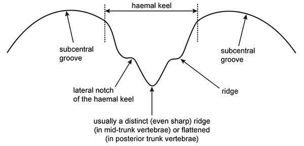
FIGURE 29. Paraxenophis spanios gen. et sp. nov. from Maramena, paratypes: posterior trunk vertebra (UU MAA 7536) in dorsal (1), anterior (2), posterior (3), ventral (4), and right lateral (5) views; posteriormost trunk vertebra (UU MAA 7533) in ventral (6), anterior (7), and left lateral (8) views. Abbreviations: cd, condyle; ct, cotyle; hk, haemal keel; lf, lateral foramen; na, neural arch; ns, neural spine; p, parapophysis; pof, postzygapophyseal articular facet; prf, prezygapophyseal articular facet; zg, zygantrum; zy, zygosphene.
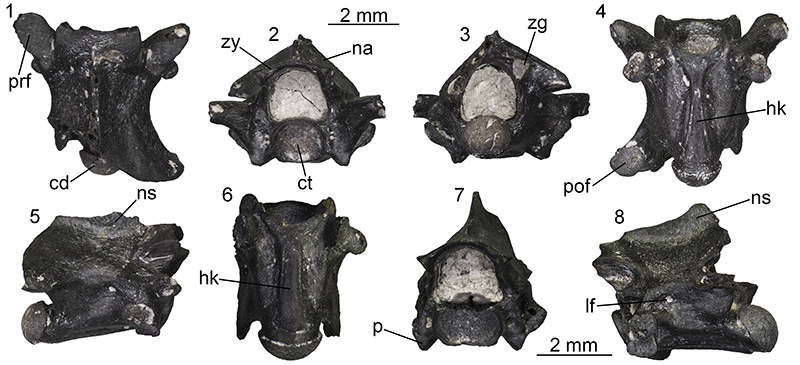
FIGURE 30. Paraxenophis spanios gen. et sp. nov. from Maramena, referred specimens: cervical vertebra (UU MAA 7870) in anterior (1) and ventral (2) views; cervical vertebra (UU MAA 7876) in ventral (3) and anterior (4) views; mid-trunk vertebra (UU MAA 7843) in left lateral view (5); mid-trunk vertebra (UU MAA 7808) in ventral view (6); mid-trunk vertebra (UU MAA 7650) in ventral (7) and right lateral (8) views; mid-trunk vertebra (UU MAA 7868) in ventral view (9); middle to posterior trunk vertebra (UU MAA 7770) in dorsal (10) and ventral (11) views; cloacal vertebra (UU MAA 7858) in left lateral (12) and anterior (13) views; anterior caudal vertebra (UU MAA 7873) in anterior (14), ventral (15), dorsal (16), and right lateral (17) views. Abbreviations: ct, cotyle; hk, haemal keel; hy, hypapophysis; lnhk, lateral notch of the haemal keel; ly, lymphapophysis; na, neural arch; pof, postzygapophyseal articular facet; prp, prezygapophyseal accessory process; zy, zygosphene.
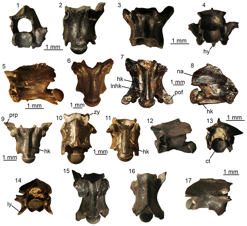
FIGURE 31. Paraxenophis spanios gen. et sp. nov. from Maramena, referred specimens: mid-trunk vertebra (UU MAA 7646) in right lateral (1), dorsal (2), ventral (3), and anterior (4) views; mid-trunk vertebra (UU MAA 7647) in right lateral (5), dorsal (6), ventral (7), anterior (8), and posterior (9) views; mid-trunk vertebra (UU MAA 7649) in dorsal (10), ventral (11), and anterior (12) views. Abbreviations: ct, cotyle; hk, haemal keel; lf, lateral foramen; lnhk, lateral notch of the haemal keel; na, neural arch; nc, neural canal; ns, neural spine; p, parapophysis; pctf, paracotylar foramen; po, postzygapophysis; scf, subcentral foramen; scg, subcentral grooves; scr, subcentral ridge; zg, zygantrum; zy, zygosphene.
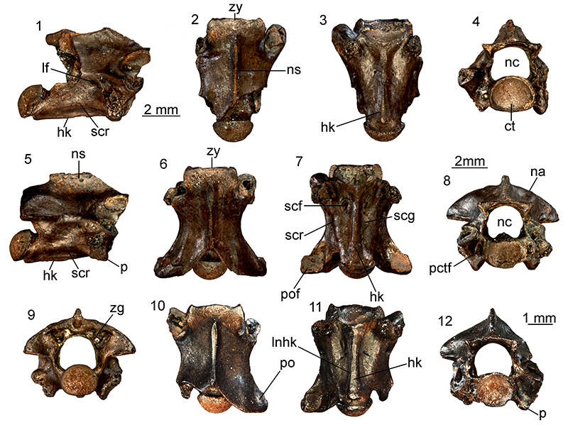
FIGURE 32. ?Paraxenophis spanios, young individual from Maramena: mid-trunk vertebra (UU MAA 7648) in dorsal (1), left lateral (2), ventral (3), anterior (4), and posterior (5)views. Abbreviations: cd, condyle; ct, cotyle; hk, haemal keel; lf, lateral foramen; nc, neural canal; ns, neural spine; scf, subcentral foramen; zg, zygantrum; zy, zygosphene.
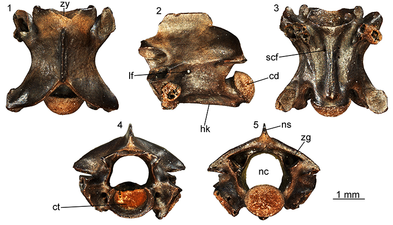
FIGURE 33. “Colubrinae” indet. (morphotype 1) from Maramena: mid-trunk vertebra (UU MAA 7638) in left lateral (1), dorsal (2), ventral (3), right lateral (4), anterior (5), and posterior (6) views. Abbreviations: cd, condyle; d, diapophysis; hk, haemal keel; lf, lateral foramen; na, neural arch; nc, neural canal; p, parapophysis; pctf, paracotylar foramen;pr, prezygapophysis; prf, prezygapophyseal articular facet; r, ridge; scf, subcentral foramen; zg, zygantrum.
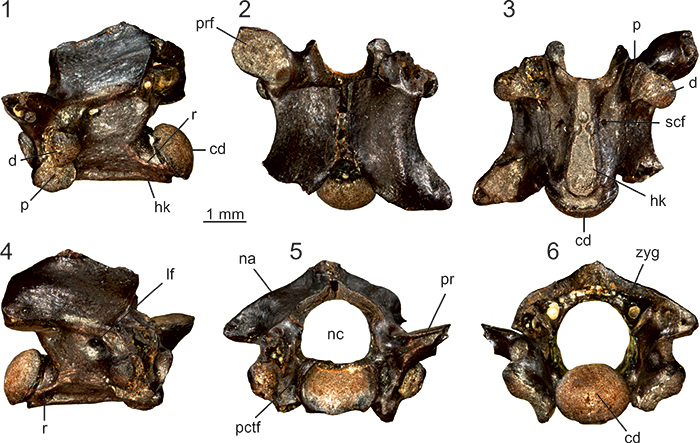
FIGURE 34. Natrix aff. rudabanyaensis from Maramena: anterior precloacal vertebra (UU MAA 7669) in left lateral (1), dorsal (2), ventral (3), anterior (4), and posterior (5) views; precloacal vertebra (UU MAA 7670) in right lateral (6), dorsal (7), ventral (8), anterior (9), and posterior (10) views; posterior trunk vertebra (UU MAA 7667) in dorsal (11) and ventral (12)views; posterior trunk vertebra (UU MAA 7668) in right lateral (13), dorsal (14), ventral (15), and anterior (16) views; caudal vertebra (UU MAA 7694) in right lateral (17), dorsal (18), ventral (19), and anterior (20) views. Abbreviations: cd, condyle; ct, cotyle; d, diapophysis; hy, hypapophysis; ir, interzygapophyseal ridge; lf, lateral foramen; na, neural arch; nc, neural canal; p, parapophysis; pctf, paracotylar foramen; pof, postzygapophyseal articular facet; pp, parapophyseal process; pr, prezygapophysis; prf, prezygapophyseal articular facet; scf, subcentral foramen; scg, subcentral groove; scr, subcentral ridge; sctt, subcotylar tubercles; zy, zygosphene.
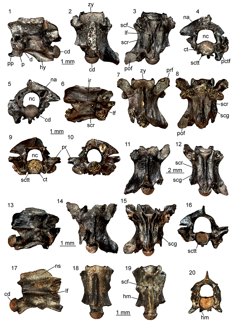
FIGURE 35. Natrix sp. from Maramena: precloacal vertebra (UU MAA 7699) in dorsal (1), left lateral (2), ventral (3), anterior (4), and posterior (5) views;posterior precloacal vertebra (UU MAA 7700) in dorsal (6), right lateral (7), and ventral (8) views. Abbreviations: cd, condyle; ct, cotyle; hy, hypapophysis; nc, neural canal; pctf, paracotylar foramen; po, postzygapophysis; prf, prezygapophyseal articular facet; prp, prezygapophyseal accessory process; scf, subcentral foramen; zg, zygantrum; zy, zygosphene.
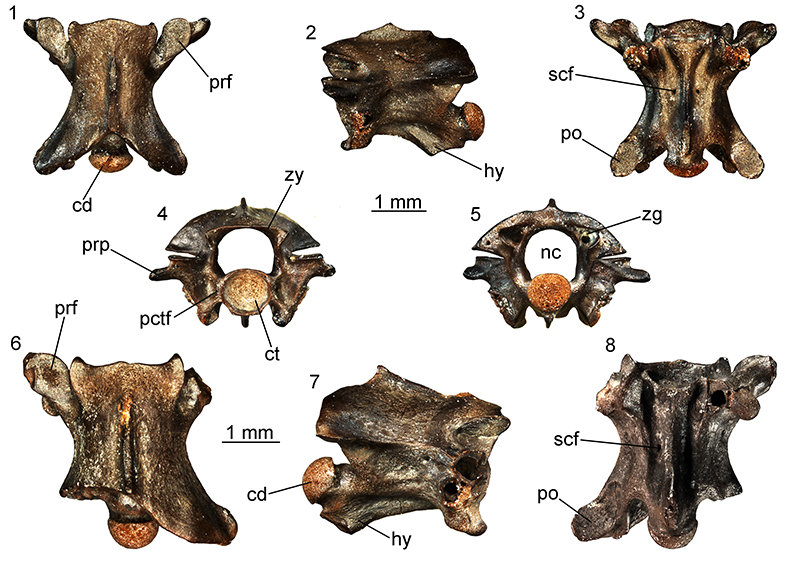
FIGURE 36. Naja sp. from Maramena: mid-trunk vertebra (UU MAA 7531) in anterior (1), posterior (2), right lateral (3), left lateral (4), dorsal (5), and ventral (6) views. Abbreviations: cd, condyle; ct, cotyle; lf, lateral foramen; nc, neural canal; ns, neural spine; prf, prezygapophyseal articular facet; scg, subcentral grooves; zg, zygantrum; zy, zygosphene.
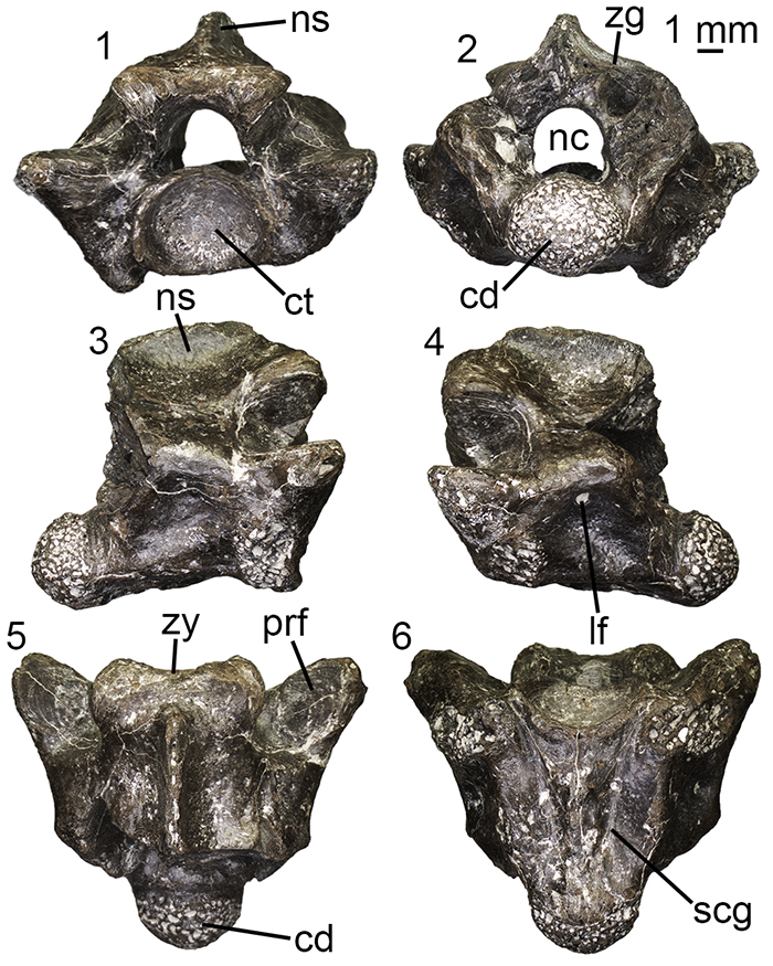
FIGURE 37. cf. Micrurus sp. from Maramena: mid-trunk vertebra (UU MAA 7535) in anterior (1), left lateral (2), dorsal (3), ventral (4), right lateral (5), and posterior (6) views. Abbreviations: cd, condyle; ct, cotyle; hy, hypapophysis; lf, lateral foramen; ns, neural spine; zy, zygosphene.
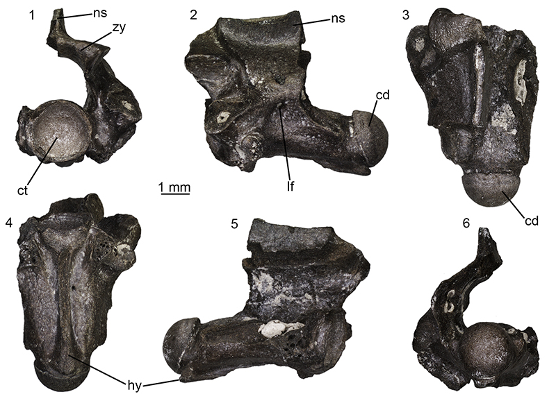
FIGURE 38. “Oriental vipers” complex indet. from Maramena: fang (UU MAA 7115) in anterior (1), posterior (2), right lateral (3), left lateral (4), and dorsal (5) views; fang (UU MAA 7281) in anterior (6), posterior (7), right lateral (8), left lateral (9), and dorsal (10) views.
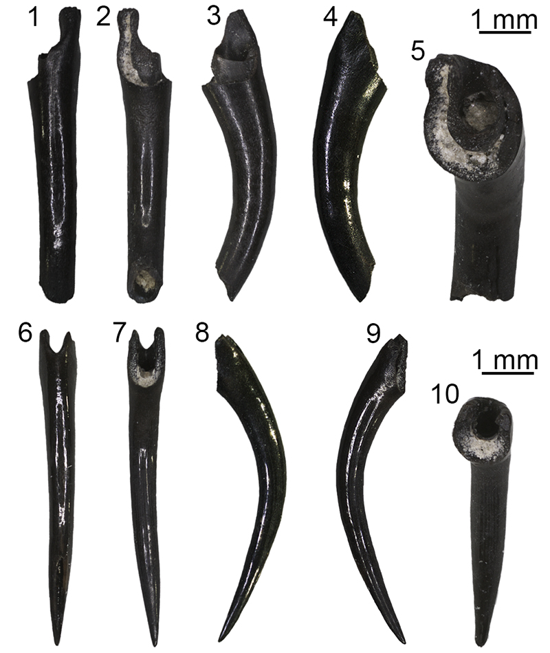
FIGURE 39. Serpentes indet. from Maramena: right dentary (UU MAA 7749) in lingual (1) and labial (2) views.