FIGURE 1. Anatomical, directional, and morphometric terminology used in this study. Figure modified from Hendrickx et al. (2015d). 1, Close up of the enamel surface of crown in 3 in labial view; 2, basal cross-section of crown in 3 in basal view showing CBL (crown-base length) and CBW (crown-base width); 3, idealized lateral theropod tooth in labial view; 4, idealized lateral theropod tooth in distal view; 5, idealized denticles of a denticulated distal carina in labial view; 6, idealized lateral theropod tooth in labial view showing the crown ornamentations; 7, idealized fluted theropod tooth in labial view showing CBL and CH (crown height); 8, U-shaped cross-section with convex lingual margin; 9, U-shaped cross-section with central ridge on the lingual margin; 10, D-shaped cross-section; 11, salinon-shaped cross-section; 12, labiolingually wide J-shaped cross-section; 13, labiolingually narrow J-shaped cross-section. Abbreviations: bst, basal striation; ca, carina; ce, cervix; co, crown; dca, distal carina; de, denticle; ent, enamel texture; flu, flute; ids, interdenticular sulcus; idsp, interdenticular space; lri, longitudinal ridge; mun, marginal undulation; mca, mesial carina; ro, root; tun, transverse undulation.
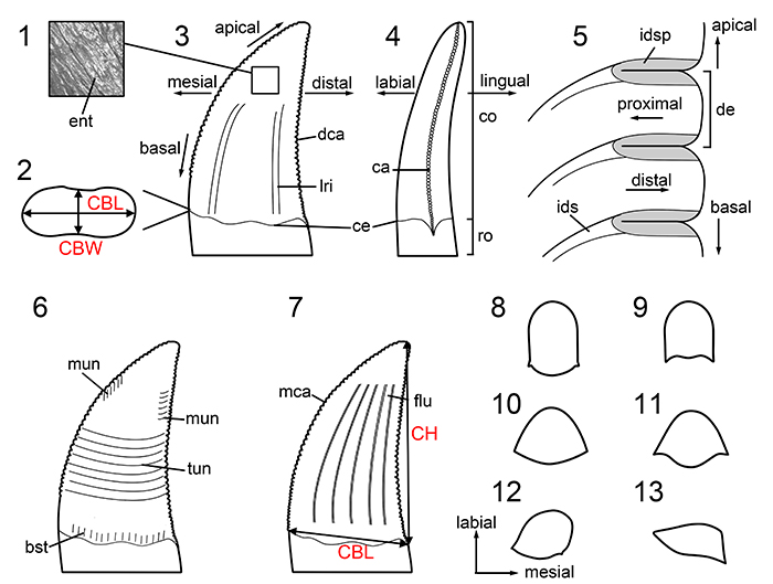
FIGURE 2. Phylogeny and stratigraphic distribution of theropod dinosaurs. Figure modified from Hendrickx et al. (2015b; see references therein). The theropod silhouettes are from Conty (Eodromaeus), the Smithsonian Institution (Daemonosaurus; modified), Julio Garza (Dilophosauridae), Gregory S. Paul (Metriacanthosauridae), T. Michael Keesey (Deinocheiridae), Funkmonk (Alvarezsauroidea and Therizinosauria), Jaime Headden (Caenagnathidae), and Scott Hartman (all other silhouettes). All silhouettes other than those from the Smithsonian Institution and Gregory S. Paul were downloaded from phylopic.org and are under a Creative Commons Attribution-NonCommercial-ShareAlike 3.0 Unported License unless stated otherwise (see Appendix 1.2).
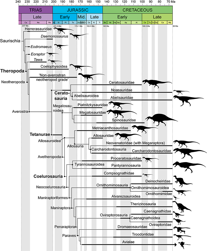
FIGURE 3. Dentition-based synapomorphies in non-coelurosaur Saurischia. Dentition-based synapomorphies on a tree following the topology obtained by Müller et al. (2018) for non-averostran Saurischia, Rauhut and Carrano (2016) and Wang et al. (2017a) for Ceratosauria, and Rauhut et al. (2016) for non-coelurosaurian Tetanurae, Brusatte and Carr (2016) for Tyrannosauroidea. The list of dental synapomorphies for each clade is provided in Table 1. Abbreviations: CBR, Crown Base Ratio; d, number of dentary teeth; de, number of denticles; DSDI, denticle size density index; m, number of maxillary teeth; pm, number of premaxillary teeth. For silhouette acknowledgements see Appendix 1.2.
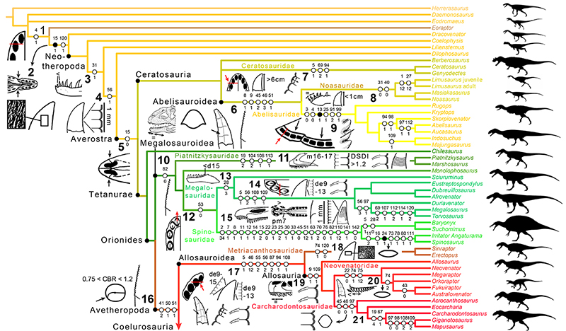
FIGURE 4. Dentition-based synapomorphies in Coelurosauria. Dentition-based synapomorphies on a tree following the topology obtained by Cau et al. (2017) based on the dataset of Brusatte et al. (2014) for non-tyrannosauroid Coelurosauria. The list of dental synapomorphies for each clade is provided in Table 1. Abbreviations: CHR, Crown Height Ratio; d, number of dentary teeth; dc, distal carina; DSDI, denticle size density index; lad, labial depression; m, number of maxillary teeth; mc, mesial carina; pm, number of premaxillary teeth. For silhouette acknowledgements see Appendix 1.2.
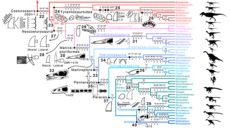
FIGURE 5. CI and RI scores obtained in eight supermatrices separated into skeleton-based, non-dental based and dental-based data matrices, and CI and RI values for each dentition sub-unit. 1, CI and RI scores in eight supermatrices combining our dentition-based data matrix to eight of the most recent datasets on non-avian theropods, ceratosaurs, non-coelurosaur tetanurans and non-avian coelurosaurs, when dental characters are included (whole skeleton), excluded (non-dental), and considered separately (dental); 2, CI and RI values measured for each dentition sub-unit in eight trees of the theropod classification (see Appendix 4.2, Table A5). Abbreviations: dent, dentition.
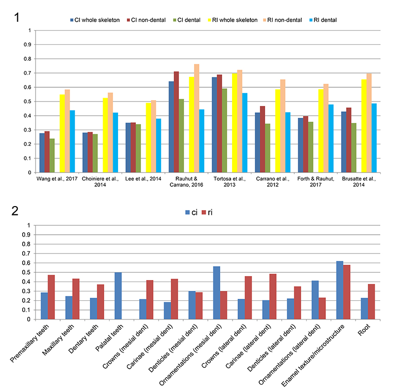
FIGURE 6. Dental variation along the tooth row in non-avian Theropoda. 1, First four right maxillary teeth of the carcharodontosaurid Acrocanthosaurus atokensis (NCSM 14345) in labial view, showing both the presence of marginal and transverse undulations on the third and fourth maxillary teeth, respectively, and a distal carina strongly displaced labially in the first maxillary tooth; 2, First six left maxillary teeth of the tyrannosaurid Alioramus altai (MPC-D 100-1844) in lingual view, showing the strong labial displacement of the distal carina in the three first maxillary teeth, a longitudinal groove, a lingual depression and transverse undulations on the first, third and sixth maxillary teeth, respectively, and the variable basal extension (represented by the horizontal red line) of the mesial carina along the maxillary tooth row; 3, Mesial left maxillary teeth of the troodontid Byronosaurus jaffei (MPC-D 100-983) in labial view, showing the presence and absence of a basal constriction in the mesial and distal maxillary teeth, respectively, a single and two longitudinal ridges on the labial surface of the second and third ziphodont maxillary teeth, respectively, and a labial depression on the third ziphodont maxillary tooth. Abbreviations: bac, basal constriction; dca, distal carina; lad, labial depression; lid, lingual depression; lgr, longitudinal groove; lri, longitudinal ridge; mca, mesial carina; mun, marginal undulation; tun, transverse undulation. Scale bars = 5 cm (1), 3 cm (2), 5 mm (3).
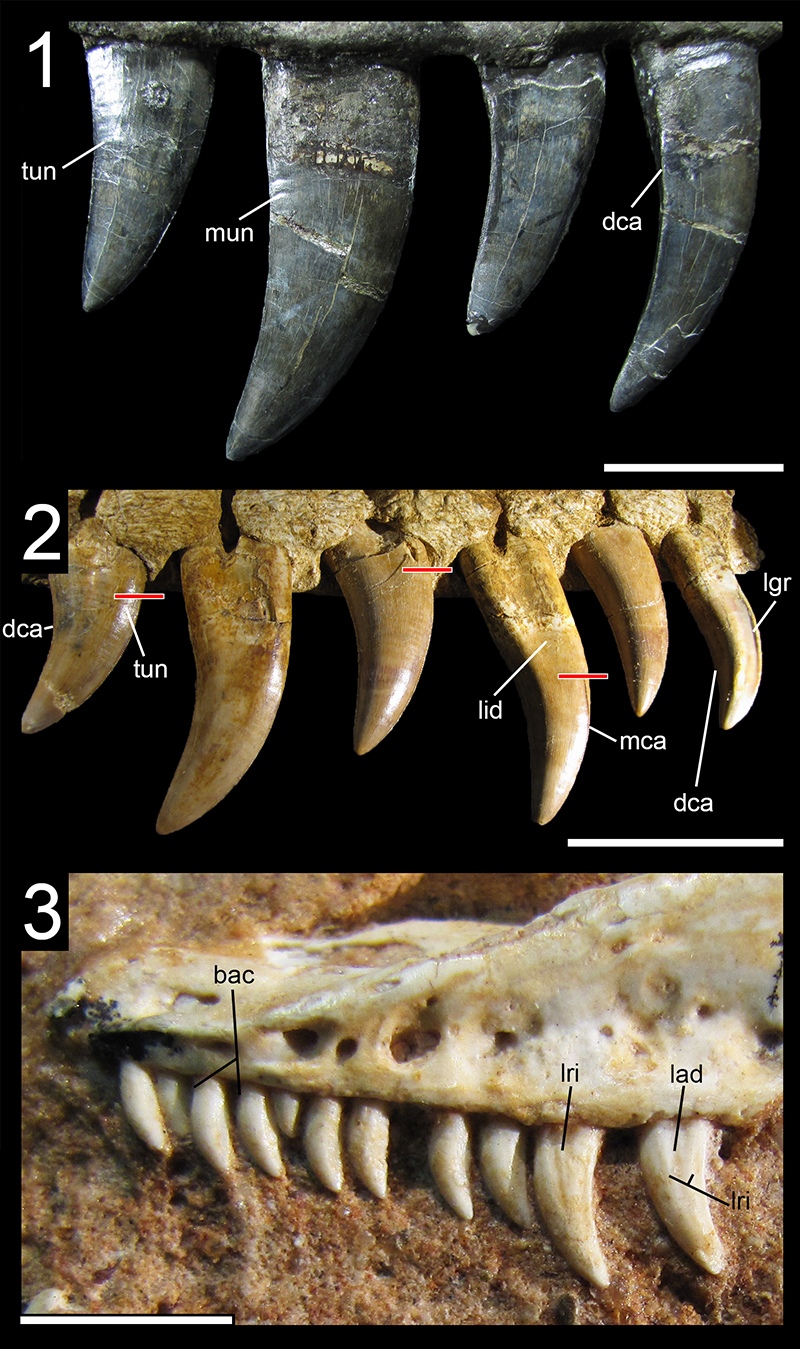
FIGURE 7. Distribution of dental features in non-neocoelurosaur Theropoda. Phylogenetic tree based on the results obtained by Langer et al. (2017), for non-neotheropod Saurischia, Ezcurra (2017) and Wang et al. (2017a) for non-averostran Neotheropoda, Rauhut and Carrano (2016) and Wang et al. (2017a) for Ceratosauria, Carrano et al. (2012) and Rauhut et al. (2016) for non-coelurosaurian Tetanurae, Brusatte and Carr (2016) for Tyrannosauroidea, and Novas et al. (2012), Rauhut et al. (2012) and Choiniere et al. (2014a) for the phylogenetic distribution of Bicentenaria, Sciurumimus and Zuolong, respectively. The branch colors represent the dentition types and the presence or absence of constricted crowns: ziphodont taxa with unconstricted crowns are in red, ziphodont taxa with a few constricted crowns are in green, conidont taxa are in turquoise, and pachydont taxa are in violet. The colors of taxa represent the presence or absence of serrations on the mesial and distal carinae for both mesial (left column) and lateral dentition (right column): toothless taxa are in grey, taxa with unserrated crowns are in green, taxa with a denticulated distal carina and a denticulated mesial carina not reaching the cervix are in red, taxa with a denticulated distal carina and a denticulated mesial carina reaching the cervix are in blue, and taxa with a denticulated distal carina and an unserrated mesial carina are in yellow. Taxa whose dentition is not known are on a white background.Taxa with distal denticles larger than mesial ones are boxed in green. Some compsognathid taxa possess a double condition in their mesial and lateral dentition: Juravenator bears mesial crowns with denticulated and unserrated distal carina, Compsognathus shows lateral crowns with unserrated and denticulated distal carina, and Sinocalliopteryx possesses denticulated and unserrated mesial carinae in the lateral teeth. Abbreviations: 8, figure-8-shaped cross-section of lateral teeth; D, D-shaped cross-section of mesial teeth; J, J-shaped cross-section of mesial teeth; O, subcircular/lanceolate cross-section of mesial teeth; S, Salinon-shaped cross-section of mesial teeth; U, U-shaped cross-section of mesial teeth.
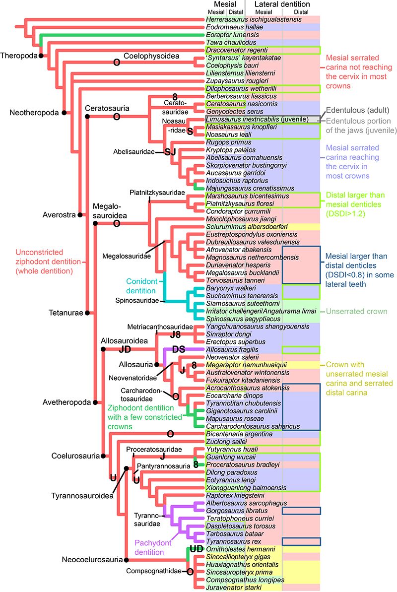
FIGURE 8. Distribution of dental features in Neocoelurosauria. Phylogenetic tree of Cau et al. (2017) based on the dataset of Brusatte et al. (2014) for Neocoelurosauria, with changes brought by Lamanna et al. (2014) for Caenagnathoidea, and Xu et al. (2018) and Pu et al. (2013) for the phylogenetic position of Aorun and Jianchangosaurus, respectively. The branch colors represent the dentition types: ziphodont taxa with unconstricted crowns are in red, ziphodont taxa with a few constricted crowns are in green, taxa with both folidont and ziphodont lateral dentition are in orange, folidont taxa with unconstricted mesial crowns are in pink, folidont taxa with constricted crowns only are in blue, and conidont taxa are in turquoise. Colors of taxa represent the presence or absence of serrations on the mesial and distal carinae for both mesial (left) and lateral dentition (right): toothless taxa are in grey, taxa with unserrated crowns are in green, taxa with a denticulated distal carina and an unserrated mesial carina are in yellow, taxa with denticulated mesial and distal carinae are in red, and taxa with both denticulated mesial and distal carinae not reaching the cervix are in blue. Taxa (Sinocalliopteryx, Compsognathus and Juravenator) showing both conditions (e.g., mesial dentition with unserrated teeth and lateral dentition with denticulated teeth) are bicolored. Taxa whose dentition is not known are on a white background. Some paravians such as Troodon, Velociraptor and Saurornitholestes possess a lateral dentition with denticulated and unserrated carinae. Taxa with distal denticles larger than mesial ones are boxed in green, and taxa with large typically hooked denticles are boxed in purple. Abbreviations: 8, figure-8-shaped cross-section of lateral teeth; D, D-shaped cross-section of mesial teeth; J, J-shaped cross-section of mesial teeth; O, subcircular/lanceolate cross-section of mesial (black ‘O’) and lateral (blue ‘O’) teeth; U, U-shaped cross-section of mesial teeth.
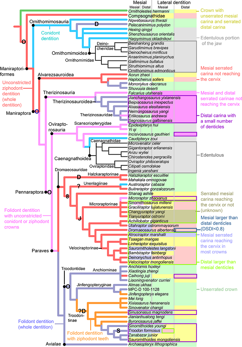
FIGURE 9. Distribution of dental features in non-avian Theropoda. Phylogenetic tree based on Hendrickx et al. (2015b) and Cau et al. (2017), with the exclusion of megaraptorans here placed among Neovenatoridae (Benson et al., 2010; Carrano et al., 2012). Letters between brackets represent polymorphic features. Clade numbers: 1, Neotheropoda; 2, Averostra; 3, Ceratosauria; 4, Tetanurae; 5, Megalosauroidea; 6, Spinosauridae; 7, Avetheropoda; 8, Allosauroidea; 9, Coelurosauria; 10, Tyrannosauroidea; 11, Neocoelurosauria; 12, Maniraptoriformes; 13, Maniraptora; 14, Paraves; 15, Dromaeosauridae; 16, Troodontidae; 17, Avialae. Abbreviations: 0, absent; 1, present at least in some teeth or some taxa; 8, figure-8-shaped cross-section at the cervix; ?, unknown; -, inapplicable; ~, medium-sized denticles (i.e., between 15 and 250 denticles on the carina); ≠, difference between mesial and distal denticles; <, distal denticles significantly larger than mesial ones (DSDI > 1.2); <<, minute denticles (more than 250 denticles on the carina); >, mesial denticles significantly larger than distal ones (DSDI < 0.9); >>, large denticles (i.e., fewer than 15 denticles on the carina); A, anastomosed texture; B, braided texture; bco, basal constriction at the cervix; bst, basal striations; C, conidonty (dentition with conical crowns); CBR, crown base ratio; CH, crown height in the largest teeth, in centimeters; codm, convex distal margin; cos, concave surface adjacent to carinae; D, D-shaped cross-section; ddca, displaced distal carina; dd, distal denticles; den, dentition type; des, denticle size; dt, present in the dentary; ent, enamel texture; edj, edentulous jaw; F, folidonty (dentition with lanceolate crowns); flu, fluted teeth; hd, hooked denticles; I, smooth or irregular non-oriented texture; ids, interdenticular sulci; J, J-shaped cross-section; L, present in all lateral teeth and, for the edentulism, edentulous posterior portion of the maxilla and/or dentary; la, present in some lateral teeth (e.g., la>, mesial denticles significantly larger than distal ones in some lateral teeth; la-lri, longitudinal ridge present in some lateral teeth); Lc, presence of laterocumbent teeth; lgr, longitudinal groove; lri, longitudinal ridges; M, present in all mesial teeth and, for the edentulism, edentulous premaxilla and anterior portion of the dentary; m, present in some mesial teeth (e.g., m<, distal denticles significantly larger than mesial ones in some mesial teeth; m-lgr, longitudinal groove present in some mesial teeth); Mcs, mesial teeth, cross-sectional outline at the cervix; md, mesial denticles; mde, mesial denticles reaching the cervix; mun, marginal undulations; mx, present in the maxilla; O, subcircular/lanceolate cross-section; P, pachydonty (dentition with particularly thick blade-shaped crowns); Pc, presence of procumbent teeth; PcRcLc, procumbent, retrocumbent and laterocumbent dentition; S, Salinon-shaped cross-section; pm, present in the premaxilla; Rc, presence of retrocumbent teeth; tmca, twisted mesial carina; tun, transverse undulations; U, U-shaped cross-section; udca, unserrated distal carina; umca, unserrated mesial carina; V, veined texture; W, present in the whole dentition and, for the edentulism, fully edentulous jaws; w, present in some mesial and lateral teeth; Z, ziphodonty (dentition with blade-shaped crowns). *, this applies to derived therizinosaurs and troodontids given that the basal members of these clades either have no serrations at all (e.g., Anchiornis) or minute denticles (e.g., Falcarius, Sinovenator). As for Oviraptorosauria, this applies to the lateral dentition of Incisivosaurus given that caudipterids have unserrated teeth; **, the dentition of basal tyrannosauroids have a DSDI > 1.2 whereas some tyrannosaurid teeth have a DSDI < 0.9. Images of unserrated tooth (udca) and unserrated mesial carina (umca) by Jaime Headden, denticle size (des) and hooked denticles (hd) from Currie et al. (1990, modified), and mesial denticles smaller than distal ones (md≠dd) from Ostrom (1969; modified).
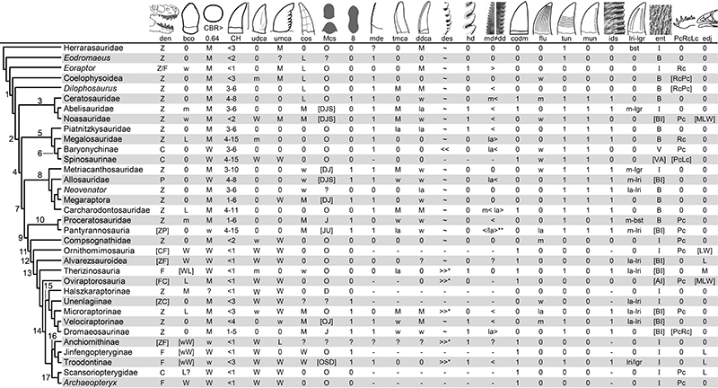
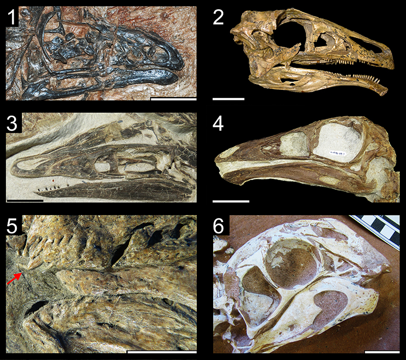
FIGURE 10. Basal constriction in non-avian Theropoda. 1, Isolated lateral tooth of the noasaurid Masiakasaurus knopfleri (FMNH PR 2476) in labial view; 2, Isolated tooth of the troodontid Troodon formosus (DMNH 22837) in labial view; 3, Thirteenth left dentary tooth of the therizinosaur Eshanosaurus deguchiianus (IVPP V11579) in lingual view; 4, Fourth right premaxillary tooth of the proceratosaurid Proceratosaurus bradleyi (NHMUK PV R.4860) in labial view. Scale bars equal 2 mm (1), 3 mm (3), 5 mm (2, 4).
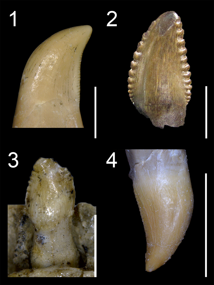
FIGURE 11. Unserrated teeth in non-avian Theropoda. 1, Second right premaxillary tooth of the coelophysoid Coelophysis bauri (DMNS 30596) in labial view; 2, First and second left premaxillary teeth of the megalosaurid Sciurumimus albersdoerferi (BMMS BK 11; courtesy of H. Tischlinger and O. Rauhut) in labial view; 3, Penultimate? right maxillary tooth of the spinosaurid Irritator challengeri (SMNS 58022) in labial view; 4, Third? right premaxillary tooth of the pantyrannosaurian Xiongguanlong baimoensis (FRDC-GS JB16-2-1; courtesy of P. Makovicky) in mesioapical view, showing the central ridge on the lingual surface of the crown; 5, Left dentary tooth of the compsognathid Compsognathus longipes (MNHN CNJ79) in lingual view; 6, Left maxillary tooth of the basal ornithomimosaur Nqwebasaurus thwazi (AM 6040) in labial view; 7, Right maxillary teeth of the alvarezsaurid Shuvuuia deserti (MPC-D 100-977) in labial view; 8, Second right? premaxillary tooth of the basal oviraptorosaur Caudipteryx zoui (IVPP V12430) in labial view; 9, Second left dentary tooth of the dromaeosaurid Buitreraptor gonzalezorum (MPCA 245) in labial view; 10, Right maxillary tooth of Almas ukhaa (MPC-D 100-1323) in labial view. Abbreviation: lri, longitudinal ridge. Scale bars equal 1 mm (1‒2, 5‒10), 5 mm (4), 1 cm (3).
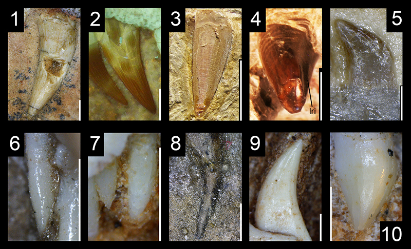
FIGURE 12. Concave surface adjacent to a carina in non-avian Theropoda. 1, Third left maxillary tooth of the dilophosaurid Dilophosaurus wetherilli (UCMP 37303) in lingual view; 2, Isolated tooth of the megalosaurid Afrovenator abakensis (MNN UBA1) in labial view; 3, Isolated tooth of the neovenatorid Neovenator salerii (MIWG 6348) in labial view; 4, Fifth left maxillary tooth of the metriacanthosaurid Sinraptor dongi (IVPP 10600) in labial view, also showing transverse undulations (courtesy of R. Benson). Abbreviation: tun, transverse undulation. Scale bars equal 1 cm.
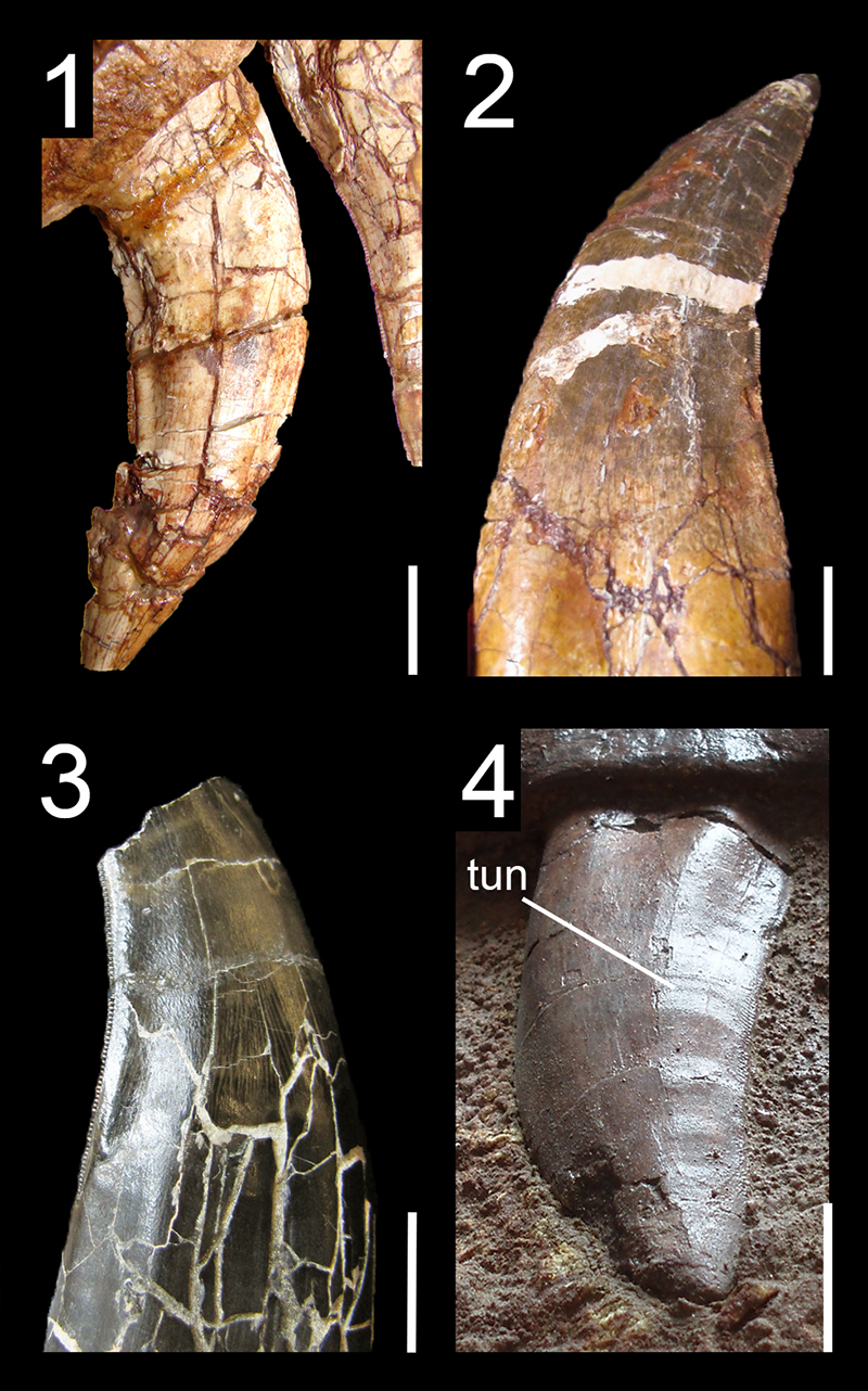
FIGURE 13. Cross-section of mesial teeth in non-avian Theropoda. 1, Salinon-shaped cross-section in the first right premaxillary tooth of the abelisaurid Majungasaurus crenatissimus (FMNH PR.2008) in apical view; 2, U-shaped cross-section in an isolated premaxillary tooth of the basal pantyrannosaurian Eotyrannus lengi (MIWG 1997.550; reversed) in apical view; 3, D-shaped cross-section in the first left premaxillary tooth of the allosaurid Allosaurus fragilis (UMNH VP 9258; reversed) in apical view; 4, J-shaped cross-section in the second right premaxillary tooth of the dromaeosaurid Atrociraptor marshalli (TMP 1995.166.01) in apical view. Scale bars equal 5 mm.
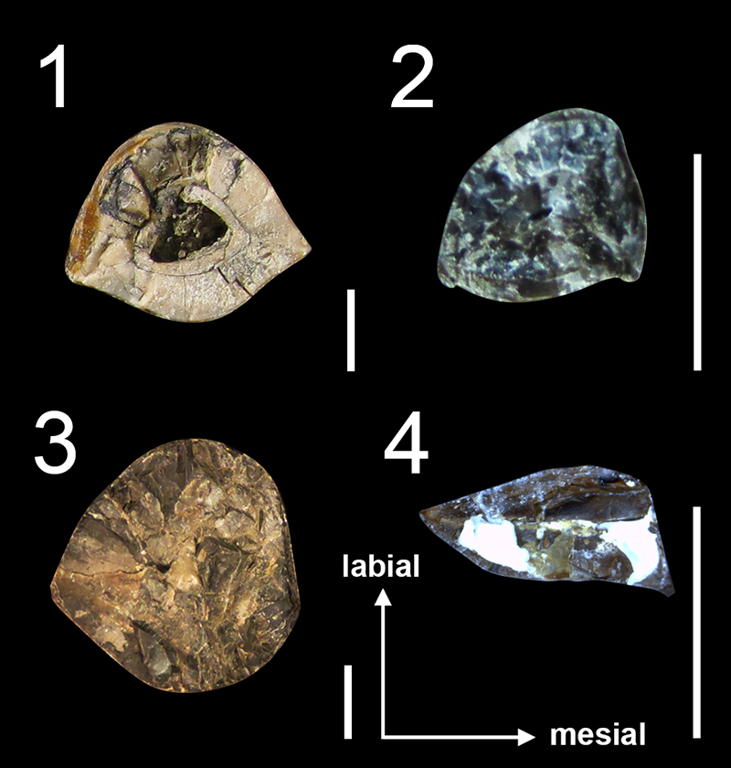
FIGURE 14. Hooked denticles in non-avian Saurischia. 1, Distal denticles of the third right premaxillary tooth of the basal sauropodomorph Eoraptor lunensis (PVSJ 512; image upside down) in labial view; 2, Distal denticles of an isolated tooth of the noasaurid Masiakasaurus knopfleri (FMNH PR.2696) in lingual view; 3, Distal carina of the second right premaxillary tooth of the abelisaurid Majungasaurus crenatissimus (FMNH PR 2008; image upside down) in lateral view; 4, Distal denticles of an isolated lateral tooth of the piatnitzkysaurid Piatnitzkysaurus floresi (PVL 4073) in lateral view; 5, Distal denticles of the sixth left dentary tooth of the therizinosaur Eshanosaurus deguchiianus (IVPP V11579) in lingual view; 6, Mesial denticles of the fifth? right dentary tooth of the therizinosaur Alxasaurus elesitaiensis (IVPP V88402; reversed) in lingual view; 7, Distal denticles of the first right maxillary tooth of the dromaeosaurid Atrociraptor marshalli (TMP 1995.166.01) in labial view; 8, Distal denticles of an isolated tooth of the troodontid Troodon formosus (DMNH 22337) in lateral view. Scale bars equal 1 mm.
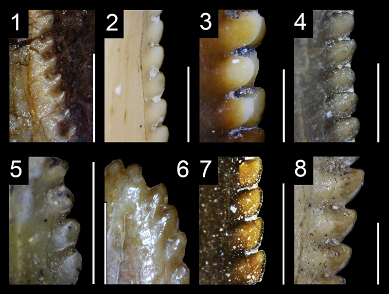
FIGURE 15. Denticles and carinae in Spinosauridae. 1, Carina and denticles of an isolated tooth of Baryonyx walkeri (NHMUK PV R.9951 R.278) in lateral view with details on the basally curving and veined enamel surface texture; 2, Carina and denticles of an isolated tooth of Suchomimus tenerensis (MNN G73-3) in lateral view; 3, Maxillary tooth of Irritator challengeri (SMNS 58022) in labial view showing the marginal undulations and the ‘beaded’ carina; 4, Isolated tooth of Spinosaurus cf. aegyptiacus (MSNM V6422) in lateral view, showing the ‘beaded’ carina and the anastomosed enamel surface texture. Abbreviations: mun, marginal undulation. Scale bars equal 5 mm (3), 1 mm (1‒2, 4).
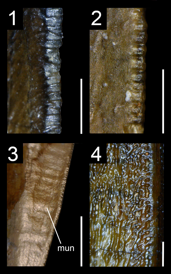
FIGURE 16. Bilobate denticles in non-avian Theropoda. 1, Mesial carina of an isolated crown of the abelisaurid Aucasaurus garridoi (MCF-PVPH 236) in lateral view; 2, Mesial carina of an isolated tooth of the megalosaurid Megalosaurus bucklandii (NHMUK PV R.234; tooth in matrix) in lateral view; 3, Mesial carina of the third left maxillary tooth of the putative metriacanthosaurid Erectopus superbus (MNHN 2001‒4) in labial view; 4, Mesial carina of the tenth maxillary tooth of the tyrannosaurid Tyrannosaurus rex (FMNH PR.2081) in labial view. Scale bars equal 1 mm.
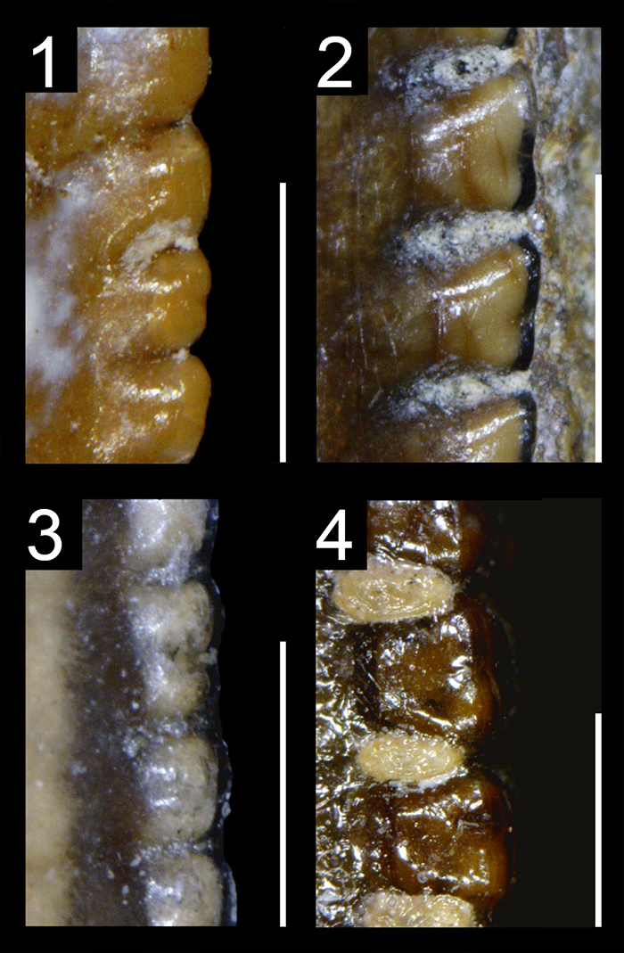
FIGURE 17. Distal larger than mesial denticles in non-avian Theropoda. 1, Isolated lateral crown of the noasaurid Masiakasaurus knopfleri (FMNH PF 2221) in labial view; 2, Fifth right dentary crown of the piatnitzkysaurid Marshosaurus bicentesimus (UMNH 6368) in lingual view; 3, First left maxillary crown of the basal pantyrannosaurian Eotyrannus lengi (MIWG 1997.550) in lingual view; 4, Distal dentary crown of the dromaeosaurid Deinonychus anthirrhopus (YPM 5232 612) in lingual view. Scale bars equal 1 mm.
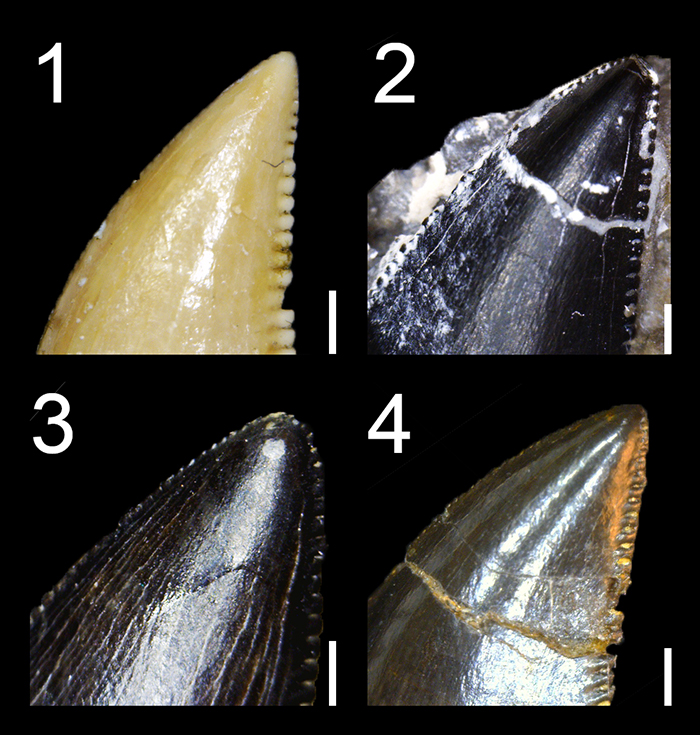
FIGURE 18. Fluted teeth in non-avian Theropoda. 1, Fifth? left maxillary tooth of the basal theropod Tawa hallae (GR 241) in lingual view (courtesy of Sterling Nesbitt); 2, Fourth? left maxillary tooth of the non-averostran neotheropod Sinosaurus triassicus (IVPP V34) in labial view, showing the transverse undulations at the base of the crown; 3, First left and right premaxillary teeth of the ceratosaurid Ceratosaurus nasicornis (UMNH VP 5278) in lingual view; 4, Third right dentary tooth of the noasaurid Masiakasaurus knopfleri (UA 8680) in lingual view; 5, Isolated tooth of the baryonychine Suchosaurus cultridens (= Baryonyx walkeri ?; NHMUK PV R.36536) in lingual view; 6, Second right maxillary tooth of the basal pantyrannosaurian Dilong paradoxus (IVPP V14242) in labial view (n.b., an apparent labial concavity delimited by two faint longitudinal ridges results from a lightning effect); 7, Fifth left premaxillary tooth of the compsognathid Scipionyx samniticus (SBA-SA 163760) in lingual view (reprinted with permission from Dal Sasso and Maganuco (2011), courtesy of Cristiano Dal Sasso & Simone Maganuco, © Soprintendenza Archeologia, Belle Arti e Paesaggio per le Province di Caserta e Benevento); 8, Seventh or eight? right maxillary tooth of the dromaeosaurid Microraptor sp. (IVPP 2008.5) in labial view; 9, Fourth left maxillary tooth of the dromaeosaurid Sinornithosaurus millenii (IVPP V12811) in labial view; 10, First right premaxillary tooth of the dromaeosaurid Velociraptor mongoliensis (AMNH 6515) in labial view. Abbreviation: tun, transverse undulation. Scale bars equal 1 cm (1‒2, 4), 1 mm (3, 5‒8).
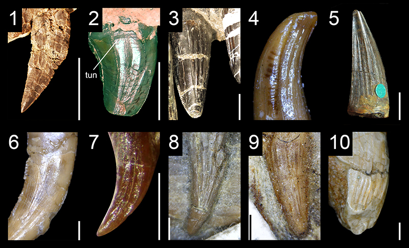
FIGURE 19. Transverse undulations in the teeth of non-avian Saurischia. 1, Fifth? left maxillary tooth of the herrerasaurid Sanjuansaurus gordilloi (PVSJ 605) in labial view; 2,Fifth right dentary tooth of the ceratosaurid Genyodectes serus (MLP 26-39) in basolabial view; 3, Isolated lateral tooth of the noasaurid Masiakasaurus knopfleri (FMNH PR 2221) in labial view; 4, Isolated lateral tooth of the abelisaurid Aucasaurus garridoi (MCF-PVPH-236) in labial? view; 5, Fourth right premaxillary tooth of the megalosauroid Monolophosaurus jiangi (IVPP 84019) in labial view; 6, Isolated lateral tooth of the piatnitzkysaurid Marshosaurus bicentesimus (DMNS 3718) in lingual view; 7, Isolated lateral tooth of the carcharodontosaurid Giganotosaurus carolinii (MUCPv-CH-1) in labial view; 8, Fourteenth left maxillary tooth of the therizinosaur Falcarius utahensis (UMNH VP 14545) in labial view; 9, Fifth right maxillary tooth of the dromaeosaurid Atrociraptor marshalli (TMP 1995.166.01) in labial view; 10, Isolated tooth of the troodontid Troodon formosus (DMNH 22337) in labiodistal view. Scale bars = 1 cm (1‒2, 4‒7), 5 mm (9‒10), 1 mm (3, 8).
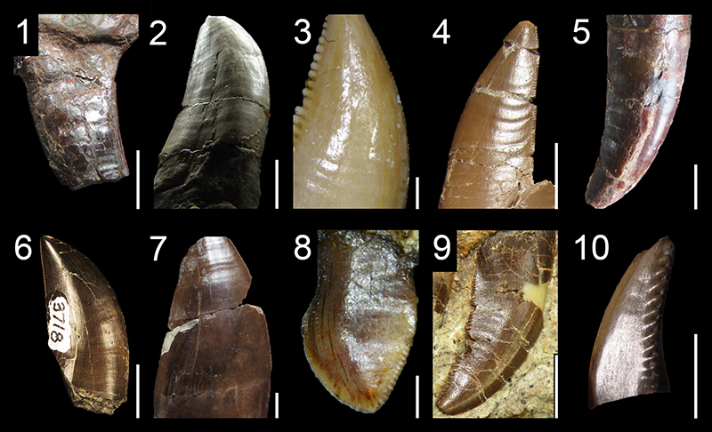
FIGURE 20. Marginal undulations in the teeth of non-avian Theropoda. 1, Fifth left maxillary tooth of the ceratosaurid Ceratosaurus nasicornis (UMNH VP 5278; reversed) in lingual view; 2, Second left maxillary tooth of the noasaurid Masiakasaurus knopfleri (FMNH PR.2696) in labiodistal view; 3, Second left maxillary tooth of the abelisaurid Majungasaurus crenatissimus (FMNH PR 2100) in basolingual view; 4,Second right maxillary crown of the megalosauroid Monolophosaurus jiangi (IVPP 84019) in labial view; 5, Isolated lateral tooth of the piatnitzkysaurid Piatnitzkysaurus floresi (MACN 895) in linguodistal view; 6, Isolated lateral tooth of the spinosaurid Suchomimus tenerensis (MNN G51) in labiodistal? view; 7, Sixth left maxillary tooth of the allosaurid Allosaurus sp. (UMNH VP 9168) in labial view and showing both the marginal and transverse undulations on the crown; 8, Distalmost isolated tooth of the carcharodontosaurid Mapusaurus roseae (MCF-PVPH-108.103) in basolabial view; 9, Isolated mesial tooth of the basal pantyrannosaurian Aviatyrannis jurassica (MG27801 D90) in labial view; 10, Isolated lateral tooth of the pantyrannosaurian Teratophoneus curriei (UMNH VP 16690) in basolabial view, showing both the marginal and transverse undulations. Abbreviation: tun, transverse undulation. Scale bars equal 1 cm (1, 3‒8), 5 mm (10), 1 mm (2, 9).
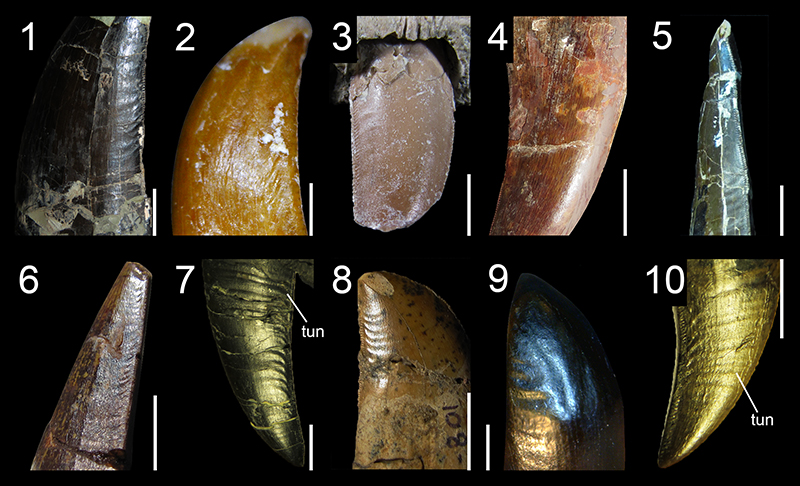
FIGURE 21. Well-developed interdenticular sulci in non-avian Theropoda. 1, Distal carina of the eighth left maxillary tooth of the ceratosaurid Ceratosaurus nasicornis (UMNH VP 5278; image upside down) in lateral view; 2, Distal carina of the sixth right maxillary tooth of the abelisaurid Majungasaurus crenatissimus (FMNH PR.2278; image upside down) in lateral view; 3, Distal carina of an isolated lateral tooth of the piatnitzkysaurid Piatnitzkysaurus floresi (PVL 4073) in lateral view; 4, Distal carina of an isolated lateral tooth of the megalosaurid Megalosaurus bucklandi (NHMUK PV R.234) in labial view; 5, Distal carina of an isolated lateral tooth of the metriacanthosaurid Sinraptor dongi (IVPP V10600) in lateral view; 6, Distal carina of an isolated tooth of the allosaurid Allosaurus sp. (UMNH VP 6177) in lateral view, also showing the transverse undulations at the crown base; 7, Distal carina of an isolated lateral tooth of the carcharodontosaurid Giganotosaurus carolinii (MUCPv CH1 L2) in lateral view; 8, Distal carina of the fifth right maxillary tooth of the tyrannosaurid Tyrannosaurus rex (FMNH PR.2081; image upside down) in lateral view; 9, Distal carina of a right mid-dentary tooth of the basal therizinosaur Falcarius utahensis (UMNH VP 14528) in labial view; 10, Distal carina of the last? right dentary tooth of the dromaeosaurid Deinonychus anthirrhopus (YPM 5232) in labial view. Scale bars equal 1 mm.
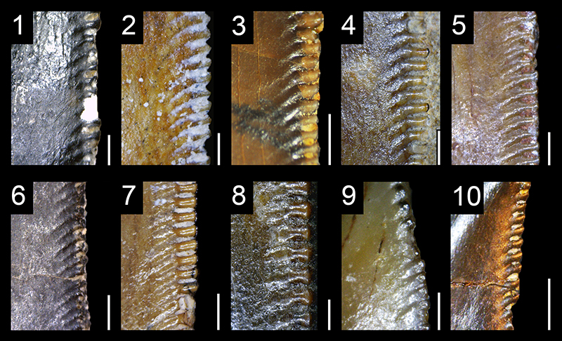
FIGURE 22. Longitudinal ridges in the teeth of non-avian Theropoda. 1, Isolated tooth of the megaraptoran Orkoraptor burkei (MPM-Pv 3458) in lateral view (courtesy of M. Ezcurra); 2, Fourth right premaxillary tooth of the pantyrannosaurian Raptorex kriegsteini (LH PV18) in mesiolabial view; 3, Fifth left maxillary tooth of the dromaeosaurid Bambiraptor feinbergi (AMNH 30556) in labial view; 4, Second? maxillary tooth of the dromaeosaurid Acheroraptor temertyorum (ROM 63777) in labial view (courtesy of D. Larson). Scale bars equal 1 cm (1, 4) and 1 mm (2‒3).
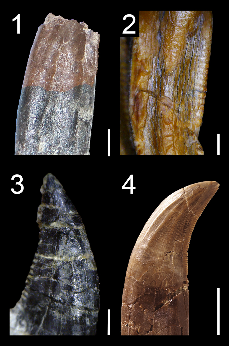
FIGURE 23. Irregular enamel texture of non-avian Theropoda. 1, Tenth left maxillary tooth of the herrerasaurid Herrerasaurus ischigualastensis (PVSJ 407) in labial view; 2, Isolated tooth of the abelisaurid Aucasaurus garridoi (MCF-PVPH-236) in lateral view; 3, Second premaxillary tooth of the allosaurid Allosaurus ‘ jimmadseni' (NHFO 455) in labial view; 4, Tenth maxillary tooth of the tyrannosaurid Tyrannosaurus rex (FMNH PR.2081) in labial view.
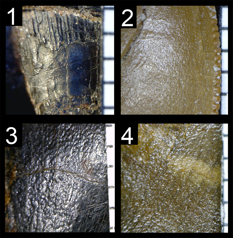
FIGURE 24. Braided enamel texture of non-avian Theropoda. 1, First right premaxillary tooth of the ceratosaurid Ceratosaurus nasicornis (UMNH VP 5278) in labial view; 2, Isolated tooth of the neovenatorid Neovenator salerii (MIWG 6348) in lateral view; 3, Third right maxillary tooth of the tyrannosaurid Lythronax argestes (UMNH VP 20 200) in labial view; 4, Isolated premaxillary tooth of the dromaeosaurid Dromaeosaurus albertensis (AMNH 5356) in lingual view. Scale bars equal 1 mm.
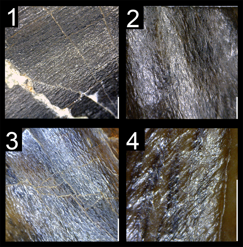
FIGURE 25. Enamel texture of spinosaurid teeth. 1, Veined enamel texture of an isolated tooth of the baryonychine Baryonyx walkeri (NHMUK PV R.9951 278) in lateral view; 2, Veined enamel texture of an isolated tooth of the baryonychine Suchomimus tenerensis (MNN G43‒4) in lateral view; 3, Smooth enamel texture of a maxillary tooth of the spinosaurine Irritator challengeri (SMNS 58022) in lateral view; 4, Anastomosed enamel texture of an isolated tooth of the spinosaurine Spinosaurus aegyptiacus (MSNM V6422) in lateral view. Scale bars equal 1 mm.
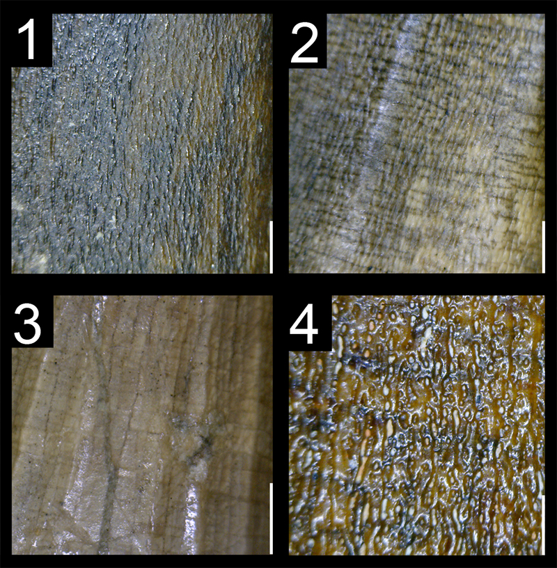
FIGURE 26. Procumbent teeth in non-avian Theropoda. 1, Procumbent premaxillary teeth of the right rostrum of the basal neocoelurosaur Ornitholestes hermanni (AMNH 619; reversed) in lateral view; 2, Procumbent premaxillary and dentary teeth of the right rostrum of the scansoriopterygid Epidexipteryx hui (IVPP V15471; reversed) in lateral view; 3, Hypothetical procumbent mesial maxillary teeth of the left maxilla of the spinosaurid Baryonyx walkeri (NHMUK PV R.9951) in lateral view; 4, Hypothetical procumbent mesial maxillary teeth of the right maxilla of the non-averostran neotheropod Dilophosaurus wetherilli (UCMP 37303; reversed) in lateral view; 5, Procumbent mesial dentary teeth of the left dentary of the noasaurid Masiakasaurus knopfleri (FMNH PR 2471) in lateral view; 6, Procumbent mesial dentary teeth of the right dentary of the proceratosaurid Proceratosaurus bradleyi (NHMUK PV R.4860; reversed) in lateral view. Scale bars equal 1 cm (1‒2, 5), 2 cm (6), and 5 cm (3, 4).
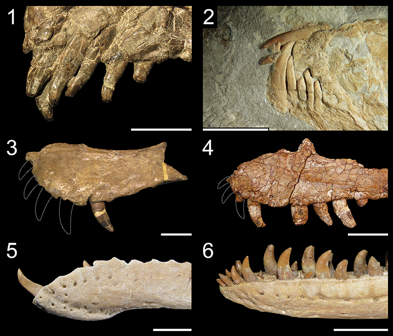
FIGURE 27. Laterocumbent and retrocumbent teeth in non-avian Theropoda. 1, Laterocumbent teeth (teeth facing ventrolabially) in the maxillae of the spinosaurid Spinosaurus aegyptiacus (MSNM V4047) in palatal view; 2, retrocumbent teeth (distally inclined teeth) in the cranium of the dromaeosaurid Deinonychus antirrhopus (YPM 5232) in labial view. Scale bars equal 10 cm (1) and 2 cm (2).
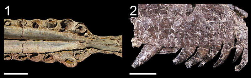
FIGURE 28. Partial and complete edentulism in non-avian Theropoda. 1, Skull of the toothless noasaurid Limusaurus inextricabilis (IVPP V15523) in right lateral view; 2, skull of the therizinosaurid Erlikosaurus andrewsi (MPC-D 100-111) with a premaxilla and anterior portions of the maxilla and dentary toothless in right lateral (cranium) and medial (mandible) views; 3, skull of the basal ornithomimosaur Shenzhousaurus orientalis (NGMC 97-4-002), with a dentulous anterior portion of the dentary, in left laterodorsal view; 4, Skull of the toothless ornithomimosaurid Ornithomimus edmontonicus (TMP 1995.110.01) in left lateral view; 5, premaxilla, maxilla and dentary of the caudipterid Caudipteryx zoui (IVPP V12430), with a toothed anterior portion of the premaxilla, in left lateral view; 6, Skull of the toothless oviraptorid Khaan mckennai (MPC-D 100-1002) in right lateral view. Scale bars equal 1 cm (5), 2 cm (3), 3 cm (1, 6), and 5 cm (2, 4).