FIGURE 1. General locality map showing the position of St. Bathans in New Zealand (bottom left) and the major localities of Blue Lake, Grey Lake, and Mata Creek.
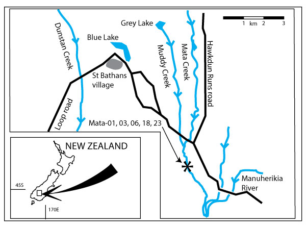
FIGURE 2. Blue Lake, St. Bathans. Showing location of samples. Greyed area is soil/no-outcrop area. Dashed lines show steep cliffs or scarps, usually a result of gold-sluicing activity. A-A’ is the line of cross-section shown in Figure 4.
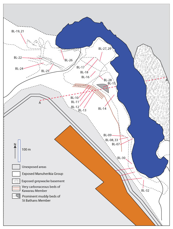
FIGURE 3. Grey Lake, to the north of St. Bathans. Showing location of samples. Greyed area is soil/no-outcrop area. Dashed lines show steep cliffs or scarps, usually a result of gold-sluicing activity. B-B’ is the line of cross-section shown in Figure 4.
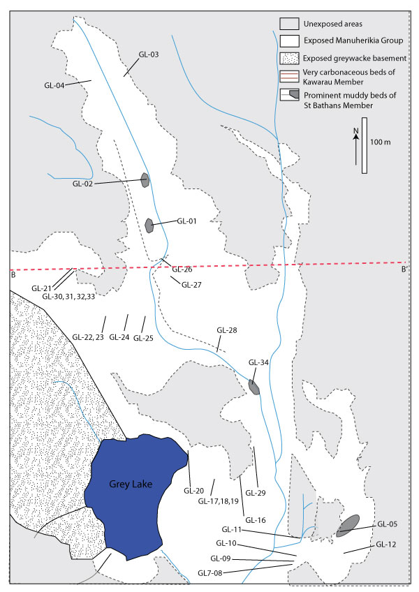
FIGURE 4. Representative cross-sections of Blue and Grey Lakes. The inferred earlier dip and extension of the St. Bathans and Kawarau Members (before erosion and mining) is shown by dashed lines.
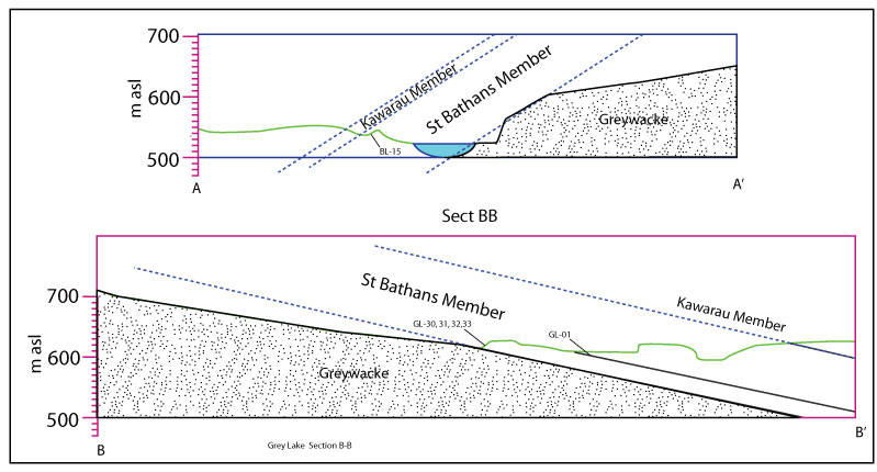
FIGURE 5. Four important plant-fossil bearing facies in the St. Bathans Palaeovalley. 1. Muddy lenses (GL-02, figure near top of bed, gives scale). 2. Beds of redeposited leaf cuticle (GL-24, rock hammer gives scale). 3. In situ deposition leaf packs. Note intact Retrophyllum shoot (GL-32, ball-point pen gives scale). 4. Redeposited leaf packs (Mata-01, ball-point pen gives scale).
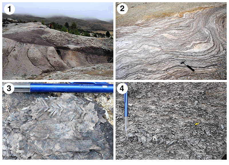
FIGURE 6. GL-01 leaf size histograms, showing length (left), width (middle) and the leaf size classes of Webb (1959) (right). The average leaf length is c. 75 mm.
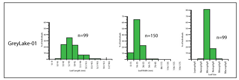
FIGURE 7. Palm frond. (LX1777, BL-30, scale bar equals 50 mm).
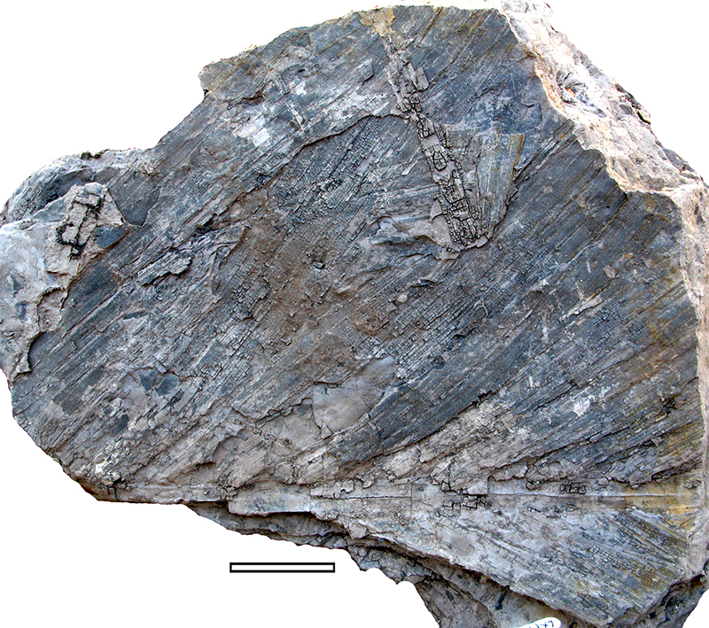
FIGURE 8. MANU-57 (GL-05). 1. In situ leaf on mudstone (LX1700, scale bar equals 10 mm). 2. TLM of cuticle showing dense trichome bases or trichome attachments (scale bar equal 50 μm).
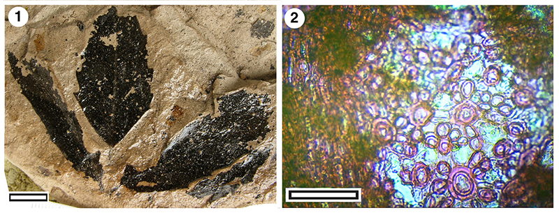
FIGURE 9. MANU-58, compound leaves in situ on mudstone (GL-03, scale bars equal 10 mm). 1. LX1784. 2. LX1786. 3. LX1399. 4. LX1785.
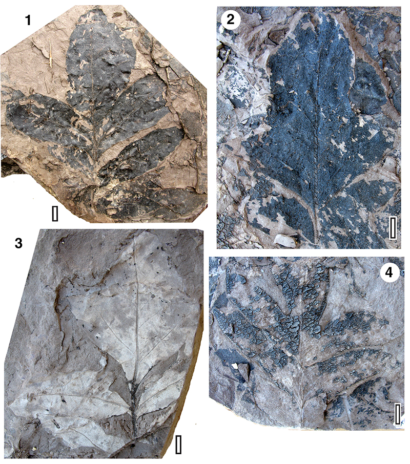
FIGURE 10. CUT-P-EHJ-cuticle of MANU-58. (all material from specimen LX1399, GL-03). 1. TLM showing several stomatal complexes and the larger, more darkly staining trichome attachment sites (scale bar equals 200 μm). 2. TLM showing several stomatal complexes and the larger, more darkly staining trichome attachment sites (scale bar = 100 μm). 3. TLM of single stomatal complex (scale bar equals 50 μm). 4. TLM of adaxial cuticle showing two multi-cellular trichome insertion bases (scale bar equals 100 μm). 5. SEM of outer cuticle surface showing radiating ridges from trichome attachment site (S-1929, scale bar equals 20 μm). 6. SEM of outer cuticle surface with single stomatal complex in centre (S-1906, scale bar equals 10 μm).
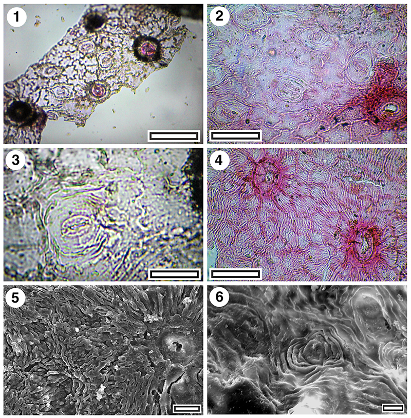
FIGURE 11. MANU-6. Nothofagus azureus. 1. LX1934. 2. LX1935. 3. LX1936. 4. LX1937 (GL-03, all scale bars equal 10 mm).
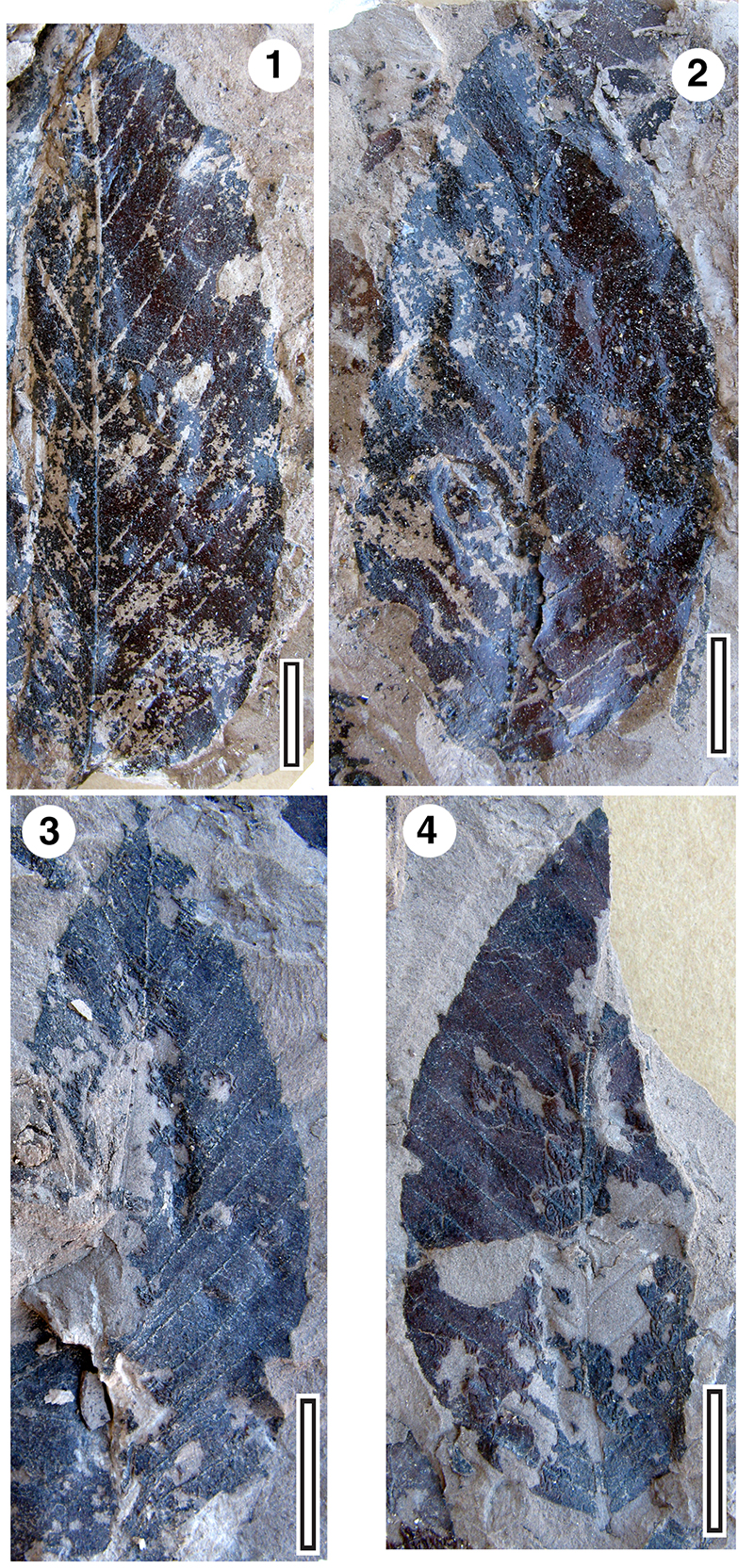
FIGURE 12. Gymnostoma sp. 1. TLM of cuticle showing a single stomatal band (SL3233, GL-30, scale bar equals 50 μm). 2. TLM detail of cuticle showing four rows of transversely oriented stomata (SL3232, GL-30, scale bar equals 20 μm).
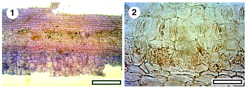
FIGURE 13. MANU-15. Elaeocarpus/Cunoniaceae leaf fossils. 1. Glycerine jelly/plastic sheet mount (SL1478, BL-30, scale bar equals 10 mm). 2. Glycerine jelly/plastic sheet mount, detail of margin (SL1478, BL-30, scale bar equals 5 mm). 3. Apparent compound leaf on mudstone matrix, (LX337, GL-01, scale bar equals 10 mm). 4. Leaf on mudstone matrix (LX1410, GL-05, scale bar equals 5 mm). 5. PVB resin mount (LX1388, scale bar equals 5 mm).
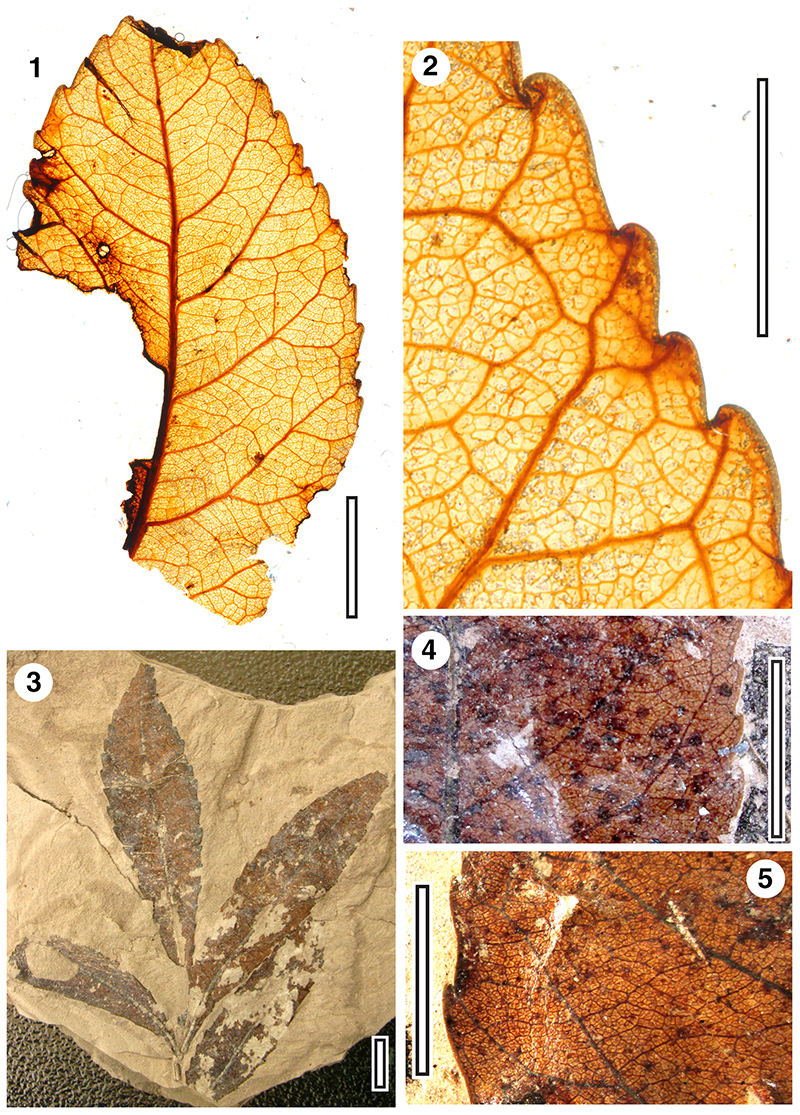
FIGURE 14. Cuticle from MANU-15-CUT-Z-FBE (BL-30). 1. TLM with a group of normal stomatal complexes at left, and a ‘giant’ stomatal complexes over a vein at upper right (SL1478, scale bar equals 100 μm). 2. TLM with two groups of normal stomatal complexes separated by a vein with a simple trichome attachment (SL1478, scale bar equals 100 μm). 3. TLM of single stomatal complex (SL1478, scale bar equals 20 μm). 4. TLM of single stomatal complex (SL1478, scale bar equals 20 μm). 5. TLM of two stomatal complexes with a trichome attachment (SL1478, scale bar equals 50 μm). 6. TLM of upper cuticle with a small ‘cork wart’ (SL1478, scale bar equals 100 μm). 7. SEM of inner cuticle with two stomatal complexes (S-1948, scale bar equals 10 μm). 8. SEM of outer cuticle with one stomatal complex in the centre (S-1948, scale bar equals 10 μm).
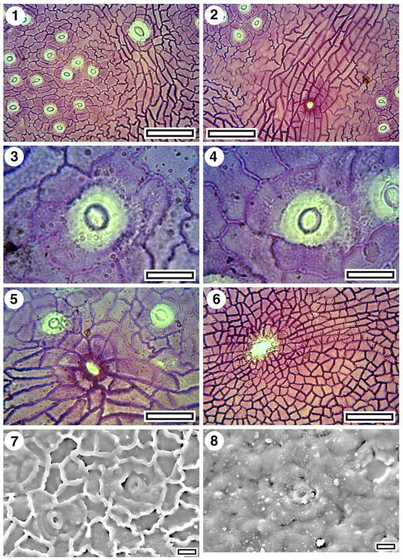
FIGURE 15. MANU-17 and CUT-Z-FCJ (SL1214, GL-01). 1. Base of leaf, glycerine jelly/plastic sheet mount (scale bar equals 10 mm). 2. TLM of cuticle with stomatal complexes and single trichome attachment (scale bar equals 50 μm). 3. TLM of cuticle with stomatal complexes and single trichome attachment (scale bar equals 50 μm).
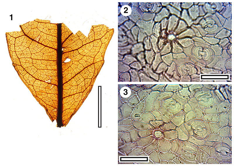
FIGURE 16. MANU-53 and (?) CUT-M-EGF (LX260, GL-01). 1. Whole leaf, in situ on mudstone (scale bar equals 10 mm). 2. TLM of cuticle with numerous stomatal complexes surrounding glandular structure in centre (scale bar equals 200 μm). 3. TLM of cuticle (scale bar equals 50 μm). 4. TLM of cuticle with stomatal complexes at top and paired lid cells below, arrowed (scale bar equals 50 μm).
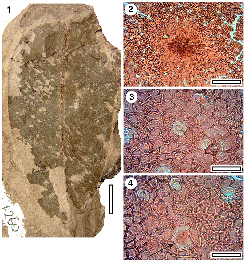
FIGURE 17. MANU-46 and CUT-Z-EDD (SL1481, BL-30). 1. A whole leaf (base was intact, but damaged during mounting), glycerine jelly/plastic sheet mount. 2. TLM of cuticle with stomatal complex to left and trichome attachment to right (scale bar equals 50 μm). 3. SEM of inner cuticle surface(S-1949, scale bar equals 20 μm). 4. SEM of outer cuticle surface with stomatal complexes surrounded by short radiating ridges (S-1949, scale bar equals 20 μm).
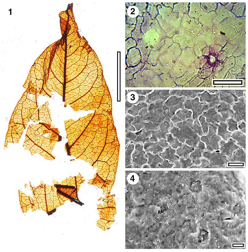
FIGURE 18. MANU-51 and (?) CUT-Z-FJI (SL6501, GL-01). 1. Whole leaf, glycerine jelly/plastic sheet mount (scale bar equals 10 mm). 2. TLM of cuticle (SL6501, scale bar equals 50 μm). 3. TLM of cuticle (SL6501, scale bar equals 200 μm). 4. SEM of inner cuticle surface(S-1951, scale bar equals 10 μm). 5. SEM of outer cuticle surface(S-1951, scale bar equals 20 μm).
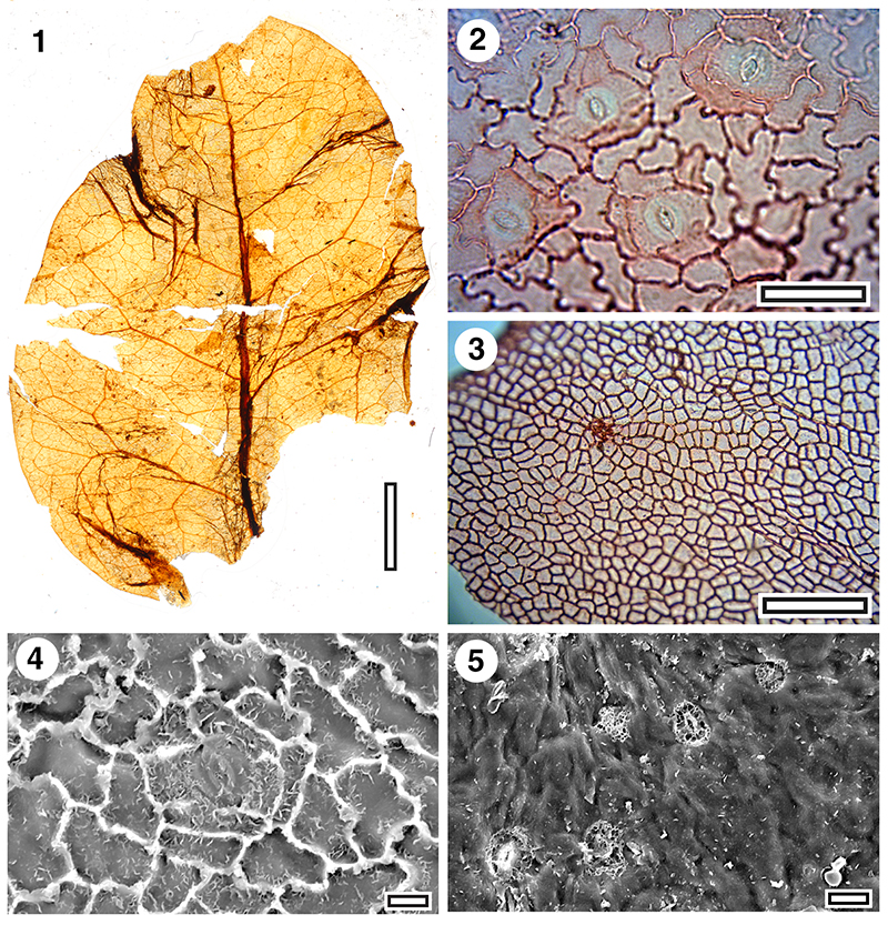
FIGURE 19. MANU-61 and CUT-Z-FJI (SL1211, GL-01). 1. Whole leaf, glycerine jelly/plastic sheet mount (scale bar equals 10 mm). 2. TLM of cuticle (scale bar equals 50 μm). 3. TLM of cuticle (scale bar equals 50 μm). 4. TLM of cuticle (scale bar equals 200 μm). 5. TLM of cuticle (scale bar equals 200 μm). 6. SEM of outer cuticle surface (S-1953, scale bar equals 10 μm). 7. SEM of inner cuticle surface(S-1953, scale bar equals 20 μm).
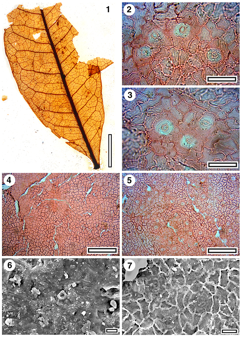
FIGURE 20. MANU-50 and CUT-Z-GFJ, Strychnos sp. (SL1510, GL-01). 1. Whole leaf, glycerine jelly/plastic sheet mount (scale bar equals 10 mm). 2. CUT-Z-GFJ, TLM (scale bar equals 50 μm). 3. CUT-Z-GFJ, TLM (scale bar equals 50 μm). 4. CUT-Z-GFJ, SEM of inner cuticle surface with a single, indistinctly outlined stomatal complex (S-1946, scale bar equals 20 μm). 5. CUT-Z-GFJ, SEM of outer cuticle surface with at least one almost cryptic stomatal complex (S-1946, scale bar equals 20 μm). 6. Leaf base of extant Strychnos lucida Brown (1810) (AQ837571, scale bar equals 10 mm). 7. Extant Strychnos minor Dennstedt (1818) cuticle, TLM (AQ481523, scale bar equals 200 μm).
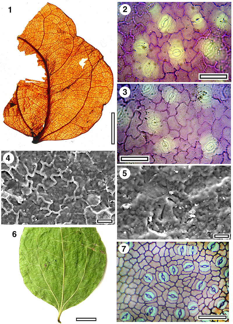
FIGURE 21. MANU-13 and its cuticle CUT-Z-CCE (Mata-01). 1. Glycerine jelly/plastic sheet mount, SL1212 (scale bar equals 10 mm). 2. Glycerine jelly/plastic sheet mount, SB1237 (scale bar equals 10 μm). 3. TLM of cuticle with six stomatal complexes (SL1212, scale bar equals 50 μm). 4. TLM of cuticle with two stomatal areas and a major venal area at bottom right. The cells in contact with the guard cells are not clearly distinct. Two possible trichome bases are at top and right (SL1212, scale bar equals 200 μm). 5. SEM of outer cuticle surface. Four stomatal complexes are in view with simple outer stomatal ledges (S-1952, scale bar equals 20 μm). 6. SEM of inner cuticle with three stomatal complexes, two of them are sharing contact cells (S-1952, scale bar equals 10 μm).
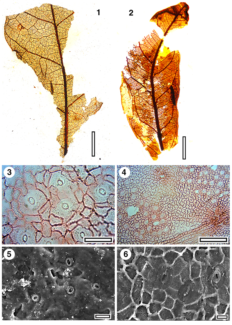
FIGURE 22. MANU-49 (BL-15). Leaves in glycerine jelly/plastic sheet mounts (scale bars equals 10 mm). 1. OU29799. 2. OU29800. 3. OU30222. 4. OU29798.
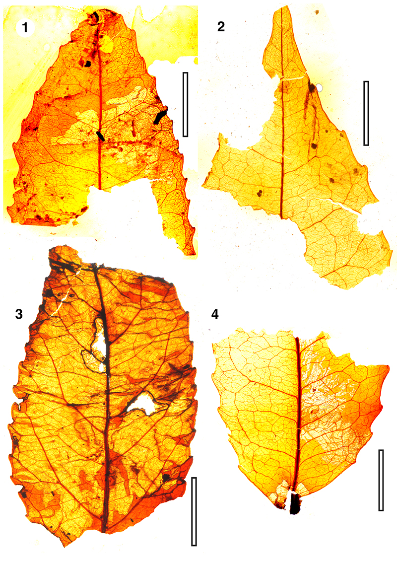
FIGURE 23. CUT-Z-ADF-cuticle of MANU-15 (BL-15). 1. TLM (OU29799, scale bar equals 50 μm). 2. TLM (OU29799, scale bar equals 50 μm). 3. SEM of outer cuticle surface with several stomatal complexes with simple outer stomatal ledges (S-1950, scale bar equals 10 μm). 4. SEM of inner cuticle surface with a single stomatal complex (S-1950, scale bar equals 10 μm).
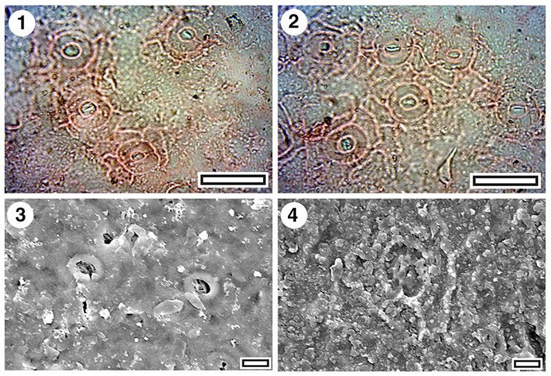
FIGURE 24. MANU-52 and CUT-Z-AJI (OU29848, BL-15). 1. Whole leaf, whole leaf, glycerine jelly/plastic sheet mount (scale bar equals 10 mm). 2. TLM of cuticle (scale bar equals 50 μm). 3. TLM of upper cuticle (scale bar equals 200 μm). 4. SEM of outer cuticle surface with several stomatal complexes and very irregular surface (S-1947, scale bar equals 20μm). 5. SEM of inner cuticle (S-1947, scale bar equals 20 μm).
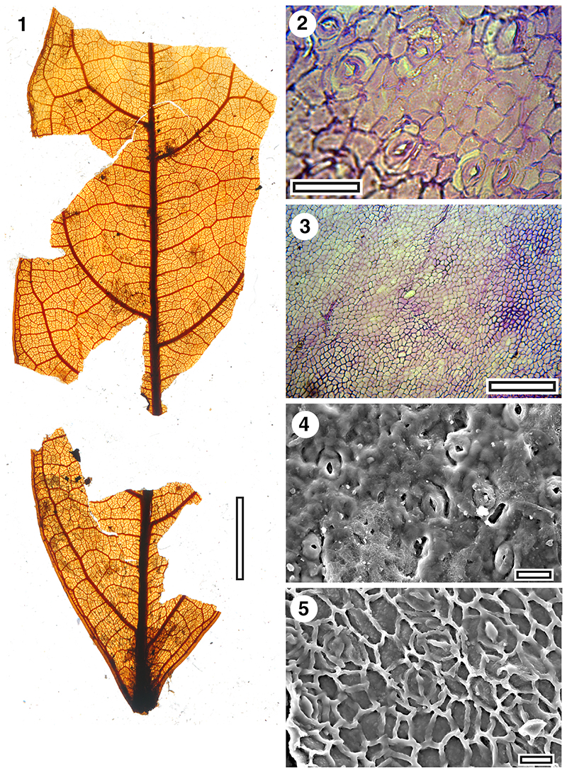
FIGURE 25. MANU-54 and CUT-Z-EHI (LX1446, GL-05). 1. Whole leaf, in situ on mudstone (scale bar equals 10 mm). 2. TLM with several stomatal complexes (scale bar equals 200 μm). 3. TLM of single stomatal complex. The outer stomatal rim is clearly visible, as is the outer bound of the subsidiary cells (scale bar equals 50 μm). 4. SEM of inner cuticle surface. Prominent T-pieces and cutinisation around the guard cells are apparent (S-1908, scale bar equals 20 μm).
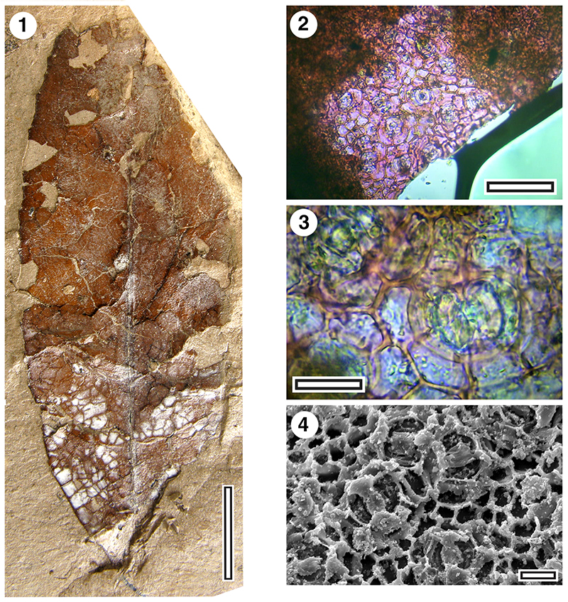
FIGURE 26. MANU-55 and CUT-Z-ABD. 1. In situ leaf on mudstone (SL4855, BL-32, scale bar equals 10 mm). 2. in situ leaf on mudstone (SL4856, BL-32, scale bar equals 10 mm). 3. TLM of cuticle showing stomatal groups separated by venal areas (SL4856, BL-32, scale bar equals 200 μm). 4. TLM of cuticle (SL4856, BL-32, scale bar equals 50 μm).
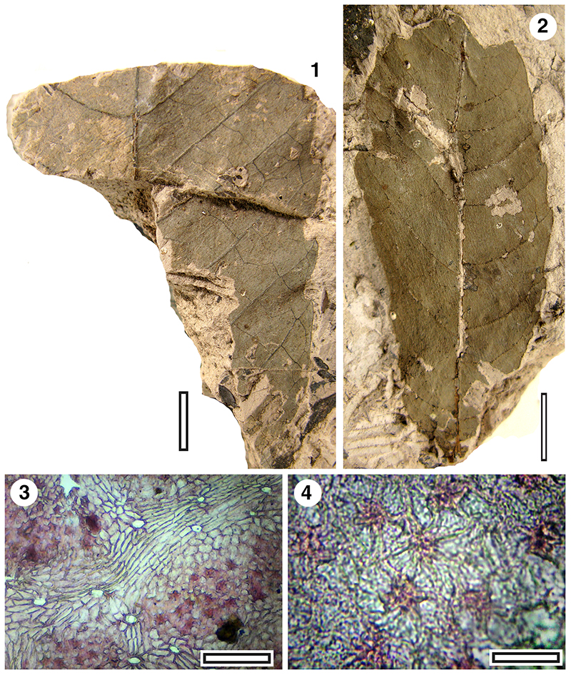
FIGURE 27. MANU-56. Digitately compound leaf, in situ on mudstone. (LX195, GL-01, scale bar equals 50 mm).
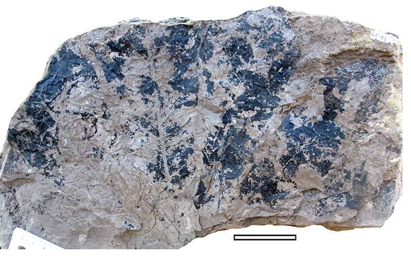
FIGURE 28. MANU-59 (LX1437, GL-05). 1. Whole leaf, in situ on mudstone (scale bar equals 10 mm). 2. SEM of outer cuticle (S-1897, scale bar equals 50 μm). 3. SEM of outer cuticle surface showing single stomatal complex (S-1897, scale bar equals 20 μm).
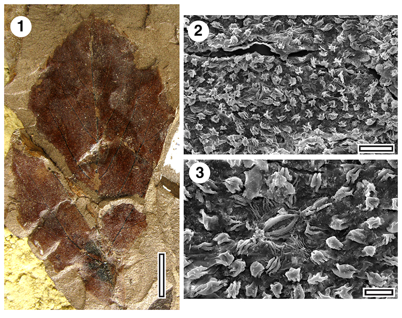
FIGURE 29. MANU-60 and CUT-Z-ECF (LX272 GL-01). 1. Whole leaf, in situ on mudstone, (scale bar equals 10 mm). 2. TLM of cuticle (scale bar equals 200 μm). 3. TLM of single stomatal complex (scale bar equals 50 μm).
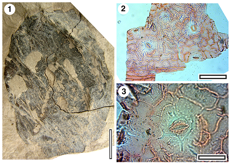
FIGURE 30. Lauraceae wood (LX1977, GL-34). TLM images of radial sections. 1. Vessels and rays (scale bar equals 0.1 mm). 2. Detail of vessel showing alternate pitting (scale bar equals 20 μm). 3. Detail of ray showing resin-filled cells (scale bar equals 20 μm).
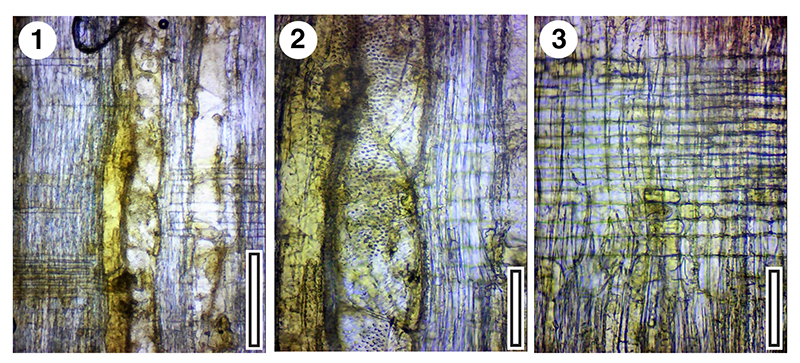
FIGURE 31. Epiphyllous fungi (TLM images). 1. Circular stromata (ascomata), cf. Phragmothyrites sp, and branched mycelia associated with elongate hyphopodia (SL3251, GL-31, scale bar equals 200 μm). 2. Branched mycelia associated with orbicular hyphopodia (SL3134, GL-02, scale bar equals 200 μm).
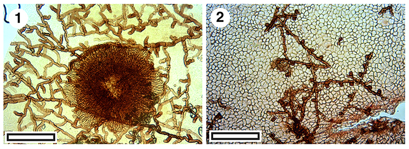
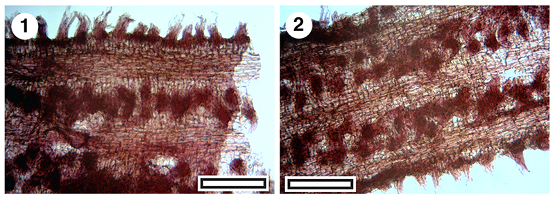
FIGURE 32. Roots, TLM images, showing parallel zones of lateral rootlets, scale bars equals 0.5 mm). 1. SL4795, GL-25. 2. SL4795, GL-25.