FIGURE 1. Map of Brazil, showing the location of the Prestosuchus chiniquensis -bearing site as indicated by von Huene (1938) within the state of Rio Grande do Sul. Map modified from Reichel et al. (2009).
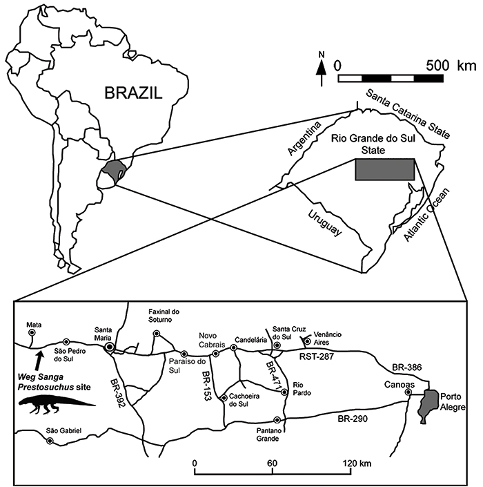
FIGURE 2. Chronostratigraphic column of the Santa Maria Supersequence, showing the Prestosuchus chiniquensis-bearing level. Modified from Horn et al. (2014).
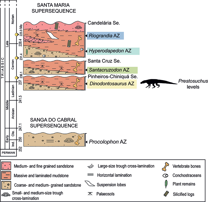
FIGURE 3. Life reconstruction (1) and preserved bones (2) of Prestosuchus chiniquensis. In red the holotype material and in white the referred material known. Drawn by Jorge Gonzales.
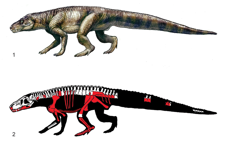
FIGURE 4. Skull material of the lectotype of Prestosuchus chiniquensis SNSB-BSPG AS XXV 28. 1-4, Right premaxilla (SNSB-BSPG AS XXV 28) in 1, lateral; 2, medial; 3, anterior; and 4, dorsal views. Abbreviations: mxpp, maxillary palatal process; pmxpp, premaxillary palatal process; pmxin, premaxillary interdental plate; pmxt, premaxillary teeth; pmxt4, premaxillary tooth number 4; 2rpmxt, replacement premaxillary tooth. Scale bar equals 1 cm.
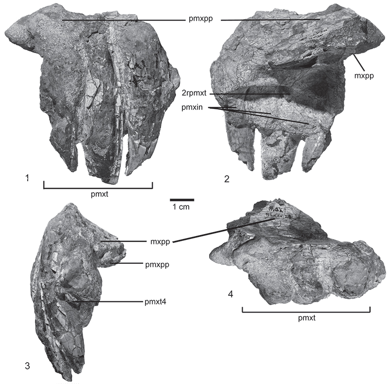
FIGURE 5. Lower jaw (SNSB-BSPG AS XXV 1) in 1, dorsal; 2, ventral; 3, lateral; and 4, medial views. 5, 2nd replacement tooth detail; 6, 11th replacement tooth. Abbreviations: an, Angular; d, dentary; de, denticles; dt3, dentary tooth 3; dt7, dentary tooth 7; fo, foramen; for, foramina; inp, interdental plate; mcf, meckelian foramen; pr, prearticular; 2rdt, replacement dentary 2 tooth; 11rdt, replacement dentary 11 tooth sa, surangular; sp, splenial. Scale bar equals 4 cm.
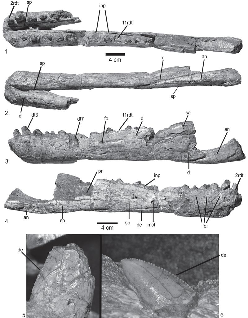
FIGURE 6. Vertebral material of the lectotype of Prestosuchus chiniquensis SNSB-BSPG AS XXV. 1-3, Anterior cervical vertebra (SNSB-BSPG AS XXV 29) in 1, left lateral; 2, ventral; and 3, posterior views. 4-6, Middle cervical vertebra (SNSB-BSPG AS XXV 30) in 4, anterior; 5, posterior; and 6, dorsal views. 7, Anterior dorsal vertebrae (SNSB-BSPG XXV 31) in left lateral view. 8-9, Anterior caudal vertebrae and chevrons (SNSB-BSPG AS XXV 3b) in 8, left lateral; and 9, ventral views. Abbreviations: acd, anterior centrodiapophyseal lamina; al, accessory lamina; chc, chevrons contact; cpr, centroprezygapophyseal lamina; cr, cervical rib; dd, deep depression; di, diapophysis; dr, dorsal rib; hc, hemal canal; if, infradiapophyseal fossa; inf, infraprezygapophyseal fossa; infp, infraposzygapophyseal fossa; lld, lateral longitudinal depression; lf, lateral fossa; mb, medial bridge; nc, neural canal; pa, parapophysis; pad, paradiapophyseal lamina; pcd, posterior centrodiapophyseal lamina; pcl, posterior centroapophyseal lamina; pd, prezygodiapophyseal lamina; pos, postzygapophysis; pr, prezygapophysis; prd, prezygodiapophyseal laminae; ps, postzygapophysis lamina; tp, transverse process; vd, ventral depression; vk, ventral keel. Arrows indicate the orientation. Scale bar equals 1 cm indicated in the figure.
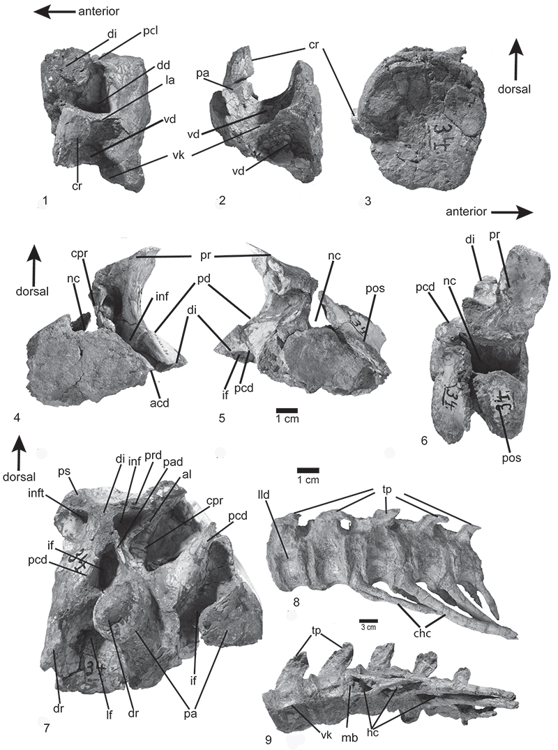
FIGURE 7. Pelvic elements of the paralectotype (1-5) and lectotype (6-8) of Prestosuchus chiniquensis SNSB-BSPG AS XXV. 1-5, Sacral vertebrae, sacral ribs, and right ilium (SNSB-BSPG XXV 7) in 1, right lateral; 2, dorsal; 3, ventral; 4, anterior; and 5, posterior views. 6-8, Left ilium and ischia (SNSB-BSPG XXV 34b/3) in 6, left lateral view; 7, Sacral centra and left ilium (SNSB-BSPG AS XXV 3a) in lateroventral; 8, dorsomedial views. Abbreviations: dv, dorsal vertebra; iac, ischial acetabulum; isp, ischial peduncle; osn, opening for the spinal nerve; pdns, posterior dorsal neural spine; po, paramedian osteoderms; pop, postacetabular process; pp, pubic peduncle; ptp, postzygapophysis; 1sns, first sacral neural spine; 2sns, first sacral neural spine; st, spine table; 1cvc, first caudal vertebra; 1sr; first sacral rib; 2sr, second sacral rib, iob: ischial obturador blate lld: lateral longitudinal depression, tp: transverse process, 2 svs: second sacral centrum. Scale bar indicate in the figure.
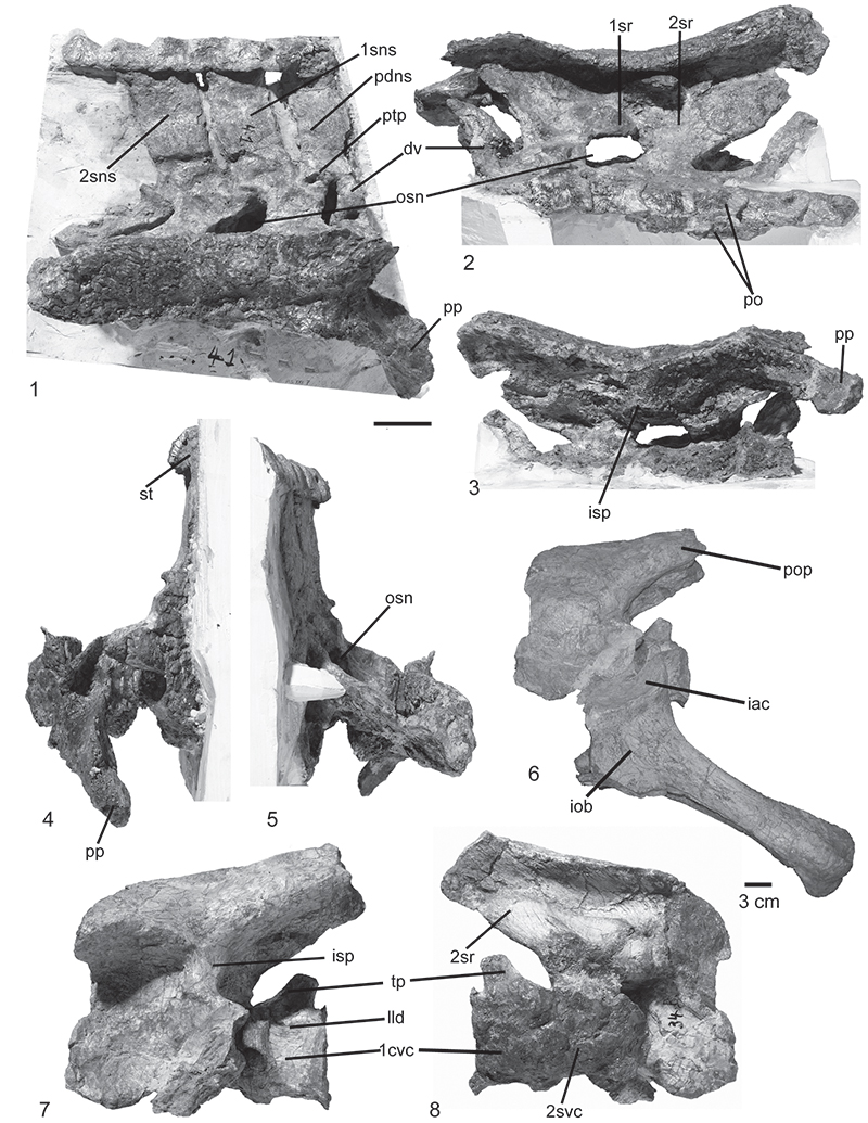
FIGURE 8. Dorsal and ventral ribs of the lectotype and dorsal osteoderm of the paralectotype of Prestosuchus chiniquensis SNSB-BSPG AS XXV. 1-2, Gastralia (SNSB-BSPG AS XXV 5a) in 1, general; 2, close-up views. 3, Dorsal ribs (SNSB-BSPG AS XXV 9, 49) in external posterodorsal view. 4, right last presacral osteoderm (SNSB-BSPG AS XXV 7) in external view. Abbreviations: ap, anterior point; c, capitulum; mc, medial contact; r, ridges; t, tuberculum. Scale bars equal 1 cm.
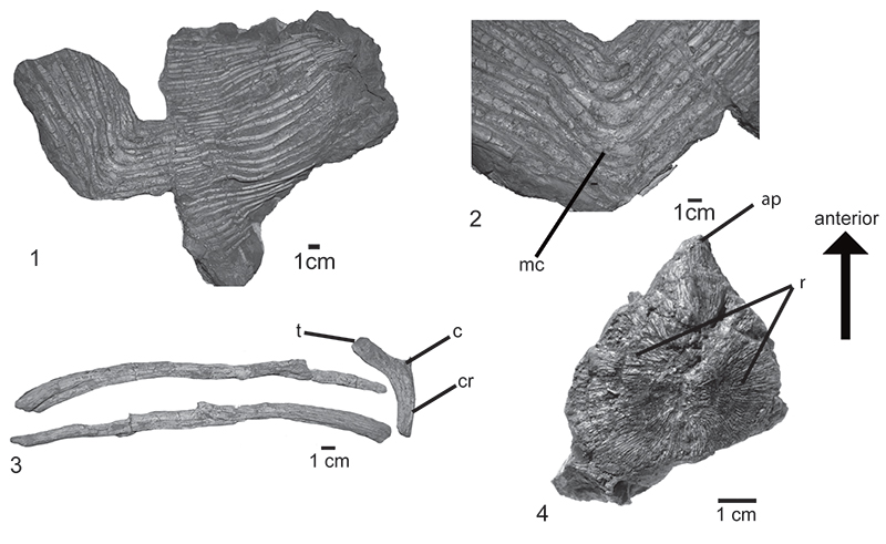
FIGURE 9. Pectoral elements of the lectotype of Prestosuchus chiniquensis SNSB-BSPG XXV 12. 1, Ventrolateral view of left scapula, coracoid, clavicle, and interclavicle; 2, lateral view; and 3, posterodorsal view. Abbreviations: cf, coracoid foramen; cl, clavicle; in, interclavicle; ld, lateral depression; oi, oval incision; pt, pathological tissue; rk, rounded keel; rt, rugose tubercle. Scale bars equal 4 cm.
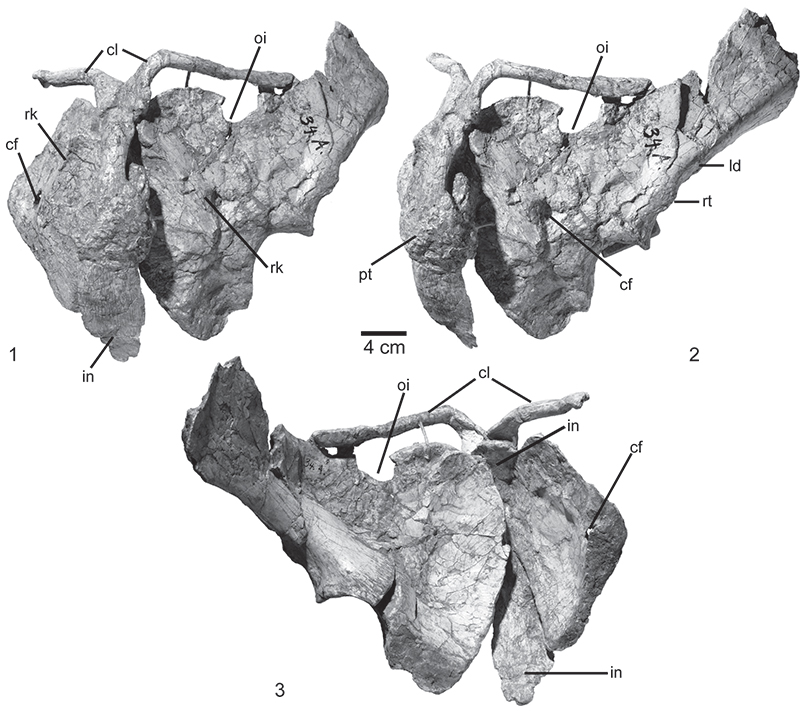
FIGURE 10. Humerus of the lectotype (1-6) of Prestosuchus chiniquensis. 1, proximal end of the left humerus (SNSB-BSPG AS XXV 33) in posterior; 2, anterior; and 3, dorsal views. 4, distal end of the right humerus (SNSB-BSPG XXV 35) in anterior view. 5, proximal end of the right humerus (SNSB-BSPG AS XXV 33) in anterior; and 6, posterior views. Abbreviations: dc, deltopectoral crest; ent, entepicondyle; hh, humeral head; it, internal tuber; tg, trochlear groove. Scale bars equal 1 cm.
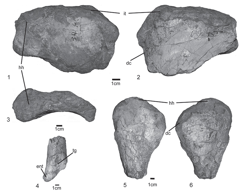
FIGURE 11. Pelvic elements of the lectotype of Prestosuchus chiniquensis SNSB-BSPG XXV. 1, distal ends of right and 2, left pubis (SNSB-BSPG AS XXV 6) in anteromedial view; 3-5, articulated ischia (SNSB-BSPG XXV 3) in 3, dorsal; 4, lateral; and 5, ventral views. Abbreviations: dlc, dorsolateral crest; iac, acetabular contribution; iob, ischial obturator blade; is, interischial suture; kde, knob-like distal end; lm, lamina of bone; vi, ventral incision. Scale bars equal 3 cm.
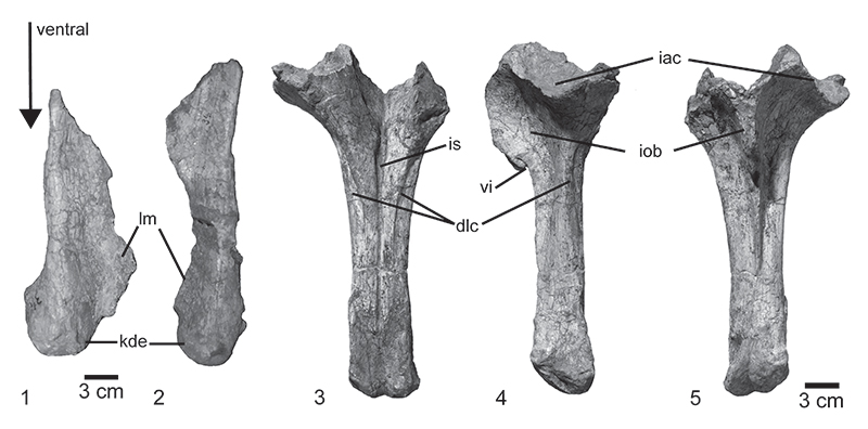
FIGURE 12. Hind limb bones of the lectotype of Prestosuchus chiniquensis SNSB-BSPG AS XXV. 1-6, left femur (SNSB-BSPG AS XXV 10) in 1, anterior; 2, posterior; 3, lateral; 4, medial; 5, proximal, and 6, distal views. 7, articulated left tibia, fibula, and proximal tarsals in anteromedial view. 8-12, left tibia (SNSB-BSPG AS XXV 11a) in 8, medial; 9, lateral; 10, posterior; 11, proximal; and 12, distal views. 13-18, left fibula (SNSB-BSPG AS XXV 11b) in 13, proximal; 14, distal; 15, lateral; 16, medial; 17, posterior; and 18, anterior views. Abbreviations: dd, deep depression; f, popliteal fossa; fc, fibular condyle; g, groove; gt, great trochanter; ilt, iliofibularis trochanter; ip; incompletely preserved surface; kn, knob; ld, longitudinal depression; lud, lunate depression; plr, posterolateral ridge; pmt, posteromedial tuber; rd, rectangular depression; rt, rugose tubercle; s, scars; t, tubercle; td, triangular depression; tc, tibial condyle; tic, tibial contact; tfc, tibiofibular crest; 4t, fourth trochanter. Scale bars are indicated in the figure.
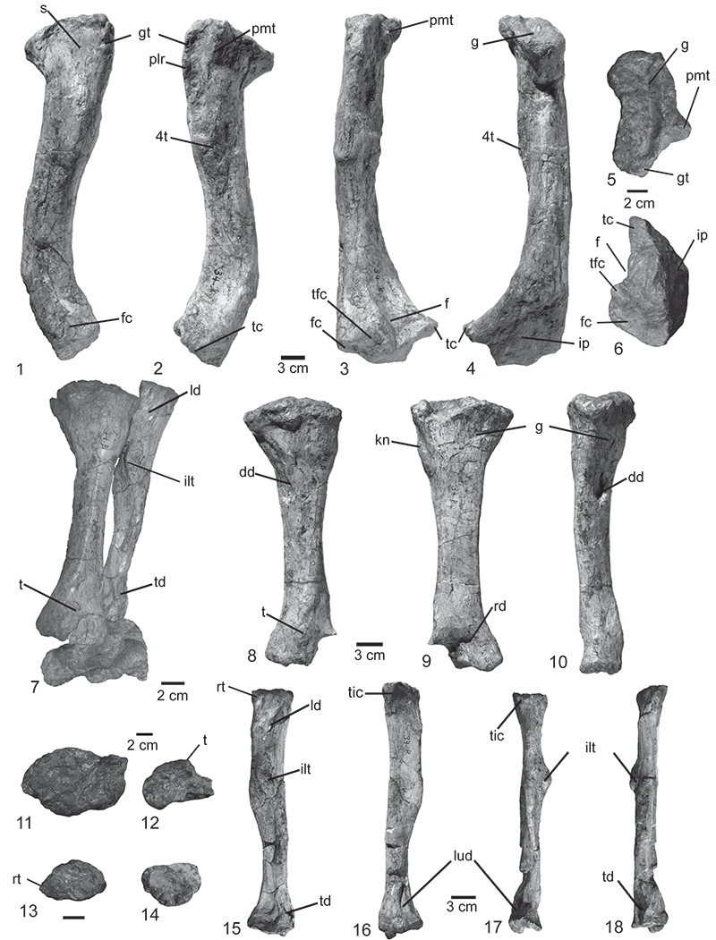
FIGURE 13. Foot bones of the lectotype of Prestosuchus chiniquensis SNSB-BSPG XXV. 1-6, left proximal tarsals (SNSB-BSPG XXV 11c) in 1, dorsal; 2, anterior; 3, ventral; 4, posterior; 5, lateral; and 6, posteroventral views. 7-8, left distal tarsal four, metatarsals, and phalanges of the five digits in articulation (SNSB-BSPG XXV 11e) in 7, dorsal; and 8, ventral views. Abbreviations: ap, anterior pit; ct, calcaneal tuber; ddd, dorsal distal depression; dp, deep pit; dt3, distal tarsal three; dt4, distal tarsal four; ff, fibular facet; lf, lateral fossae; md, median depression; p, pit; tde, tibial distal end; I-V, digits 1 to 5. Scale bars indicate in the figure.
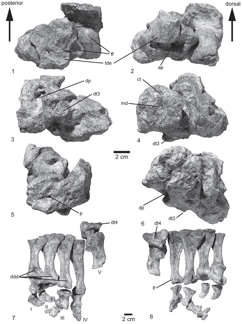
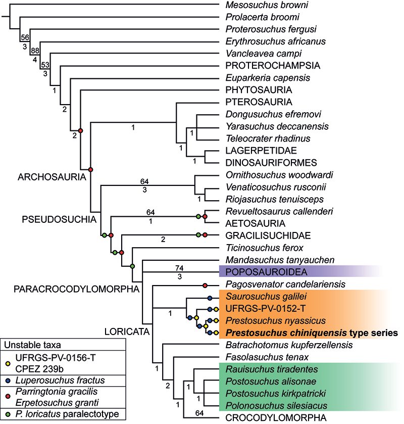
FIGURE 14. Reduced consensus tree of the 22815 most parsimonious trees of 1425 steps; bootstrap values over 50% indicated above branches and Bremer support indicated below branches. Orange: Prestosuchidae, green: Rauisuchidae, purple: Poposauroidea. Prestosuchus chiniquensis type series indicated in bold. Position of unstable taxa indicated with coloured circles.