FIGURE 1. Scanning electron microscopic images of Parviformosus wohlrabeae Barling et al., 2013. (1) Complete habitus. (2) Mesosoma. (3) Metasoma excluding ovipositor. (4) Ovipositor (arrow) engulfed in mineral ridge. (5) Metasoma, broken posterior tip with individual layers of Mt7 and Mt8 (arrow) visible. (6) Anterolateral view of mesosoma, head to right, dashed line marks posterior margin of pronotum. Scales (1) 1 mm; (2, 3) 0.1 mm; (4-6) 0.02 mm. Abbreviations: msc = mesoscutum, Mt6/7 = metasomal tergite 6/7, no1 = pronotum, pl2 = mesopleuron.
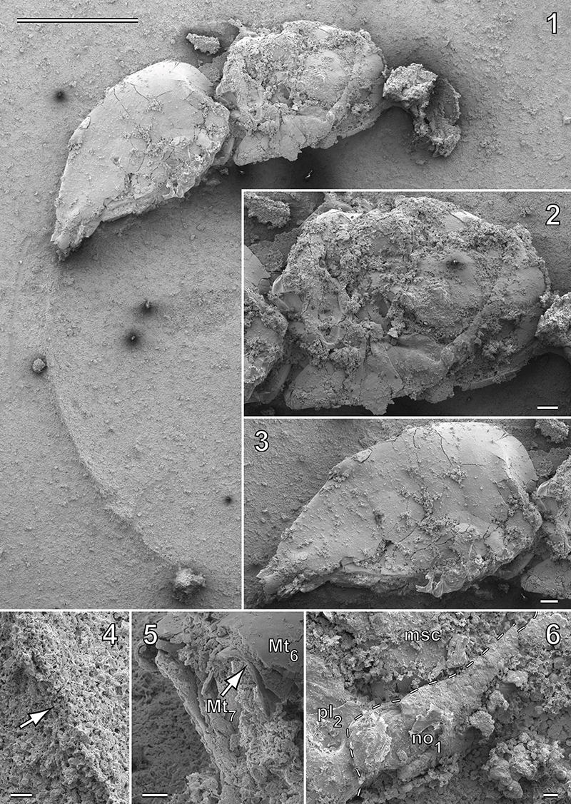
FIGURE 2. Scanning electron microscopic images of the mesosoma of Parviformosus wohlrabeae. (1) Posterior part of mesosoma with potential forewing fragment (arrow), right lateral view. (2-4) Metanotum and propodeum, (2) dorsal view, arrow indicates metanotum submedially overlapping propodeum; (3) left dorsolateral view; (4) right dorsolateral view. Scales (1-3) 0.1 mm; (4) 0.05 mm. Abbreviations: lti2/3 = left meso- or metatibia, no3 = metanotum, ppd = propodeum, rfe3 = right metafemur, rfw? = probably remnant of right forewing, scta = mesoscutellar arm, spr = spiracle, wf = wing fragment.
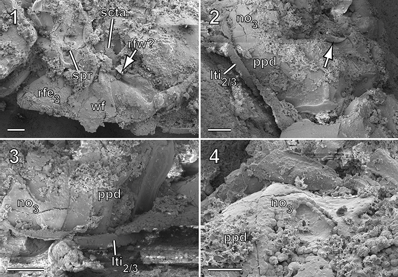
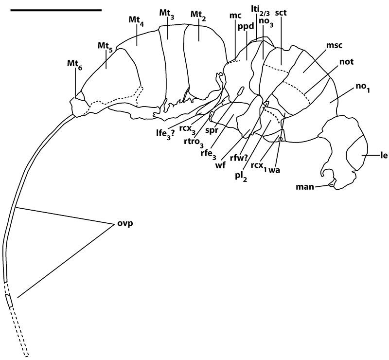
FIGURE 3. Parviformosus wohlrabeae, habitus, lateral view; Abbreviations: le = left eye, lfe3 ? = probable left metafemur, lti2/3 = left meso- or metatibia, man = mandible, mc = median carina, msc = mesoscutum, Mt = metasomal tergite, no1 = pronotum, no3 = metanotum, not = notaulus, ovp = ovipositor, pl2 = mesopleuron, ppd = propodeum, rcx1 = right procoxa, rcx3 = right metacoxa, rfe3 = right metafemur, rfw? = probably remnant of right forewing, rtro3 = right metatrochanter, sct = mesoscutellum, spr = spiracle, wa = wing articulation, wf = wing fragment; Scale: 1 mm.