FIGURE 1. General geological settings. 1, Tectonic map of Bulgaria (after Ivanov, 2017); 2, Geological scheme for part of the Western Srednogorie zone and position of the dinosaur bone locality (after Sinnyovsky et al., 2012; simplified).
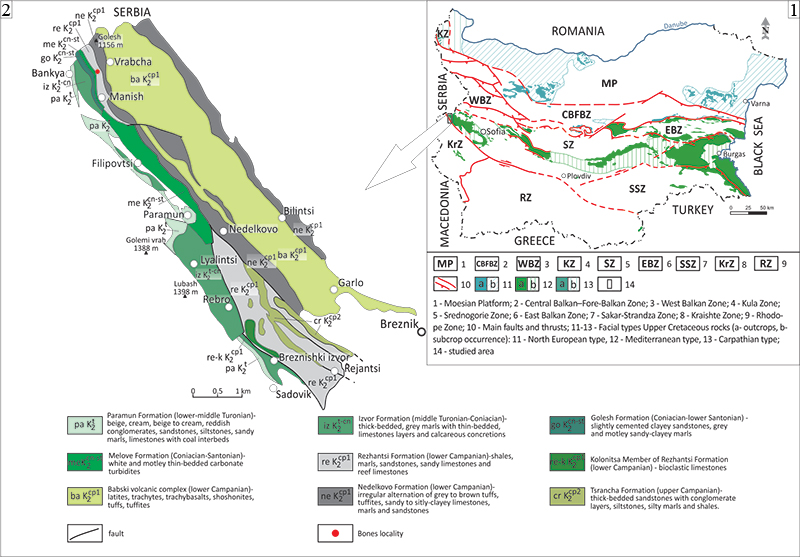
FIGURE 2. Geology, lithology and stratigraphy of the dinosaur bone locality at Vrabchov dol. 1, General view of the fossil-bearing sediments in the gully of Vrabchov dol yielding the dinosaur material. The yellow rectangle marks the sedimentary section illustrated on Figure 2.3; 2, Detail of the outcrop with position of one of the studied bone fragments (A; specimen NMNHS FR-16) and additional tetrapod remains (B). Scale bar equals 1 m; 3. Lithostratigraphic column of the Late Cretaceous sedimentary succession outcropping at the locality.
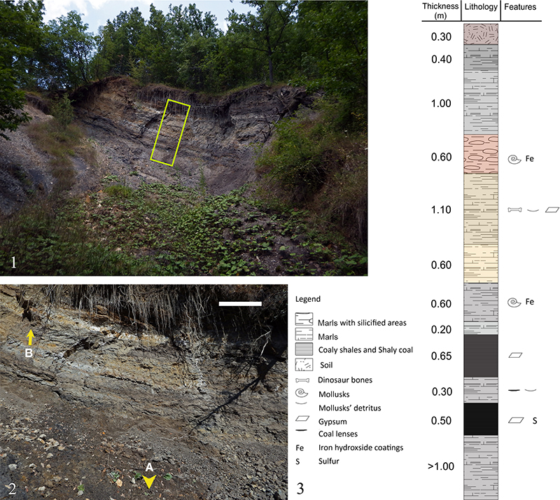
FIGURE 3. Fossilized bone fragments for the Vrabchov dol. 1, Multiple views of specimen U.S., K2 1586, an undetermined long bone diaphyseal fragment; 2. Specimen NMNHS FR-16, possibly a partial diaphysis of undetermined long bone. Scale bar equals 3 cm.
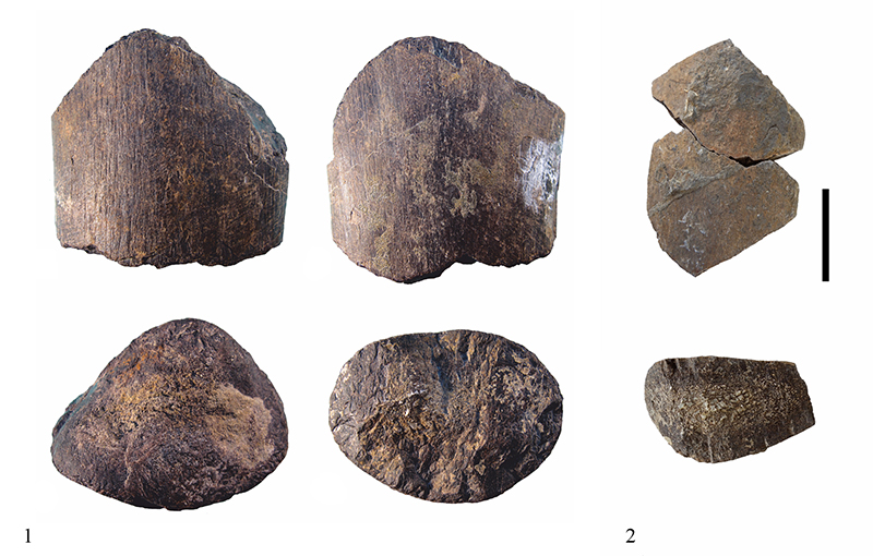
FIGURE 4. Scanned images of studied thin-sections prepared from the Vrabchov dol’s fossil fragments, illustrating the degree of cortical loss during preparation, the size of medullary region, and the extent and spatial distribution of cancellous bone tissue. Solid black line illustrates the original cross-sectional form of the fossils. 1, Thin-section U.S., K2 1586-1; 2, Thin-section U.S., K2 1586-2; 3, Thin-section U.S., K2 1586-3; 4, A single thin-section from specimen NMNHS FR-16. Abbreviations: c-cortex/compact bone tissue; cb-cancellous bone tissue; mr-medullary region. Scale bar equals 2 cm.
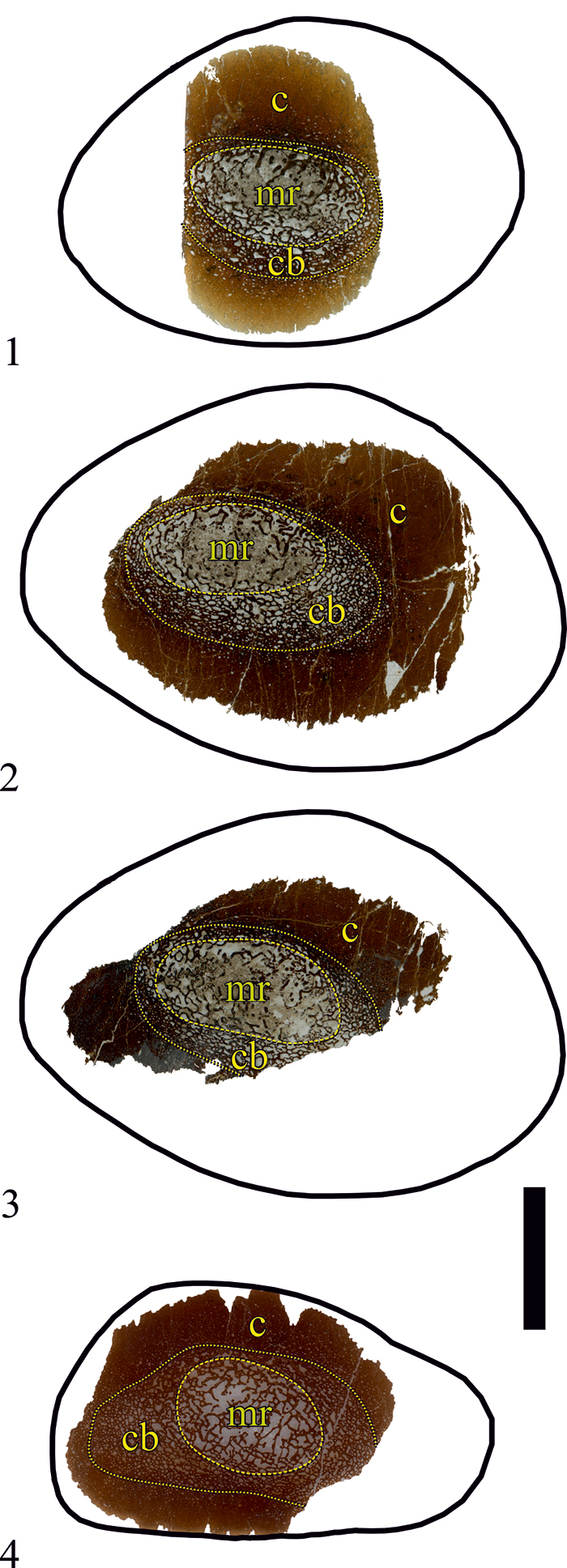
FIGURE 5. Scanned images of the processed cross-section surface of Vrabchov dol’s fossil bone fragments and osteohistology of specimen U.S., K2 1586 in reflected light. The dashed line in Figures 5.2-3 illustrates missing parts of the cortex. Blue arrow-heads mark a line of arrested growth (LAG). 1, The original whole bone fragment polished section of specimen U. S., K2 1586 after sectioning for thin-section U. S., K2 1586-1; 2, The whole bone section of U. S., K2 1586 after the further sectioning; 3, Partial bone fragment polished section of specimen NMNHS FR-16; 4-6, Different sections of specimen U. S., K2 1586 bone wall periphery following a single LAG formed sub-periosteally. Some large primary osteons with wide lumina are easily observed above the LAG. Abbreviations: c-cortex/compact bone tissue; cb-cancellous bone tissue; mr-medullary region; po-primary osteon; so-secondary osteon. Scale bar equals 2 cm (Figures 5.1-3); 1 mm (Figures 5.4-6).
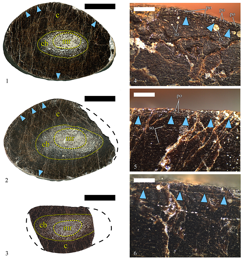
FIGURE 6. Osteohistology of specimen U. S., K2 1586, a diaphyseal fragment of a putative titanosaurian long bone, in transmitted cross-polarized light. Arrow in the lower right corner of each image indicates the direction of the periosteum. 1, Primary bone in the deep cortex consisting of highly vascularized tissues of the woven-parallel complex (sensu Prondvai et al., 2014), with strong presence of woven component in the bone scaffolding and laminar organization of the primary osteons. Secondary osteons of various size and degree of infilling are developed over the primary tissues; 2, Primary bone tissues in the mid-cortex. The tissue is clearly anisotropic and indicates high spatial organization of the bone scaffolding; 3, High magnification image of the primary bone tissues in the mid-cortex, showing optically highly anisotropic laminar bone; 4, Same bone tissue type as 3 but illustrating different area of the mid-cortex; 5, Primary bone tissues in the outer half of the cortex in thin-section U.S., K2 1586-2. Primary osteons exhibit slightly more irregular organization but it can still be described as laminar; 6, Numerous secondary osteons of at least two generations at mid-cortical levels in area less affected by Haversian remodeling; 7, Four generations of secondary osteons in the outer half of the cortex; 8, Five generations of secondary osteons in the deep cortex. Notice the formation of numerous generations of secondary osteons inside the boundaries of earlier, much larger secondary osteon. Abbreviations: cal-calcite; po-primary osteon; so-secondary osteon; wpc-woven-parallel complex (sensu Prondvai et al., 2014). Scale bar equals 1-2, 6-7-500 µm; 3-4, 8-200 µm; 5-100 µm.
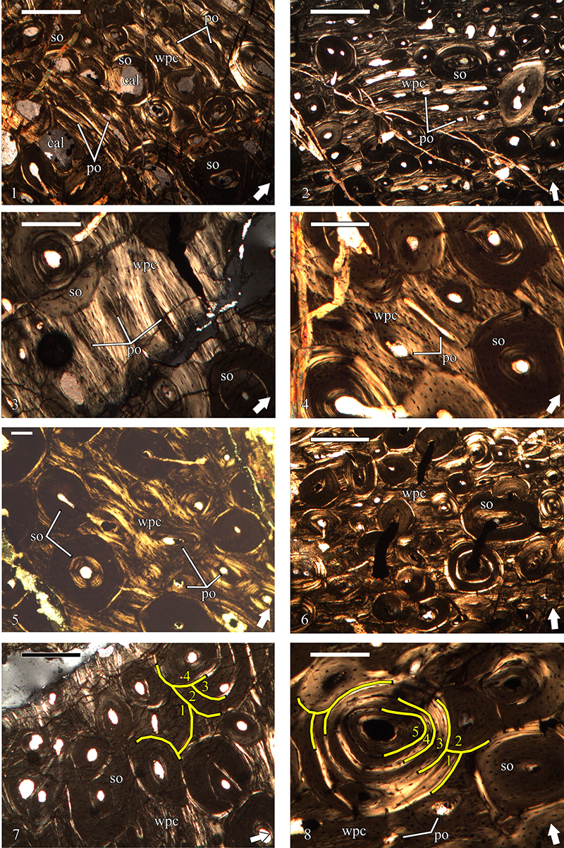
FIGURE 7. Osteohistology of specimen NMNHS FR-16, a putative titanosaurian long bone fragment, in transmitted cross-polarized light. Arrow in the lower right corner of each image indicates the direction of the periosteum. 1, Heavily remodeled outer cortex with anisotropic primary bone tissues preserved as interstices between secondary osteons; 2, Same area with preserved primary cortex as 1, but at higher magnification. Primary osteons appear to be longitudinal; 3, Dense Haversian bone with 4 generations of secondary osteons in the outer third of the cortex. Abbreviations: po-primary osteon; so-secondary osteon; wpc-woven-parallel complex. Scale bar equals 1-200 µm; 2-3 -100 µm.
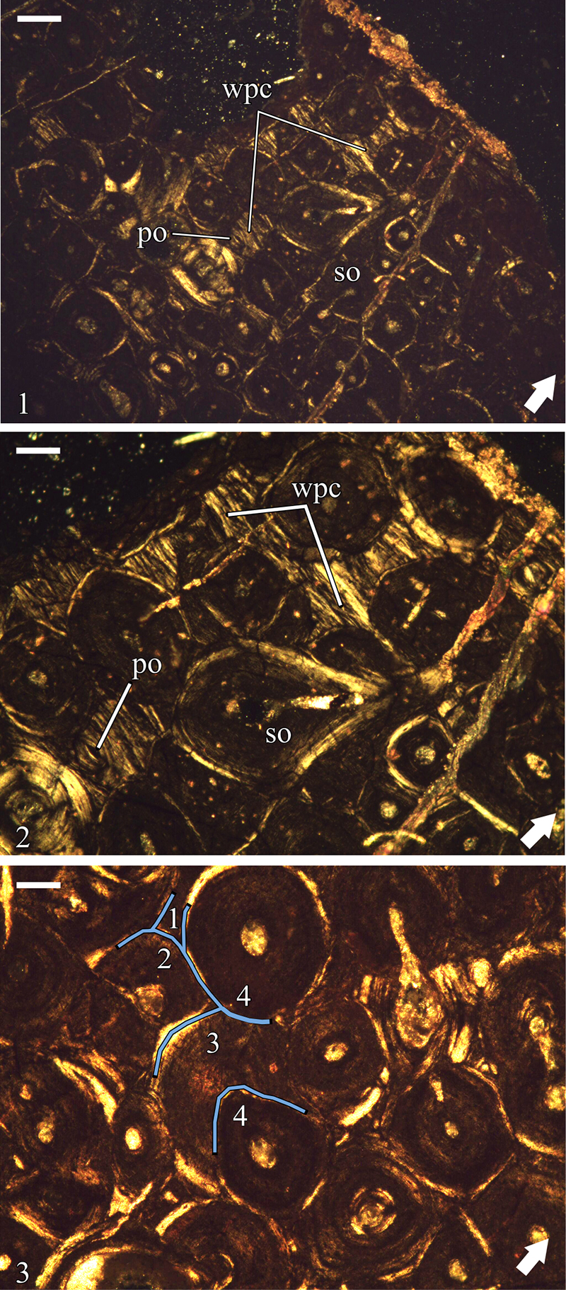
FIGURE 8. Osteohistology of the Bulgarian ornithomimosaur, specimen NMNHS F-31436-a partial left humerus, in transmitted cross-polarized light. Arrow in the lower right corner of each image indicates the direction of the periosteum. 1, Endosteal bone lining the medullary cavity and dense Haversian bone in the inner cortex; 2, Dense Haversian tissue extending up to the outermost cortex and sub-periosteally deposited lamellar primary bone, which probably represents incipient external fundamental system; 3, Interstices of laminar primary bone tissue with relatively organized bone scaffolding and longitudinal primary osteons preserved between the numerous densely packed secondary osteons. Up to 4 generations of secondary osteons can be recognized on the image; 4, Interstices of preserved primary bone tissues. Abbreviations: eb-endosteal bone; efs-external fundamental system; lb-lamellar bone; po-primary osteon; so-secondary osteon; wpc-woven-parallel complex. Scale bar equals 1-500 µm; 2-4-200 µm.
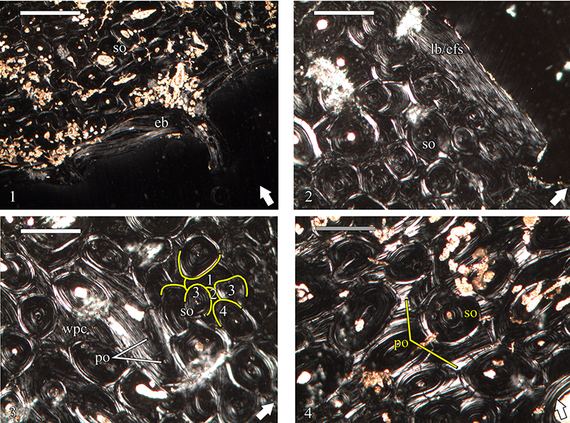
FIGURE 9. Osteohistology of the Bulgarian hadrosauroid in transmitted cross-polarized and plane-polarized light. Arrow in the lower right corner of each image indicates the direction of the periosteum. 1, Laminar bone tissue in the deep cortex of specimen NMNHS Mos19-1. The primary osteons are mostly of the longitudinal variety, or are short circumferential. The woven component dominates the bone matrix. Secondary osteons are scattered through the cortex; 2, Laminar bone tissue in the mid-cortex of specimen NMNHS Mos19-1. Note the much brighter appearance of the bone scaffolding, which is indicative of significant increase of the parallel-fibered component; 3, Laminar bone tissue in the mid-cortex of specimen NMNHS Mos19-3. The bone matrix is predominantly parallel-fibered in nature, as evidenced by the highly anisotropic optical character of the bone tissue. The degree of histological preservation is high enough to allow observation of the morphology of the osteocyte lacunae at medium magnifications; 4, Laminar bone tissue with large amounts of parallel-fibered tissue in the bone matrix in the deep cortex of specimen NMNHS Mos19-4. Locally, radially oriented primary osteons connect consecutive laminae giving the bone tissue a sub-plexiform appearance. The presence of erosional rooms indicates ongoing process of bone resorption; 5, Laminar bone tissue in the mid-cortex of specimen NMNHS Mos19-6; 6, Numerous large and irregularly shaped osteocyte lacunae in the woven bone scaffolding of the laminar bone in the deep cortex of NMNHS Mos19-6. The canaliculi system and its characteristics are easily observed at higher magnifications. Abbreviations: er-erosional room; pfb-parallel-fibered bone; po-primary osteon; so-secondary osteon. Scale bar equals 1, 4-5-500 µm; 2-3-200 µm; 6-50 µm.
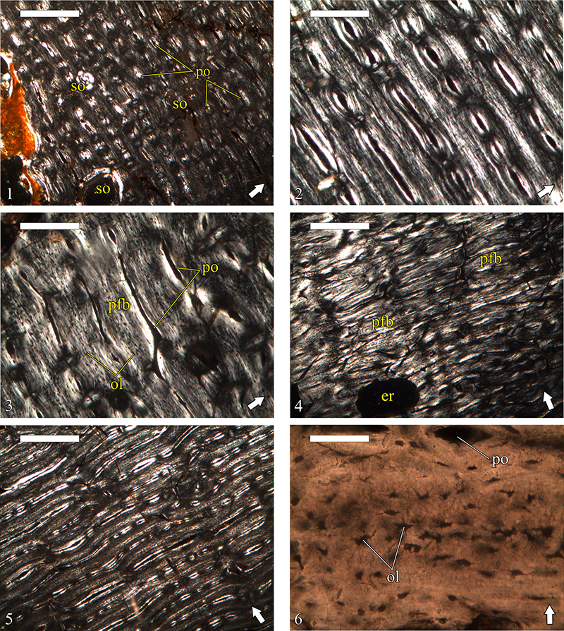
FIGURE 10. Osteohistology of the Bulgarian hadrosauroid in transmitted cross-polarized light. Arrow in the lower right corner of each image indicates the direction of the periosteum. 1, Locally restricted bone remodeling in the cortex of NMNHS Mos19-1-formation of secondary osteons in reticular primary bone at the transition towards primary bone tissue of laminar type; 2, Scattered secondary osteons formed over the reticular primary bone tissue of the deep cortex of NMNHS Mos19-2; 3, Haversian bone tissue in the inner cortex of NMNHS Mos19-3; 4, Haversian bone tissue in the cortex of NMNHS Mos19-4; 5, Detail of the dense Haversian bone of NMNHS Mos19-4 with four generation secondary osteons. Some of the osteons differ in appearance and osteocyte lacunae characteristics; 6, Numerous secondary osteons extending up to the sub-periosteal portions of the cortex of NMNHS Mos19-5; 7, Coarse compacted cancellous bone in the deep cortex of NMNHS F-31442; 8, Dense Haversian bone in the outer cortex of NMNHS F-31442. Abbreviations: pfb-parallel-fibered bone; so-secondary osteon; wpc-woven-parallel complex. Scale bar equals 1-4, 6-500 µm; 5, 7-8-200 µm.
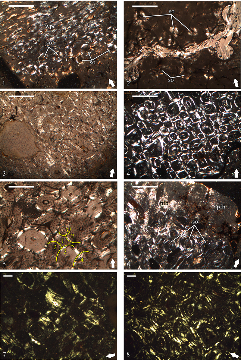
FIGURE 11. Comparison of secondary osteon radial microcracks and diagenetic alterations in the bones of Bulgarian non-avian dinosaurs. Arrow-heads point at selected microcracks which best illustrate the abundance of these structures and differences across specimens. Arrow in the lower right corner of each image indicates the direction of the periosteum. 1, Gross appearance of the Haversian bone in the cortex of NMNHS F-31436 (ornithomimosaurian humerus) in cross-polarized light. Bright spots and areas mark diagenetic mineralization, while dark brown/rusty colored areas indicate diagenetic alterations; 2, Detail of the Haversian bone of NMNHS F-31436 in cross-polarized light. Secondary osteons show numerous, but short and, more or less, “closed” radial microcracks; 3, Detail of the Haversian bone of specimen NMNHS Mos19-3 (unidentified cortical fragment) in cross-polarized light. Diagenetic mineralization, in the form of opaque optically isotropic mineral phase, is infilling diagenetic fractures, osteocyte lacunae and the vascular canal of some secondary osteons. Radial microcracks are numerous, well expressed, and sometimes widened; 4, Detail of the Haversian bone of specimen NMNHS Mos19-4 (unidentified cortical fragment) in cross-polarized light. Virtually identical in its characteristics to the Haversian tissue of NMNHS Mos19-3; 5, Secondary osteons in the deep cortex of U.S., K2 1586 (?titanosaurian partial diaphysis) in plane polarized light. Opaque mineral of diagenetic origin infills bone tissue’s osteocyte lacunae and some vascular canals. Other diagenetic alterations include fracturing. Osteonal radial microcracks are relatively rare, short, and “closed”; 6, Part of the same cortical area illustrated in 5, but in cross-polarized light, with some larger diagenetic fractures, partially filled with mineral phase, visible on the right side of the image; 7, Detail of the Haversian bone of NMNHS FR-16 (?titanosaurian long bone fragment) in cross-polarized light. Similarly to U.S., K2 1586 the bone wall is affected by diagenetic fracturing and opaque mineral phase is deposited in osteocyte lacunae and vascular canals. Radial microcracks are slightly more frequent, but identically “closed” and short in length; 8, Detail of the Haversian bone of NMNHS FR-16 in cross-polarized light. Scale bar equals 1-500 µm; 2-6-200 µm; 7-8-100 µm.
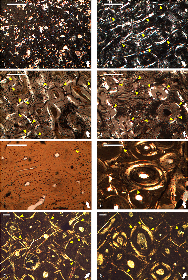
FIGURE 12. X-ray diffraction patterns of fossil bones from Vrabchov dol and Labirinta cave. The Miller indices of different Bragg reflections of fluorapatite are given on the top diffractogram. Abbreviations: Cal-Calcite; Mrc-Marcasite; Qz-Quartz.
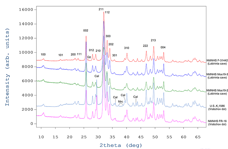
FIGURE 13. Average bone apatite crystallite size (in Å) for the fossil bones from Vrabchov dol and Labirinta cave.
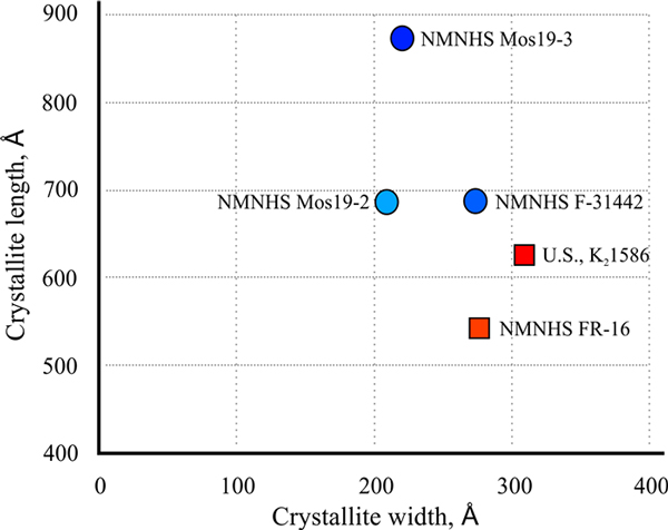
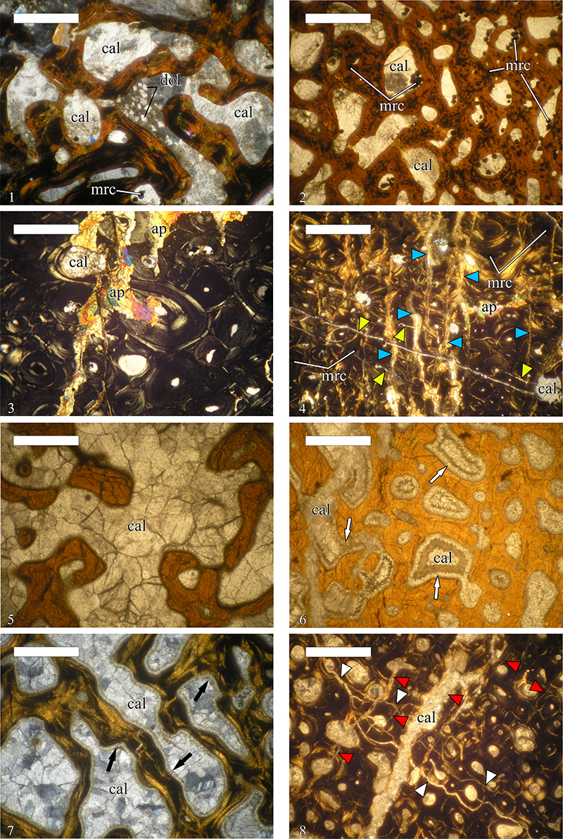
FIGURE 14. Mineralogy of specimens U.S., K2 1586 and NMNHS FR-16, two putative titanosaurian long bone fragments. 1, Large monocrystals of calcite infilling the intertrabecular spaces in the medullar region of U.S., K2 1586. Note that calcite crystals encompass several adjacent intertrabecular spaces. Small dolomite crystals are developed over the calcite; 2, Perimedullary, the intertrabecular spaces of U.S., K2 1586 are filled with calcite monocrystals and mosaics. Marcasite mineralization is infilling smaller voids and is locally developed over the calcite; 3, Locally developed later apatite mineralization overprinting the original histological features of specimen U.S., K2 1586; 4, Two systems of diagenetic fractures (marked with differently colored arrow-heads) in the cortex of U.S., K2 1586. Voids in the bone tissue (luminae and lacunae) are filled with calcite, or pyrite; 5, Calcite mosaics in the medullar region of specimen NMNHS FR-16; 6-7, Calcite syntaxial overgrowths (arrows) encrusting the bone trabeculae of NMNHS FR-16 in the perimedullar region at the transition to the compacta and in the medullar region, respectively; 8, Systems of diagenetic fractures (marked with differently colored arrow-heads) in the cortex of NMNHS FR-16. Abbreviations: ap-apatite; cal-calcite; dol-dolomite; mrc-marcasite. Scale bar equals 1-250 µm; 2-8-1000 µm.