FIGURE 1. Type material of Cheiracanthus murchisoni Agassiz, 1835 and its junior synonym C. microlepidotus Agassiz, 1844. 1-3, Holotype MHNN FOS 39 from Gamrie: 1, specimen in present condition; 2, closeup of midbody scales; 3, image in Agassiz (1833-43, plate 1c, figure 3). 4-9: Syntypes of the junior synonym C. microlepidotus from Lethen Bar: 4, 5, NHMUK PV P544; 6, 7, MHNN FOS 40; 8, 9, MHNN FOS 41. Scale bars equal 20 mm.
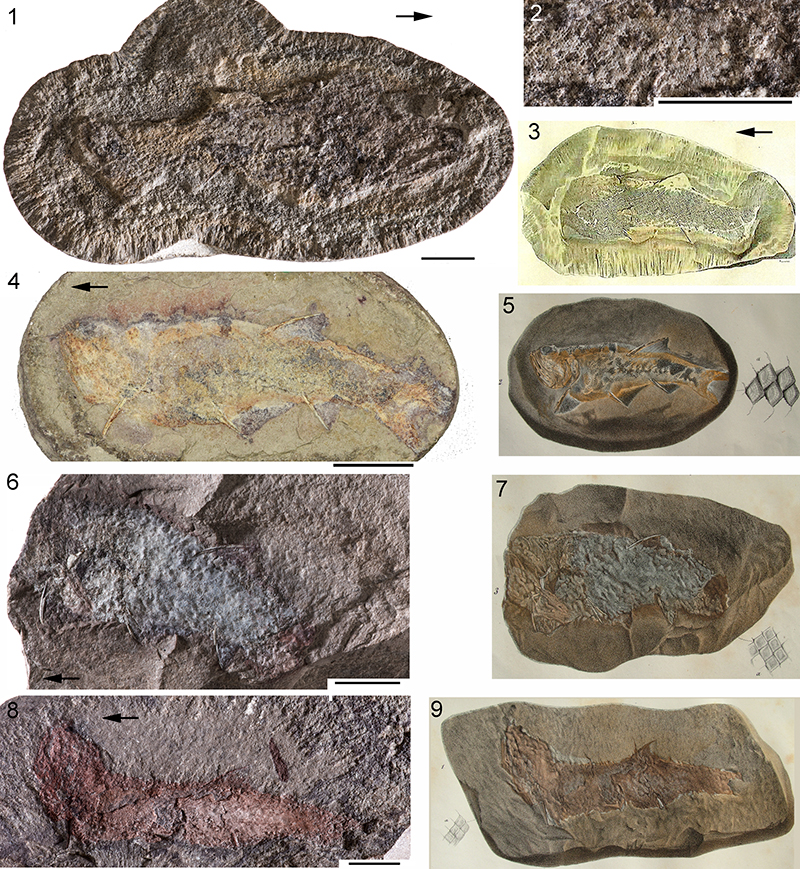
FIGURE 2. Syntypes of Cheiracanthus pulverulentus M’Coy, 1855 from Orkney, a junior synonym of Cheiracanthus murchisoni. 1, SM H4424, ‘syntype’ specimen in present condition; 2, SM H4424 specimen and scales as illustrated by M’Coy (1855, plate 2.B, figure 2), showing a mirror image of the specimen; 3, SM H4425, second ’syntype’, present condition. Scale bars equal 20 mm.
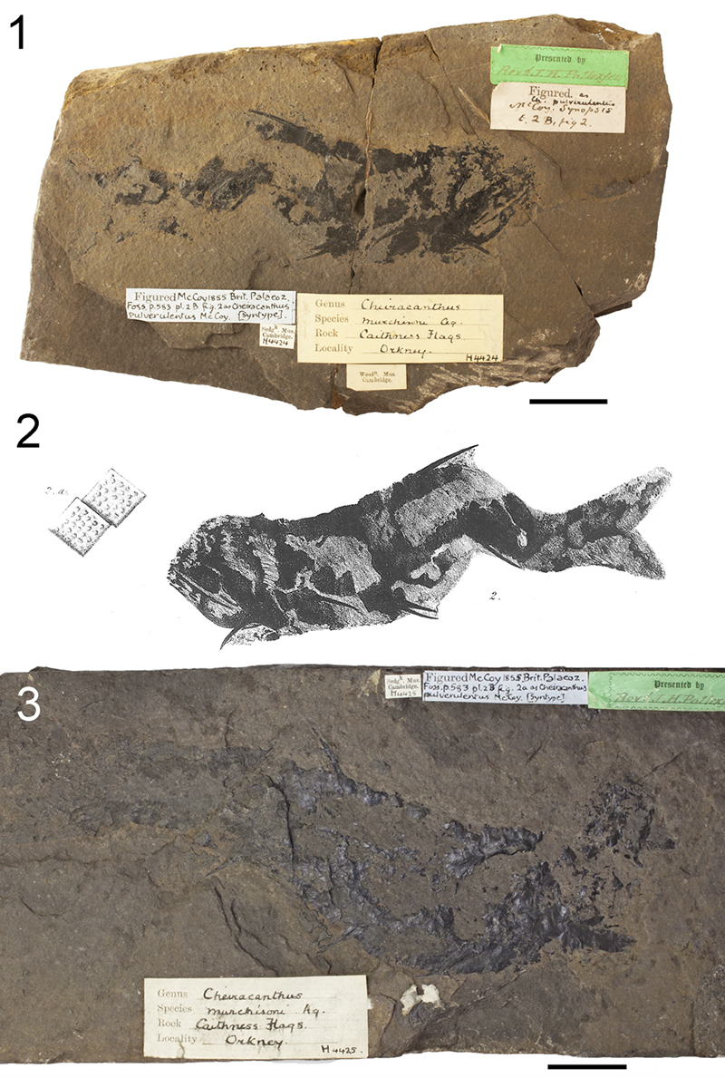
FIGURE 3. Cheiracanthus grandispinus holotype SM H4423 from Orkney. 1, mirrored illustration of specimen and scales from M’Coy (1855, plate 2.B, figure 1); 2, specimen in present condition. Scale bar equals 20 mm.
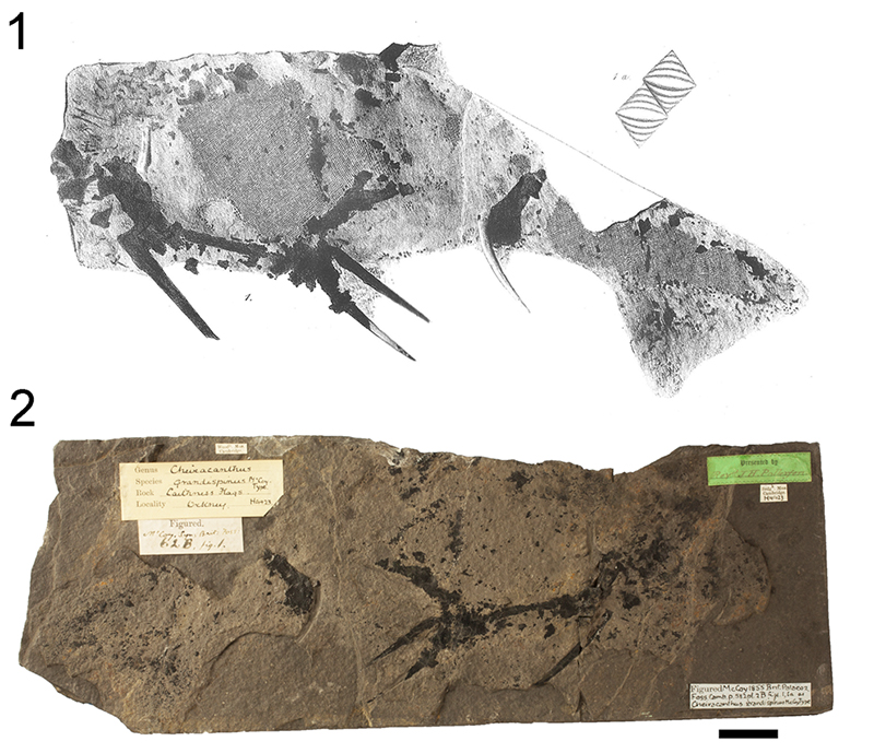
FIGURE 4. Cheiracanthus latus Egerton, 1861 holotype from Tynet Burn. 1, NHMUK PV P3253, half of specimen not previously figured; 2, half of the holotype specimen illustrated by Egerton (1861, plate 10, figure 1, 2), now lost. Scale bar equals 20 mm.
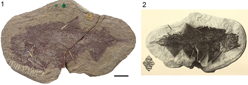
FIGURE 5. 1, Site map indicating the most important localities in northern Scotland where Middle Devonian Cheiracanthus specimens have been collected, land in white, current shoreline in blue, alluvial plains in yellow, alluvial fans in ochre; main lake area in blue; 2, biostratigraphic table of Cheiracanthus species occurrences in the Orcadian Basin of northern Scotland. Modified from Burrow et al. (2016, figure 2).
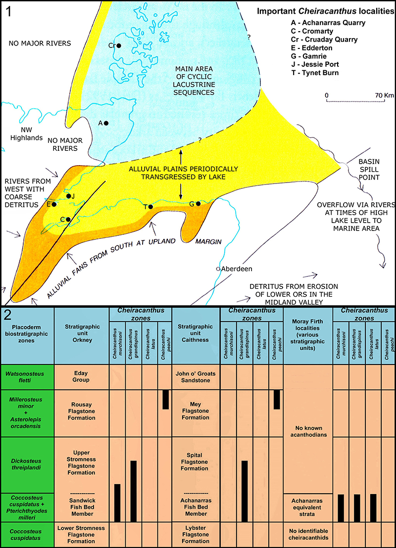
FIGURE 6. Specimens sacrificed for thin sectioning; numbered slices are vertical unless indicated. 1, 2, Cheiracanthus murchisoni NMS G.2019.3.6 from Gamrie; slices 36-38 horizontal. 3, 4, Cheiracanthus grandispinus, NMS G.2019.9.7 from Achanarras. 5, 6, Cheiracanthus latus NMS G.2019.3.3 from Jessie Port; slices 18, 19 horizontal. 7, 8. Cheiracanthus latus NMS G.2019.3.7 from Gamrie. Scale bars equal 20 mm. a.fs, anal fin spine; d.fs, dorsal fin spine; gr, gular rays; Mc, Meckel’s cartilage; pect.fs, pectoral fin spine; pelv.fs, pelvic fin spines; sc, scapulocoracoid; SEM, area where scales were removed for scanning electron microscopy; small numbers indicate suffix on NMS G specimen number for each thin section.
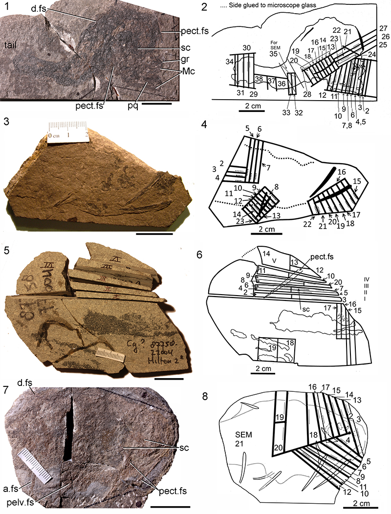
FIGURE 7. Cheiracanthus murchisoni head and branchial region morphology. 1, 2, NMS G.1884.60.3 from Tynet Burn. 3, NMS G.2019.9.24 from Cruaday Quarry, Orkney. 4, NRM P1651 from Gamrie, sclerotic ring. 5, NRM P1654 from Tynet Burn, sclerotic ring. 6, NMS G.2000.65.2 from Tynet Burn. 7, NMS G.2019.14.2 from Tynet Burn. 8, 9, NRM P1560 from Gamrie: 8, detail of branchiostegal rays; 9, impression of spiracular valve. Scale bars equal 10 mm in 1, 3, 6, 7; 5 mm in 4, 5; 2 mm in 8, 9. addf adductor muscle fossa; artc, articular cotylus; br, branchiostegal rays; ch, ceratohyal; gr, gular rays; Mc, Meckel’s cartilage; pdbr, posterodorsal branchiostegal rays; pq, palatoquadrate; pqv, palatoquadrate vacuity; pregl, preglenoid process; scl, sclerotic plate; t-l, tooth-like elements. Arrows indicate anterior.
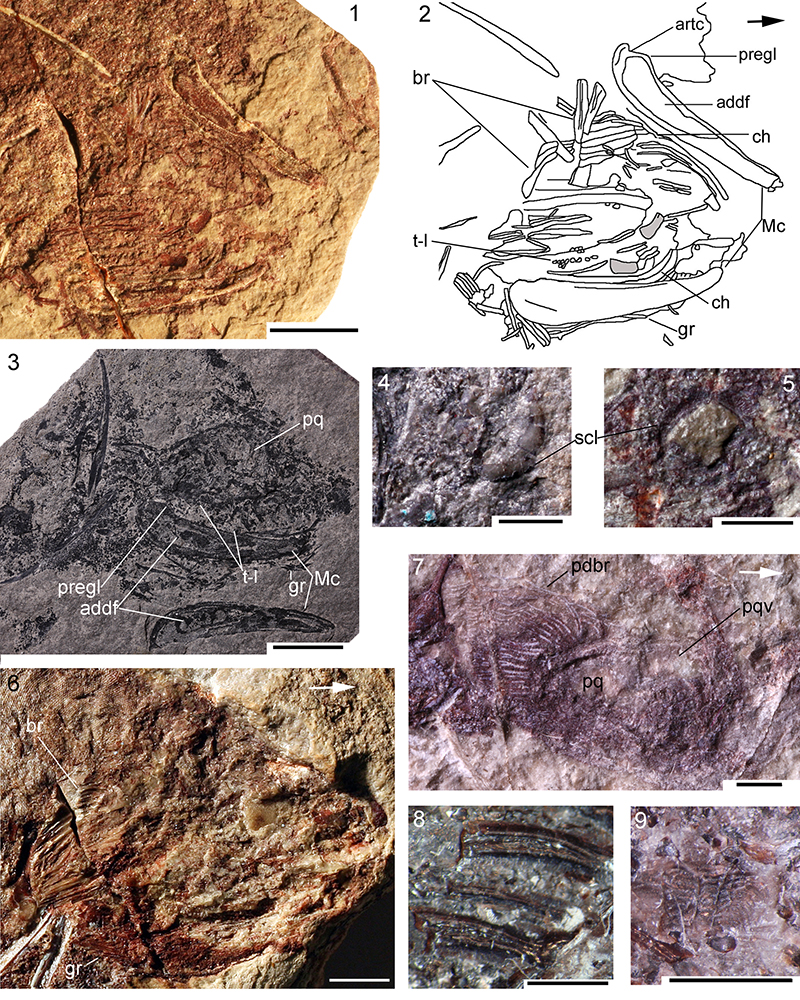
FIGURE 8. Cheiracanthus murchisoni head and branchial region histology. Thin sections of NMS G.2019.3.6 (see Figure 6.2 and text for position of sections). 1, NMS G.2019.3.6.2, Meckel’s cartilage; 2, NMS G.2019.3.6.7, calcified cartilage block showing rings of Liesegang; 3, NMS G.2019.3.6.3, separated calcified cartilage blocks overlain by dermal tessera; 4, NMS G.2019.3.6.4, cc and ?subcylindrical sensory line scale; 5, cc of palatoquadrate with dermal tesserae and pair of scales in vertical longitudinal section, edging sensory line; 6, NMS G.2019.3.6.6, ridge on dorsal edge of palatoquadrate cartilage, calcite infilling; 7, NMS G.2019.3.6.9, pair of sensory line scales, oblique section through base and lower crown; 8, NMS G.2019.3.6.12, dermal tesserae, vertical transverse sections; 9, NMS G.2019.3.6.9, pair of sensory line scales, vertical transverse section; 10, NMS G.2019.3.6.7, ceratohyal and another branchial arch; 11, NMS G.2019.3.6.6, branchial arches; 12, 13, NMS G.2019.3.6.9, ?tesserae and ?denticle of the branchial region; 14, 15, NMS G.2019.3.6.5, branchiostegal plates and calcified cartilage, with closeup of branchiostegal transverse section; 15, tessera with polyodontode crown on globular calcified cartilage; 16, NMS G.2019.3.6.4, branchiostegal plates and gular rays; 17, NMS G.2019.3.6.15, spiracular valve. Scale bars equal 1.0 mm. ba, branchial arches; br, branchiostegal plate/ray; cc, calcified cartilage; ch, ceratohyal; d, denticle; gr, gular rays; t, tessera; ?, indeterminate element, possibly subcylindrical sensory line scale.
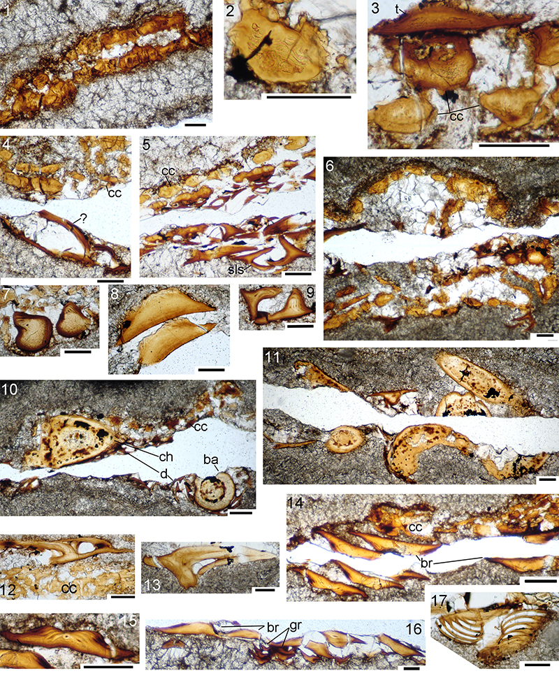
FIGURE 9. Cheiracanthus murchisoni spines and scapulocoracoid morphology. 1, NMS G.1891.92.307 from Gamrie, specimen showing disposition and relative sizes of fin spines and scapulocoracoid. 2, NMS G.1973.12.120 from Lethen Bar, articulated scapulocoracoid and pectoral fin spine. 3, QMF60005 from Tynet Burn, scapulocoracoid ‘shell’ with calcified cartilage core and calcified separation between molds of scapula and coracoid elements. 4, 5, NMS G.2019.9.27 from Edderton, pectoral spine. 6, NMV P29287 from Gamrie, anal fin spine, web, and basal cartilage. 7, NRM P1654 from Tynet Burn, dorsal fin spine with basal cartilage. 8, NMS G.1972.23.1 from Lethen Bar, dorsal fin spine with basal cartilage. Scale bars equal 20 mm in 1, 2, 6-8, 2 mm in 3-5. a.fs, anal fin spine; bc, basal cartilage; cor.m, coracoid mold; d.fs, dorsal fin spine; pect.fs, pectoral fin spine; pelv.fs, pelvic fin spines; sc.m, scapula mold. Arrow indicates anterior.
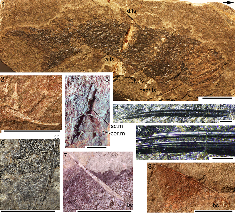
FIGURE 10. Cheiracanthus murchisoni NMS G.2019.3.6 from Gamrie, scapula shaft and pectoral spine histology. 1, NMS G.2019.3.6.27, transverse section of scapula shaft. 2-6, transmission microscope images of NMS G.2019.3.6.14, 15, 16, 18, 19; 7, drawings of main histological features in thin sections 19-14, from tip to near the base of the exserted part. Scale bars equal 0.1 mm in 1-3, 0.5 mm in 4-6. co, canal opening; en, enameloid; r, sharp crested lateral ridges.
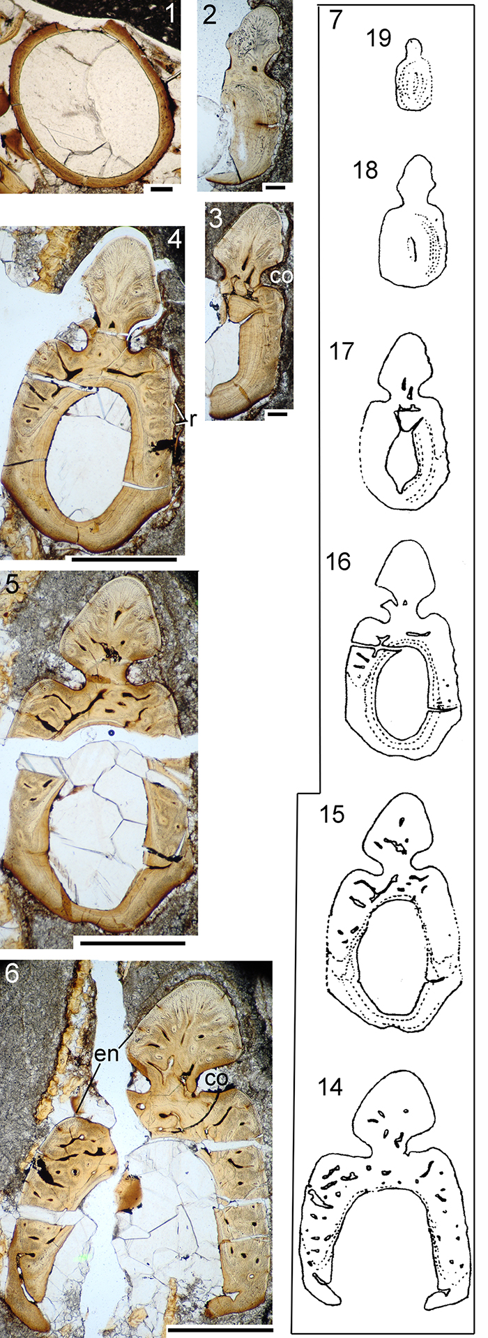
FIGURE 11. Cheiracanthus murchisoni squamation: light microscope images, all mid-body scales except 9. 1, NMS G.1892.8.1 from Gamrie. 2, NMS G.1891.92.323 from Lethen Bar. 3, NMS G.2019.9.25 from Cromarty, Sutors. 4, NMS G.2019.9.26 from Tarbat. 5, NMS G.1882.16.13 from Cromarty. 6, NMS G.1899.83.5 from Hooveth, Orkney. 7, NMS G.1877.30.2 from Tynet Burn. 8, NMS G.1968.5.3 from Edderton. 9, NMS G.2019.9.27 from Blackpark, tail. 10, NMS G.2019.9.28 from Blackpark, scales with crown pits. Scale bars equal 1.0 mm in 1, 2, 7, 8; 0.5 mm in 3-6, 9, 10. p, pit on posterior scale crown.
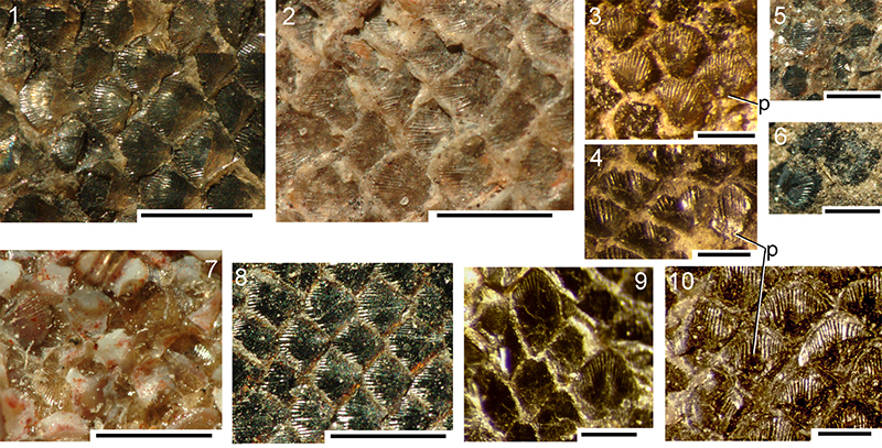
FIGURE 12. Cheiracanthus murchisoni scale morphology. 1-11, NMS G.2019.3.6 from Gamrie: 1-6, SEM images of scale patch NMS G.2019.3.6.35 removed from midflank of counterpart; 7-11, drawings of scale crowns from: 7, behind the head; 8, midbody; 9, near the dorsal fin spine; 10, in front of the tail; 11, on the tail. 12-15, SEM images of detached midbody? scales of NMS G.2019.9.29 from Cromarty Sutors: 12, NMS G.2019.9.29.11.5, crown view; 13, NMS G.2019.9.29.11.2, laterocrown view; 14, NMS G.2019.9.29.11.19, lateral view; 15, NMS G.2019.9.29.11.1, anterolateral view. 16, 17, NMS G.2019.9.29.12 from Cromarty Sutors: 16, NMS G.2019.9.29.12.3, base showing concentric lines of Sharpey’s fibre bundles; 17, NMS G.2019.9.29.12.2, patch of articulated scales, crown view. 18, 19, ventral mid-body squamation patches of NMS G.2019.9.31.2 from Den of Findon. 20, NMS G.2019.9.30.2 from Edderton, scale in squamation patch. Scale bars equal 1.0 mm in 1-3, 0.3 mm in 4-11 (7-11 all at same scale), 17, 18, 20; 0.1 mm in 12-16, 20.
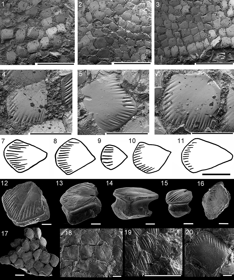
FIGURE 13. Cheiracanthus murchisoni scale histology. 1-5, 9-11: thin sections of scales from NMS G.2019.3.6 from Gamrie: 1, NMS G.2019.3.6.20, slightly oblique vertical section of mid-body scale; 2, NMS G.2019.3.6.32, vertical transverse section of ventral scale in the shoulder girdle region; 3, NMS G.2019.3.6.19, vertical transverse section of scale behind the head; 4, NMS G.2019.3.6.33, vertical longitudinal section of two contiguous scales in the midbody flank region; 5, NMS G.2019.3.6.36, subhorizontal section through base and lower crown; 9-11, NMS G.2019.3.6.28, vertical longitudinal section through fin web and body scales near pectoral fin spine: 9, distal fin scales; 10, fin web and body scales; 11, fin web scale and fin rays=ceratotrichia. 6, NMS G.2019.9.29.5 from Cromarty, vertical oblique section. 7, NMS G.2019.9.29.10 from Cromarty Sutors, vertical transverse section through posterior crown;8, NMS G.2019.9.33.6, from Jessie Port, vertical transverse section. Scale bars equal 0.1 mm. ac, ascending canals; bl, base lamellae; cir, circular canals; cW, canals of Williamson; en, enameloid; ez, embryonic zone; fr, fin ray; lac, canal lacunae; p, protuberance; rc, radial canal; Sf, Sharpey’s fibre bundles. Arrows indicate anterior.
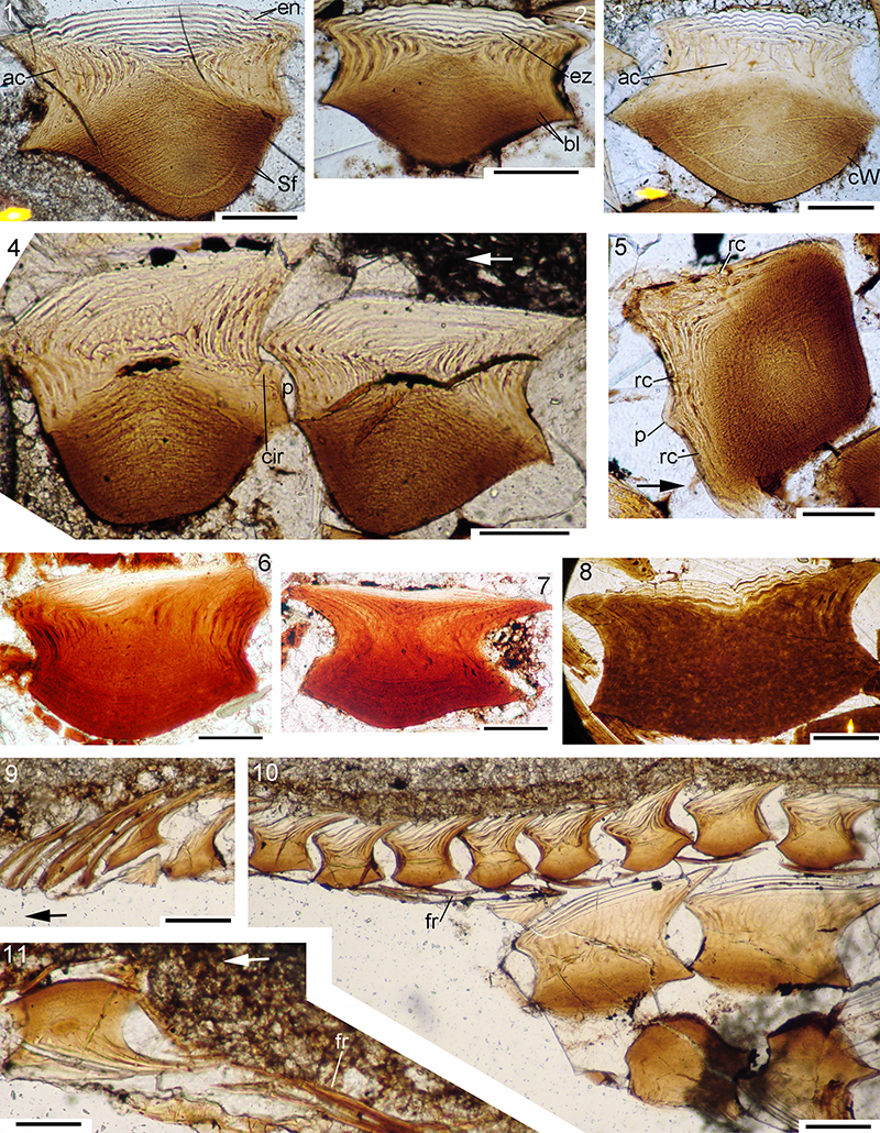
FIGURE 14. Cheiracanthus murchisoni head and branchial region reconstruction.
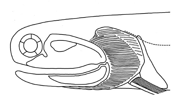
FIGURE 15. Cheiracanthus grandispinus head and branchial region, morphology. 1-2, NMS G.1966.40.24 from Lethen Bar. 3, NMS G. FR1603 from Achanarras Quarry, Caithness. 4, NMS G.2002.26.1481 from Achanarras Quarry. 5, NMS G.1903.130.19 from Achanarras Quarry, dorsoventrally flattened. 6, NHMUK PVP.1363 from Orkney. Scale bars equal 10 mm. addf, adductor muscle fossa; artc, articular cotylus; br, branchiostegal rays; cr.epq, extrapalatoquadrate ridge; gr, gular rays; Mc, Meckel’s cartilage; pdbr, posterodorsal branchiostegal rays; pq, palatoquadrate; pqv, palatoquadrate vacuity; pregl, preglenoid process. Anterior to right in 1-3, 6, to left in 4-5.
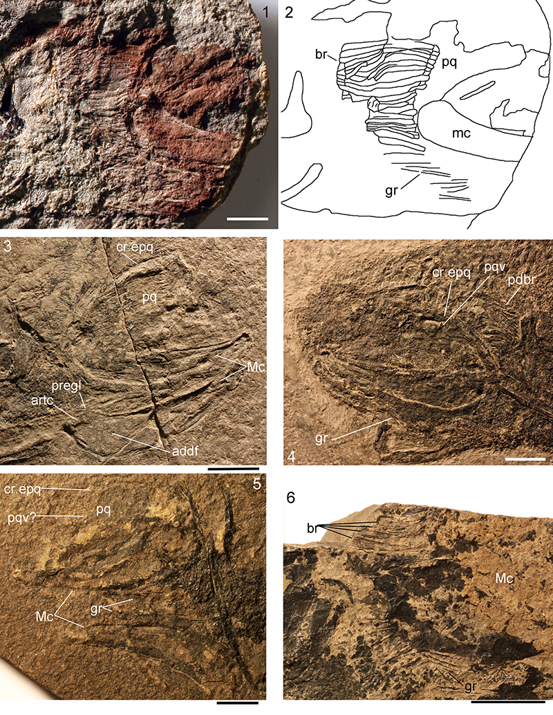
FIGURE 16. Cheiracanthus grandispinus head and branchial region, histology. 1-3, NMS G.2019.9.8: 1, 2, NMS G.2019.9.8.8 series of endoskeletal branchial rays, some showing gill raker projections; 3, NMS G.2019.9.8.14 calcified cartilage blocks; 4, NMS G.2019.9.35.1, calcified cartilage of Meckel’s cartilage? Scale bars equal 0.1 mm. gb, gill bars; gbp, gill bar projections; gcc, globular calcified cartilage; s, scale.
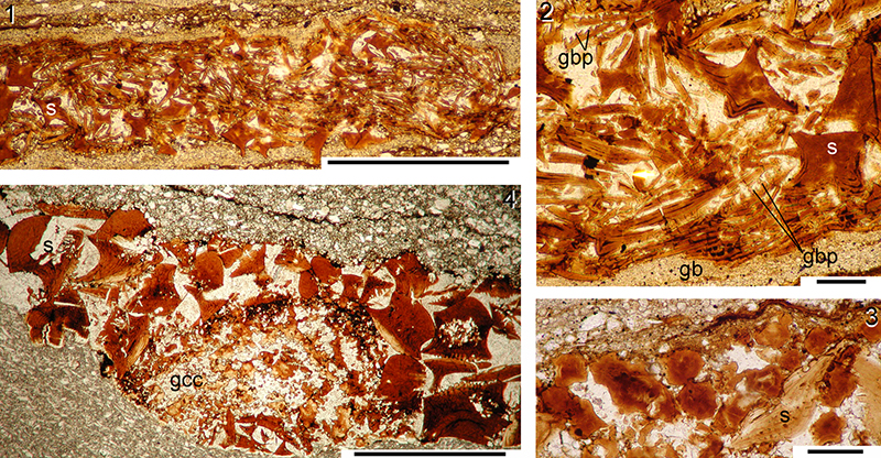
FIGURE 17. Cheiracanthus grandispinus pectoral region, spines morphology. 1, USCP F00130a from Achanarras, whole fish. 2, NMS G.FR1810 from Achanarras, scapulocoracoids and pectoral fin spines. 3, NMS G.2019.9.12, isolated scapulocoracoid from Achanarras. 4, disarticulated incomplete fish NMS G.2019.9.10.1 from Cromarty, ?two pectoral, one median (?dorsal), one pelvic spine. Scale bars equal 50 mm in 1, 10 mm in 3, 4, 2 mm in 2. cor, coracoid; fs, indeterminate fin spine, probably a pelvic; median fs, dorsal or anal fin spine; pect.fs, pectoral fin spine; pect. fw, pectoral fin web; sc.s, scapular shaft. Anterior to right.
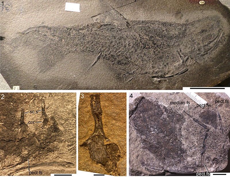
FIGURE 18. Cheiracanthus grandispinus spines histology. 1-5, NMS G.2019.9.7 from Achanarras: 1-3, NMS G.2019.9.7.21, thin section of distal end of pectoral spine, with closeups of the structure of the leading edge ridge and boundary between the main spine osteodentine and the areas bounding the trailing edge groove; 4, NMS G.2019.9.7.11, TS pelvic spine; 5, NMS G.2019.9.7.12, TS through both pelvic fin spines. 6, 7, NMS G.2019.9.8 from Jessie Port, median spine, ?anal: 6, TS NMS G.2019.9.8.8 at proximal end of exserted part; 7, NMS G.2019.9.8.15, towards mid spine. 8-10, NMS G.2019.9.35.1-7 from Achanarras, pectoral spine: 8, 9, NMS G.2019.9.35.2, midspine; 10, NMS G.2019.9.35.3, more distal. Scale bars equal 0.5 mm in 1, 4-8, 10, 0.1 mm in 2, 3, 9. bc, basal cartilage.
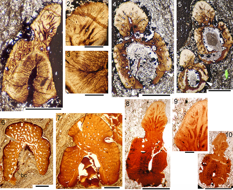
FIGURE 19. Cheiracanthus grandispinus NMS G.2019.9.7, serial sections of pectoral (22-15) and pelvic (9-14, 23) spines, thin section drawings, numbered as on map in Figure 6.4.
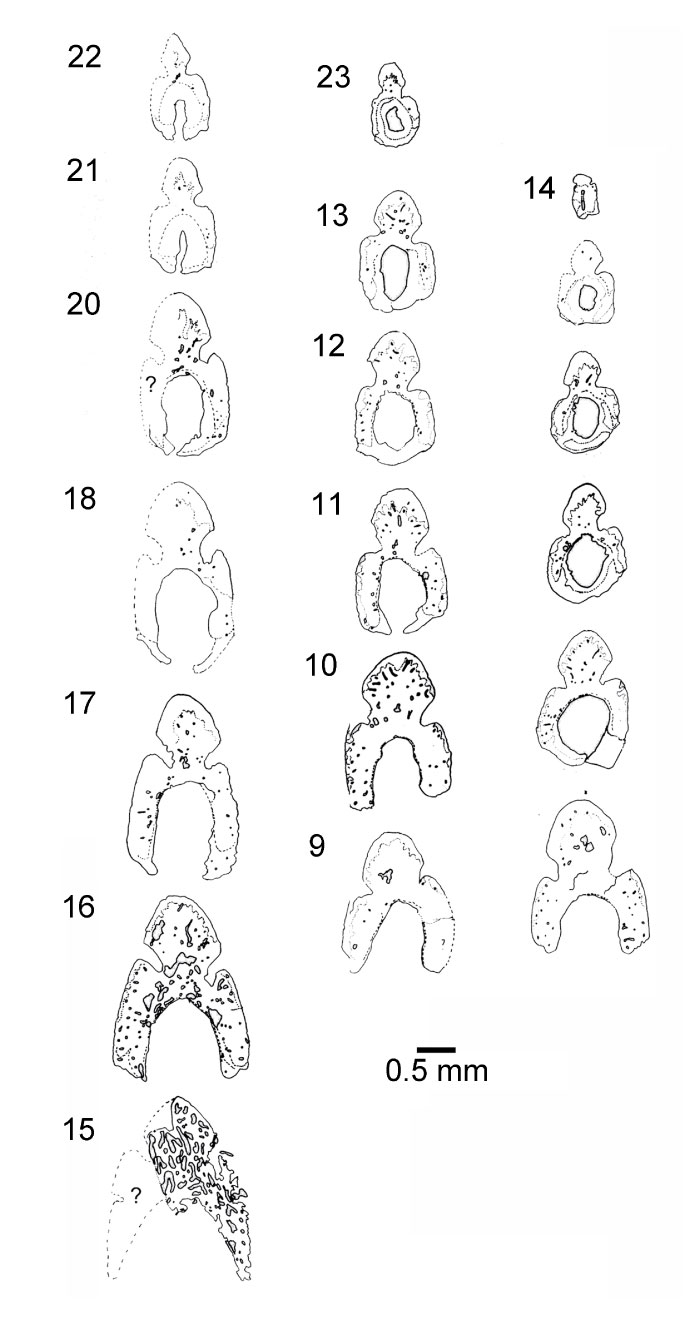
FIGURE 20. Cheiracanthus grandispinus scale morphology. 1-3, 5-9, light microscope images; 4, 10-18, SEM images; midbody scales unless detailed otherwise. 1, NMS G.1891.92.317 from Lethen Bar. 2, NMS G.2019.9.8 from Jessie Port, Ross and Cromarty. 3, 4, USCP F00115b from Edderton. 5, NMS G.2019.9.9 from Achanarras, primordial scale revealed after abrasion/flaking of overlying crown growth zones. 6-9, NMS G.2019.9.10.1 from Cromarty: 6, mid-body scales; 7-9, pectoral fin web: spine and web; 8, distal scales; 9, scales near fin base. 10-12, NMS G.2019.9.11 from Marwick: 10, view of crown before removal of scale from matrix; 11, anterocrown view; 12, posterior view. 13, NMS G.2019.9.3.27.1 from Gamrie Den of Findon, laterocrown view. 14-18, NMS G.2019.9.13.8 from Cromarty, Eathie to Navity: 14, NMS G.2019.9.13.8.16, anterocrown view; 15, NMS G.2019.9.13.8.2 pair of scales in posterocrown view; 16, NMS G.2019.9.13.8.8, laterocrown view; 17, NMS G.2019.9.13.8.6, lateral view; 18, NMS G.2019.9.13.8.17, posterobasal view. Scale bars equal 0.5 mm in 1-9, 0.2 mm in 10-18. p, neck protuberance; ps, primordial scale. Arrows indicate anterior.
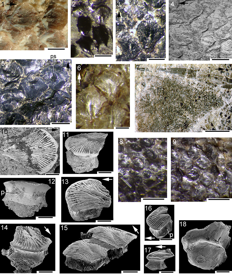
FIGURE 21. Cheiracanthus grandispinus scale histology. 1, NMS G.2019.9.3.9 from Gamrie, vertical longitudinal section through posterior protuberance. 2, 9, 12, NMS G.2019.9.8 from Jessie Port, Ross and Cromarty: 2, NMS G.2019.9.8.14, anterior crown vertical transverse section; 9, NMS G.2019.9.8.10, crown vertical transverse section; 12, NMS G.2019.9.8.13, crown horizontal section. 3, NMS G.2019.9.35.6, from Achanarras, midscale vertical transverse section. 4, 5, 10, 11, from Achanarras: 4, NMS G.2019.9.2.4, posterior half of scale, vertical transverse section; 5, NMS G.2019.9.2.2, vertical longitudinal section; 10, NMS G.1893.107.9.2, horizontal section through crown base; 11, NMS G.1893.107.9.2, horizontal section through midcrown. 6-8, NMS G.2019.9.10 from Cromarty: 6, NMS G.2019.9.10.3, off-centre vertical longitudinal section; 7, NMS G.2019.9.10.5, vertical transverse section; 8, NMS G.2019.9.10.9, oblique vertical section through side of scale. Scale bars equal 0.1 mm. Arrows indicate anterior.
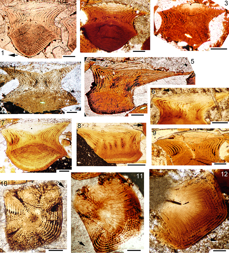
FIGURE 22. Cheiracanthus grandispinus comparison of scale structure in different sized fish. 1, NMS G.2019.9.5.2, from Achanarras, small fish c. 10 cm long, vertical transverse section through scale anterior. 2, NMS G.2019.9.6.1, from Achanarras, medium sized fish less than 30 cm long, vertical transverse section through scale anterior. 3, NMS G.2019.9.8.12, from Jessie Port, medium sized fish, vertical transverse section through scale anterior. 4, NMS G.2019.9.9.14, from Achanarras, fish 30 cm or longer, vertical transverse section through scale anterior. 5, NMS G.2019.9.9.7, from Achanarras, fish 30 cm or longer, vertical longitudinal section. NMS G.2019.9.35.4, medium sized incomplete fish, vertical section of scale showing Sharpey’s fibre bundles through base. Scale bars equal 0.1 mm.
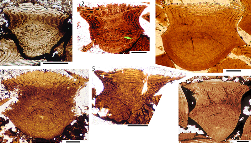
FIGURE 23. Cheiracanthus grandispinus head and branchial region reconstruction.

FIGURE 24. Cheiracanthus latus head and branchial region, morphology. 1-2, NHM PV P 3253 from Tynet Burn. 3, NMS G.1891.92.320 from Gamrie. 4, NHMUK PVP43273 from Tynet Burn, branchial region with ridges visible on lowest branchiostegal plates. Abbreviations: br.l, left branchiostegal plates; br.r, right branchiostegal plates; cop, circumorbital plate; gr, gular rays; Mc.l, left Meckel’s cartilage; Mc.r, right Meckel’s cartilage; pect.fs, pectoral fin spines; pq.l, mineralised cartilage along ventral edge of left palatoquadrate; proc, procoracoid; sc, scapulocoracoids; tess, head tesserae. Scale bars equal 10 mm.
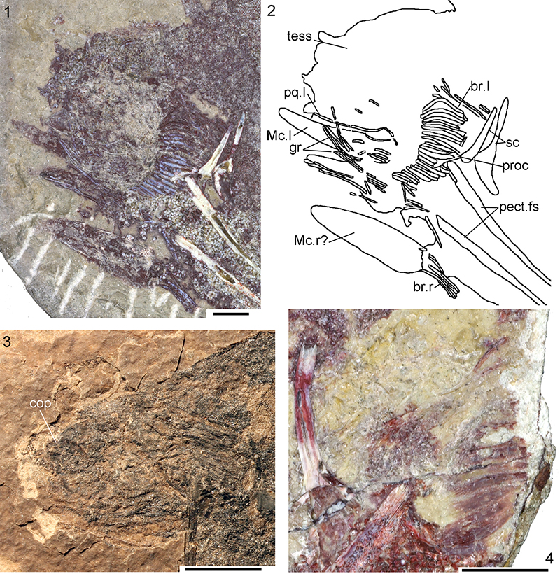
FIGURE 25. Cheiracanthus latus head and branchial region, histology, NMS G.2019.3.3; position of sections shown in Figure 6.6. 1, 2, thin section NMS G.2019.3.3.3: 1, ?Meckel’s cartilage; 2, closeup of calcified cartilage blocks; 3, 4, NMS G.2019.3.3.5, indet. jaw cartilage and branchial arches; 5, NMS G.2019.3.3.2, branchiostegal plate in oblique section, body scales, branchial denticles, indet. cartilage; 6, NMS G.2019.3.3.4, body scales, gill bars; 7, 8, NMS G.2019.3.3.17: 7, jaw cartilage, head tesserae, gill bars, branchiostegal plates; 8, head tessera vertical section, stellate mineralisation artifacts. Scale bars equal 1.0 mm in 1, 3, 5, 7, 0.1 mm in 2, 4, 6, 8. ba, branchial arch; br, branchiostegal plate; ccb, calcified cartilage blocks; d, denticle; gb, gill bar; gcc, globular calcified cartilage; gz, growth zones in crown; s, body scale; t, tessera.
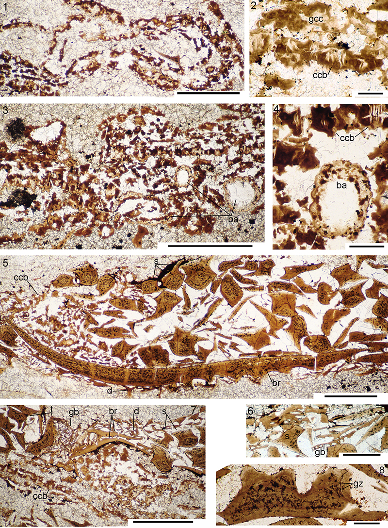
FIGURE 26. Cheiracanthus latus pectoral region and spines, morphology. 1-3, QMF60004 from Tynet Burn: 1, left and right scapulocoracoids, white arrows indicate separation between scapula and coracoid; 2, close up of right coracoid shape (dotted line); 3, dorsal fin spine insertion and basal cartilage, white arrow indicates insertion/exertion boundary. 4-6, NMS G.2018.28.26 from Tarrel Bay: 4, left and right pectoral spines as collected; 5, closeup of thin ridges on lateral or medial? side of right spine; 6, insertion area of right spine. 7, NMS G.1968.19.18, anal and pelvic spines. Scale bars equal 5.0 mm in 1-3, 6, 20 mm in 4, 7, 0.5 mm in 5. a.fs, anal fin spine; bc, basal cartilage; calc, calcite infill; pelv.fs, pelvic fin spines.
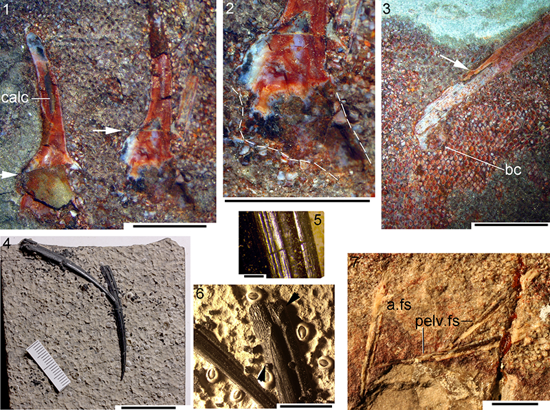
FIGURE 27. Cheiracanthus latus pectoral region and spines, histology. 1-6, NMS G.2019.3.31 (see Figure 6.6 for map of sections): 1, slice II before grinding (corresponds to sections 6, 7), arrow indicates dorsal direction; 2, NMS G.2019.3.3.7, scapulocoracoid, long section mid-shaft; 3, NMS G.2019.3.3.12, TS pectoral spine at insertion/exertion boundary; 4, NMS G.2019.3.3.10, TS of spine close to insertion; 5, NMS G.2019.3.3.6, close up of leading edge ridge structure of pectoral spine, midspine; 6, NMS G.2019.3.3.2, TS pectoral spine near the tip. 7-10, NMS G.2018.28.26, TS pectoral spines: 7, 2018.28.26.9, close to insertion; 8, 2018.28.26.7, midspine; 9, 10, 2018.28.26.5, towards tip, with closeup of infilling dentine. 11-14, NMS G.2019.3.7 TS pectoral spine, near insertion, and flange of scapulocoracoid: 11, NMS G.2019.3.7.8; 12, NMS G.2019.3.7.9; 13-14, NMS G.2019.3.7.10, outer side of spine with thin sharp ridges. Scale bars equal 10 mm in 1; 0.1 mm in 2, 5, 10; 0.5 mm in 3, 4, 6, 7-9, 11-14. c.cav, central cavity; ccb, calcified cartilage blocks; dt, dentine tubules; en, enameloid; l.pect.fs, left pectoral fin spine; min, stellate mineralisation; ost, osteodenteon; pect.fs, pectoral fin spine; r, ridges; s, scale; sc, scapulocoracoid.
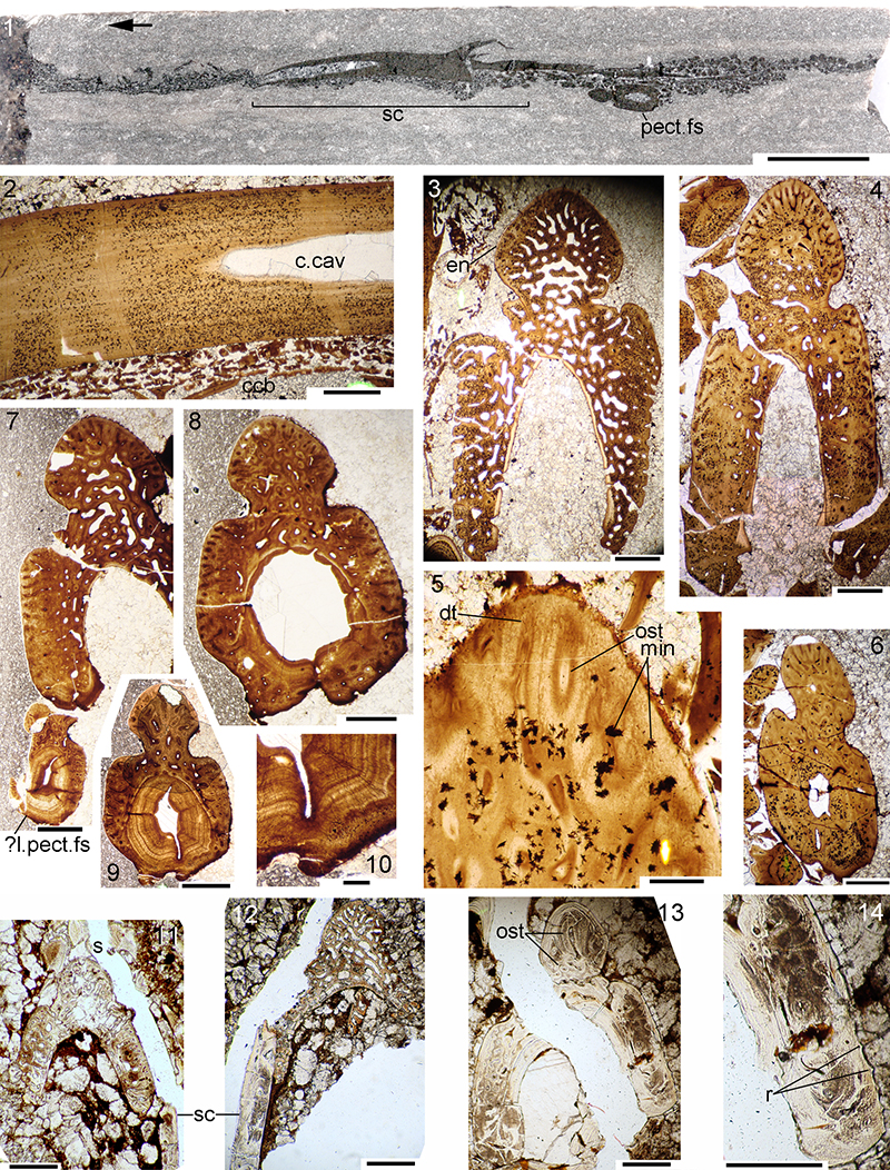
FIGURE 28. Cheiracanthus latus pectoral spines, serial transverse section drawings. 1, NMS G.2018.28.26 (see Figure 27): sketch map and drawings of each section. 2, NMS G.2019.3.7 (see Figure 6.7, 8): sections NMS G.2019.3.7.6-12 through pectoral spines and scapulocoracoid.
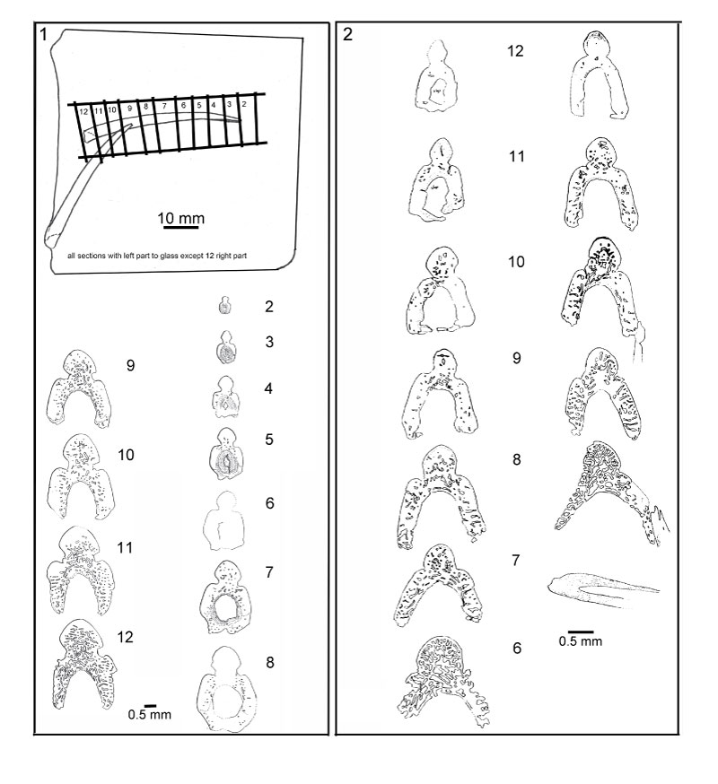
FIGURE 29. Cheiracanthus latus squamation, light microscope images. 1, 2, QMF60004 from Tynet Burn: 1, midflank scales; 2, caudal peduncle, scale impressions. 3, 4, NMS G.2018.28.26 from Tarrel Bay. 5-7, NMS G.2019.3.7 from Den of Findon, Banffshire 8, NMS G.2019.3.3 from Jessie Port. 9, NMS G.1870.14.145 from Cromarty. 10, NMS G.2019.9.17 from Geanies Point. Scale bars equal 0.5 mm. p, pit. Arrows indicate anterior.
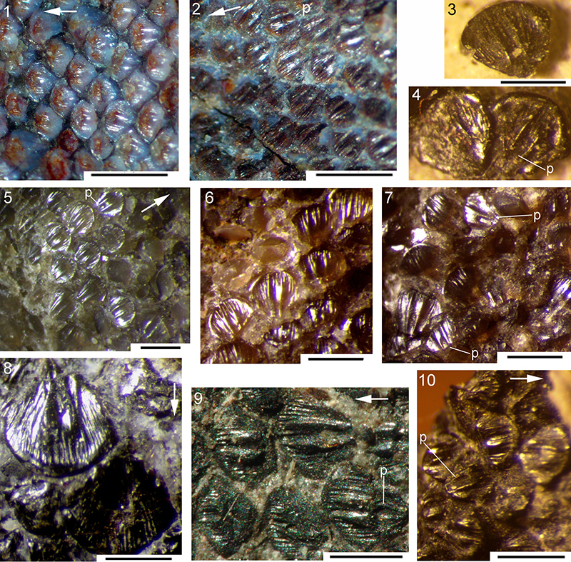
FIGURE 30. Cheiracanthus latus squamation, SEM images. 1-5, NMS G. 2019.3.7.21 from Den of Findon: 1, NMS G. 2019.3.7.21.2, crown view; 2, NMS G. 2019.3.7.21.5, crown view; 3, NMS G. 2019.3.7.21.13, anterior view; 4, NMS G. 2019.3.7.21.7, laterocrown view; 5, NMS G. 2019.3.7.21.14, laterocrown view. 6-7: NMS G.2019.14.9.2 from Geanies Point: 6, NMS G.2019.14.9.2.8, anterocrown view; 7, NMS G.2019.14.9.2.5, crown view. 8-10: NMS G.2019.14.4.2 from Hilton or Cadboll: 8, NMS G.2019.14.4.2.15, crown view; 9, NMS G.2019.14.4.2.5, crown view; 10, NMS G.2019.14.4.2.16, laterocrown view. Scale bars equal 0.2 mm. p, pit; pr, neck protuberance.
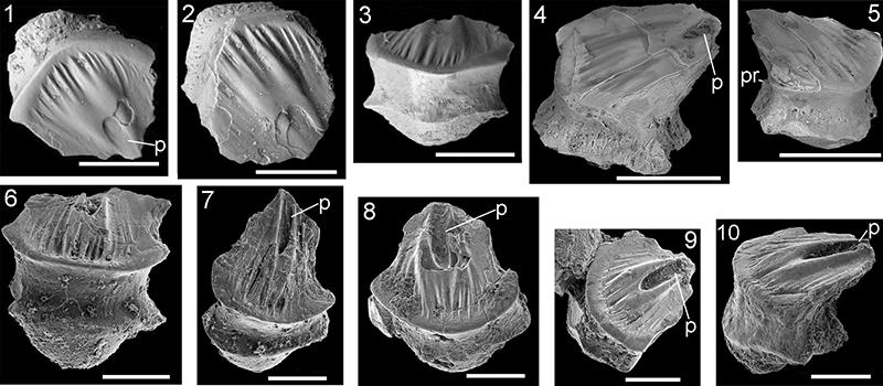
FIGURE 31. Cheiracanthus latus scale histology. 1, NMS G.2019.9.21.5, vertical transverse section, anterior half of scale. 2, NMS G.2019.9.19.2, vertical transverse section, anterior half of scale. 3, NMS G.2019.9.20.3, vertical transverse section, midscale. 4, 5, NMS G.2019.3.3.14, vertical transverse section, midscale, with closeup of central region. 6, NMS G.2019.9.21.3, vertical transverse sections of two scales, posterior half of scale. 7, NMS G.2019.3.3.12, vertical transverse section towards posterior end of scale. 8, NMS G.2019.3.7.6, subhorizontal section through anterior base and posterior crown. 9, NMS G.2019.3.7.6, vertical oblique section through posterior crown and base. 10, NMS G.2019.9.19.7, horizontal section low in crown. 11, NMS G.2019.9.22.5, horizontal section high in crown. 12, 13, NMS G.2019.9.23.7, section through fin web scales including tiny distal scales with a deep neck and flat base, closeup of distal fin web scales in 13. Scale bars equal 0.1 mm in 1-11,13, 0.5 mm in 12. Arrows indicate anterior.
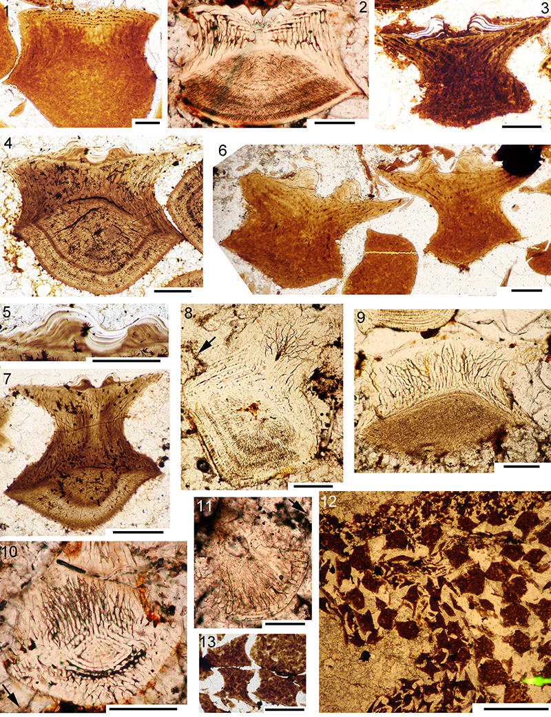
FIGURE 32. Cheiracanthus latus head and branchial region reconstruction.
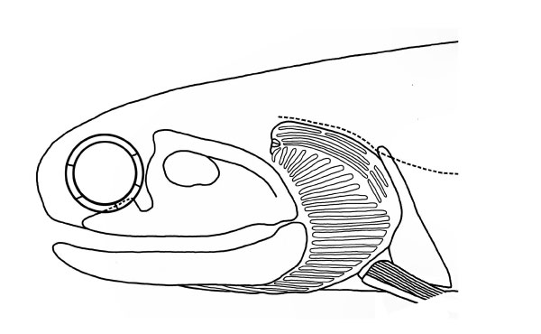
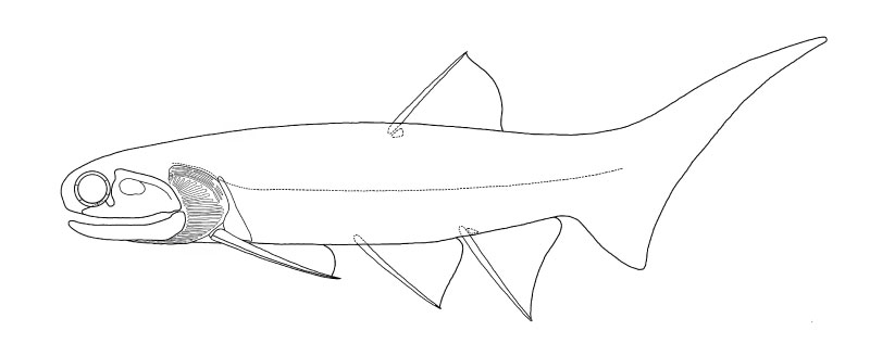
FIGURE 33. Reconstruction of a whole Cheiracanthus latus in lateral view.