FIGURE 1. (A) Single x-ray projection. Schematic representation of positioning of the investigated object inside x-ray device. (B) Multiple x-ray projections as the object rotates. Positioning of investigated object inside micro-CT device. (C) Example of 3D dataset, i.e., a group of 2D slice images acquired by the MicroCT scanner. (D) Examples of Volume rendering; technique in visualization and computer graphics, used to display object from 3D data set in different aspects and orientations.
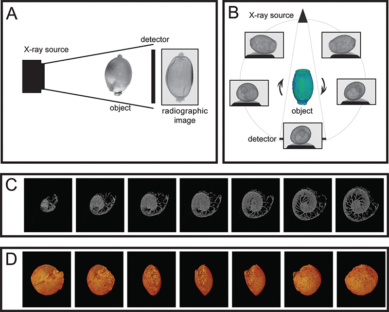
FIGURE 2. Custom-made holders specially adapted for each scanned specimen. (A) Plastic cup. (B) Polystyrene holder. (C) Aluminum holder for small specimens. (D) Plastic tube filled with polystyrene.
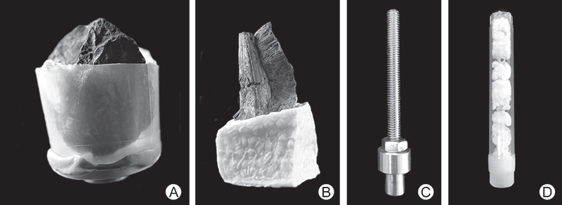
FIGURE 3. Siliceous nodules of the Šárka Formation. (A, B) Pricyclopyge binodosa, complete trilobite, no. NMP L 35055, locality Praha-Šárka, Middle Ordovician (Darriwilian), (A) Enrolled trilobite coated with ammonium chloride, exterior of objects. (B) Micro-CT image showing dense burrows, interior of objects. (C, D) Rostrum with eyes of a trilobite P. binodosa, no. NMP L46892, locality Praha-Šárka, Middle Ordovician (Darriwilian). (C) Rostrum coated with ammonium chloride, exterior of objects. (D) Micro-CT visualization of tunnels, interior of objects. (E) Bivalve Redonia deshayesi, micro-CT image showing trace fossils, interior of objects, no. NMP L 51722, locality Osek, Middle Ordovician (Darriwilian). All scale bars equal 5 mm.
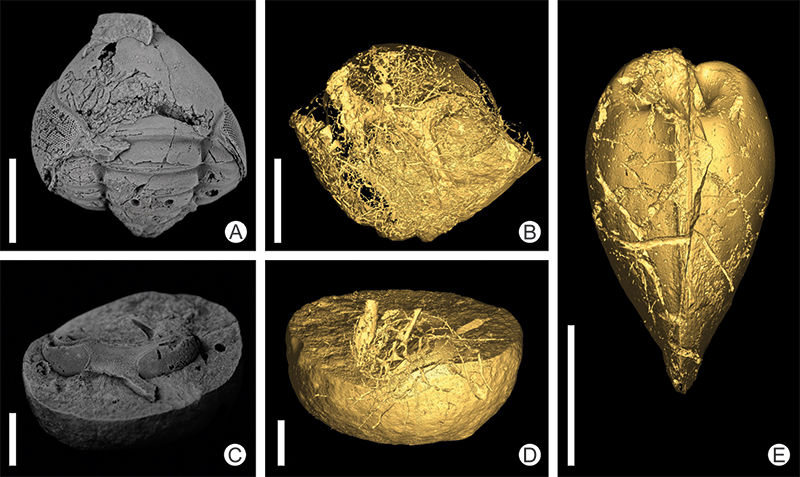
FIGURE 4. Ordovician bryozoan colony. One-half of hemispherical bryozoan, interior of object, bearing probably oldest boring attributable to ichnogenus Entobia Bronn, 1837. Besides semi-radial tunnels and exploratory threads, three bulbous chambers discovered near the center of the hemisphere. Darriwilian (middle Ordovician), Khrevitsa locality, St. Petersburg Region, Russia. Scale bar equals 1 cm.
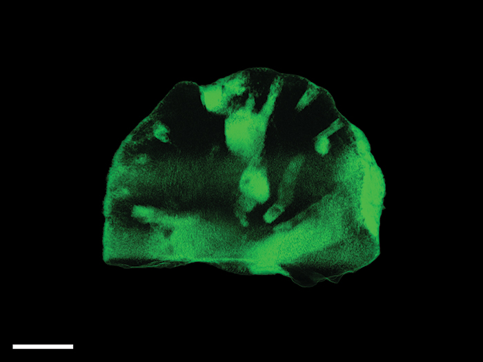
FIGURE 5. Minute conulariid specimen. (A) Conulariid specimen of Archaeoconularia fecunda and trepostome bryozoan colony; coated with ammonium chloride, no. NMP L21990, locality Loděnice, Upper Ordovician, Zahořany Formation (lower Katian) (B) Micro-CT visualizing of inner surfaces. Scale bar equals 5 mm.
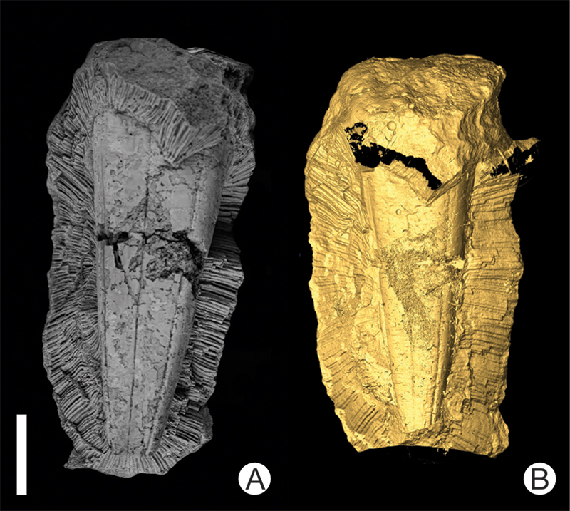
FIGURE 6. Tube fragments of the serpulid polychaete Pyrgopolon (Pyrgopolon) deforme. Left images show exterior of objects; right images show interior of objects. (A) Specimen encrusted with bryozoan colonies and serpulid worms, boreholes assigned to Entobia Bronn, 1837, representing the most common ichnogenus in the examined serpulid tubes, no. MHNLM EMV 2016.3.14. (B) Intensely bored specimen preserving tunnels of ichnogenera Entobia and Trypanites Mägdefrau, 1932, no. MHNLM EMV 2016.3.44. (C) Serpulid tube with Entobia boreholes and encrusting juvenile oyster, no. MHNLM EMV 2016.3.40. Scale bar equals 1 cm.
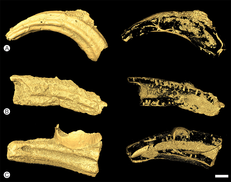
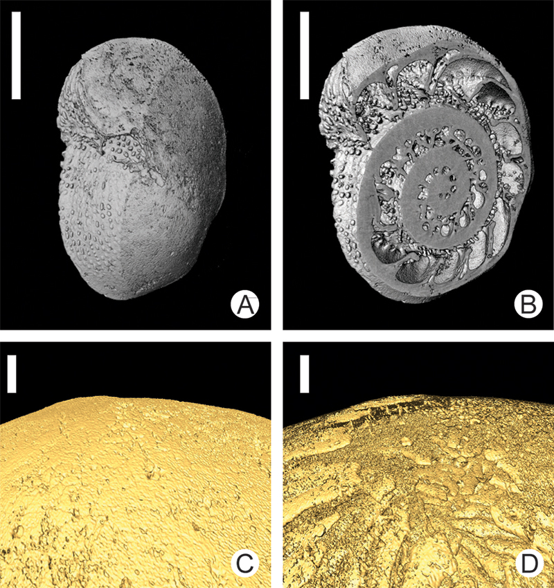
FIGURE 7. Three-dimensional visualization of a shell of the recent Foraminifera Amphistegina sp. illustrating the potential of micro-CT in investigations of recent marine shelled organisms. (A) A surface view of the whole shell. (B) A transversal section through the whole shell (C, D) Details of the shells´s surface.