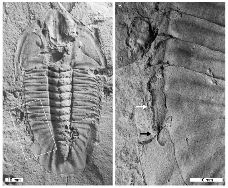
FIGURE 1. Gabriellus kierorum specimens from the (?)Rosella Formation. A, B. Gabriellus kierorum with truncation of left pleural spines on the fifth and sixth thoracic segment (TMP.1983.021.0034). A: Complete specimen. B: Close up of abnormality (white arrows). C-E: Gabriellus kierorum with bilaterally expressed thoracic abnormalities (TMP.1983.021.0039). C: Complete specimen. D: Close up of abnormality on the left side of the specimen (white arrows) and the partly recovered spine (black arrow). E: Close up of abnormality on the right side of the specimen (white arrows).
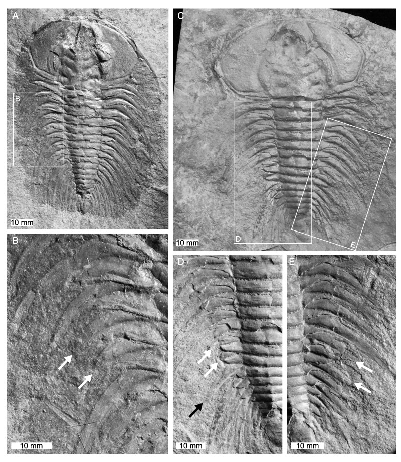
FIGURE 2. Ogygopsis klotzi specimens from the Stephen Formation with thoracic abnormalities. A, C: Ogygopsis klotzi showing truncation of the first four pleural spines of the right thoracic pleural lobe (TMP.1983.142.0002). A: Complete specimen. C: Close up of truncation of the right pleural spines. Note the ‘L’-shaped injury formed by 1st-3rd thoracic segments (white arrows), and the more notable truncation of the 4th segment (black arrow). B, D: Ogygopsis klotzi with an abnormality on the right pleural lobe that impacts the 4th-7th pleural spines (TMP. 1983.257.0001). B: Complete specimen. D: Close up of abnormality on the right pleural spines, showing pinching of the 5th and 6th segments (white arrows) and warping of the 4th and 7th segments (black arrows).
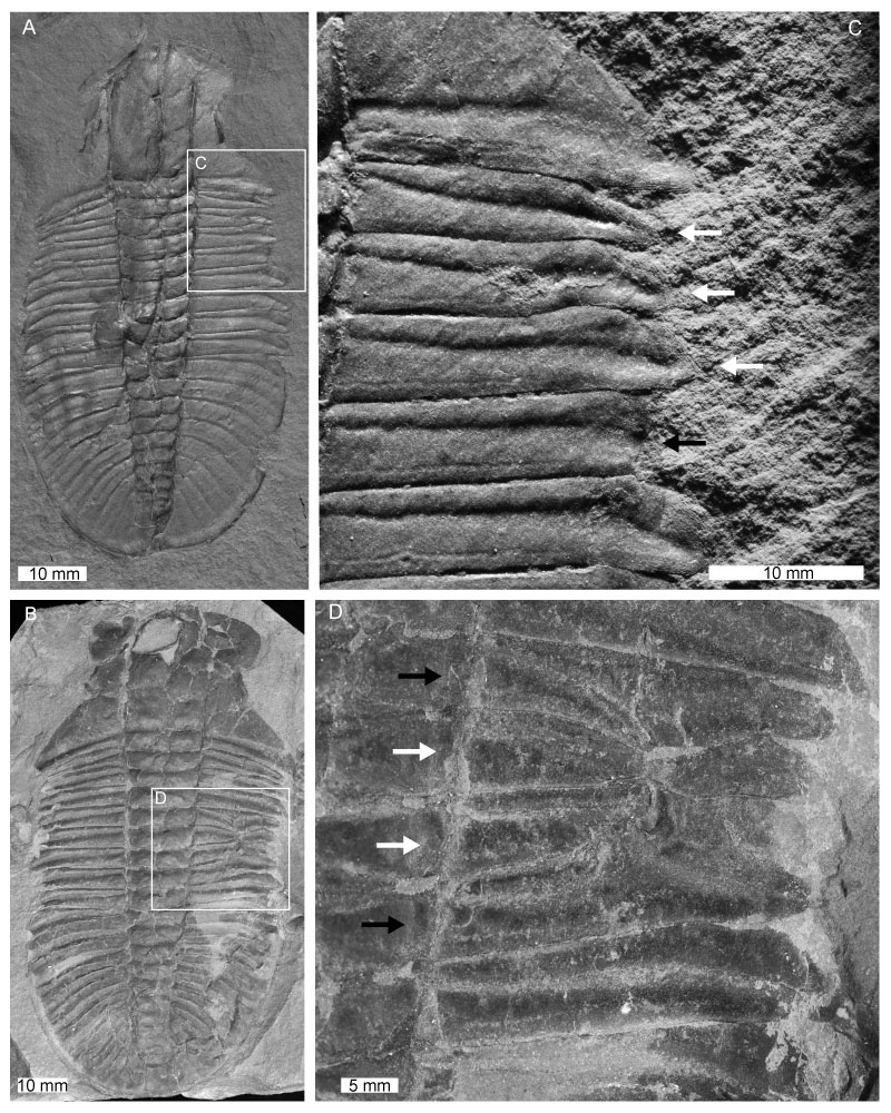
FIGURE 3. Modocia typicalis specimen with an abnormality on the right 11th thoracic segment (TMP1984.50.0004). A: Complete specimen. B: Close up of the truncated pleural spine on the right 11th thoracic segment (white arrow).
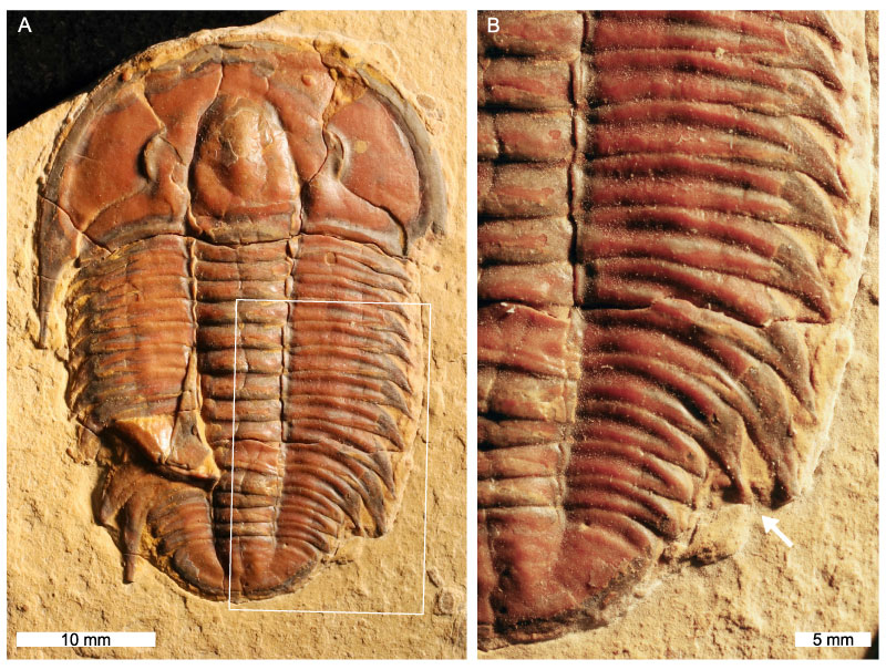
FIGURE 4. The first record of a Hemirhodon amplipyge specimen with an abnormality (TMP.2006.036.0102). A: Complete specimen. B: Close up of the abnormality on left side of pygidium, showing a truncated ‘W’- (white arrows) and ‘V’-shape (black arrow) on pygidial margin.